CARTILAGE (GENERAL HISTOLOGY)
4.1
Elastic cartilage (ear)
Specimen:
Specimen Details:
Organ: Ear
Origin: Human
Staining: Elastin-Resorcin- Carmine
Method and Specimen Description:
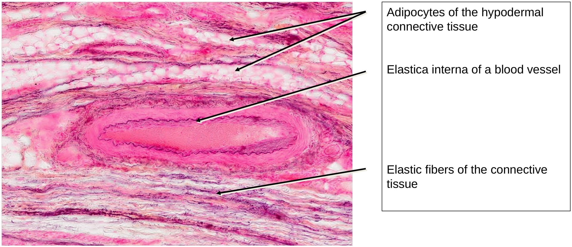
The specimen was stained using resorcin–fuchsin, which colors elastic fibers violet. Carmine was used as a counterstain, coloring nuclei bright red, while the cytoplasm and collagen fibers appear pale red.
Objective of the Examination:
To study the microscopic structure of elastic cartilage, and to identify the arrangement and course of elastic fibers within the cartilage matrix.
Specifics of the Specimen:
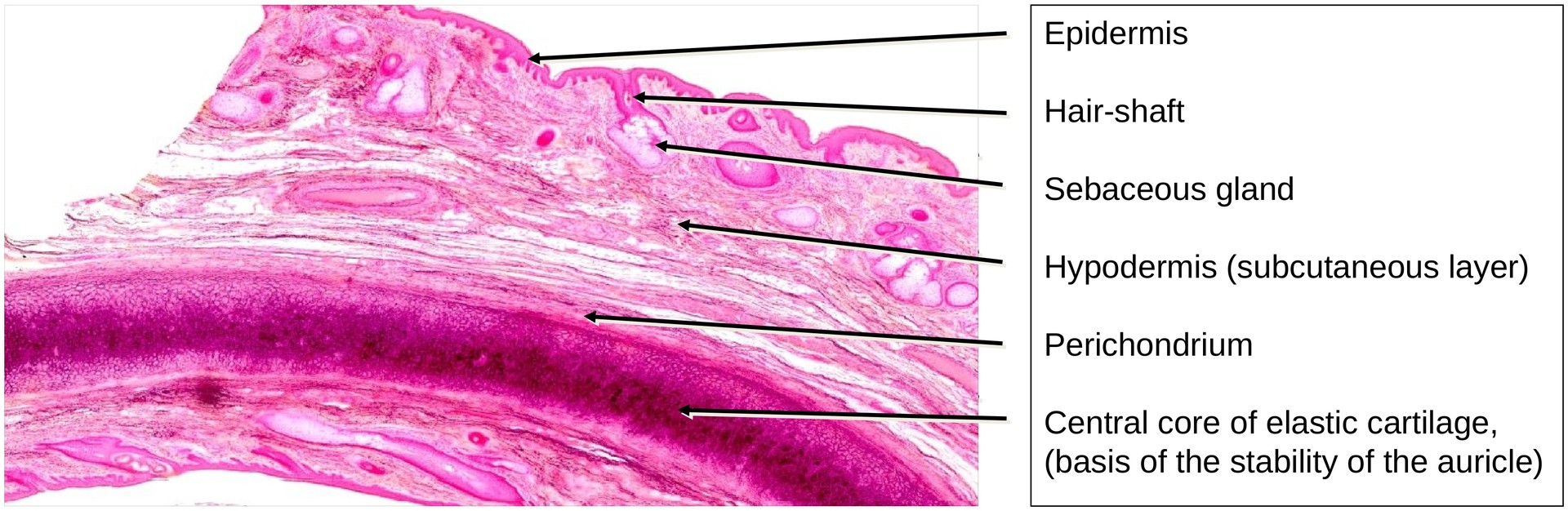
The auricle (pinna) of the external ear is a skin fold whose shape and flexibility are determined by a core of elastic cartilage. The focus of this specimen is the cartilage itself, though the overlying skin layers can also be examined (see Stratified squamous epithelium specimen for comparison).
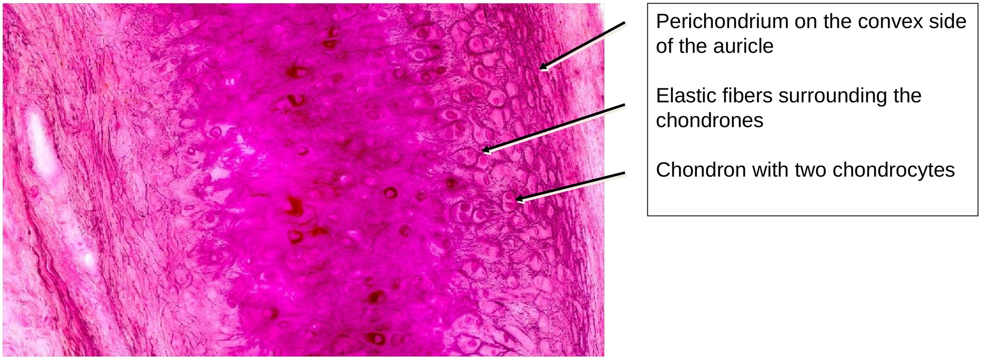
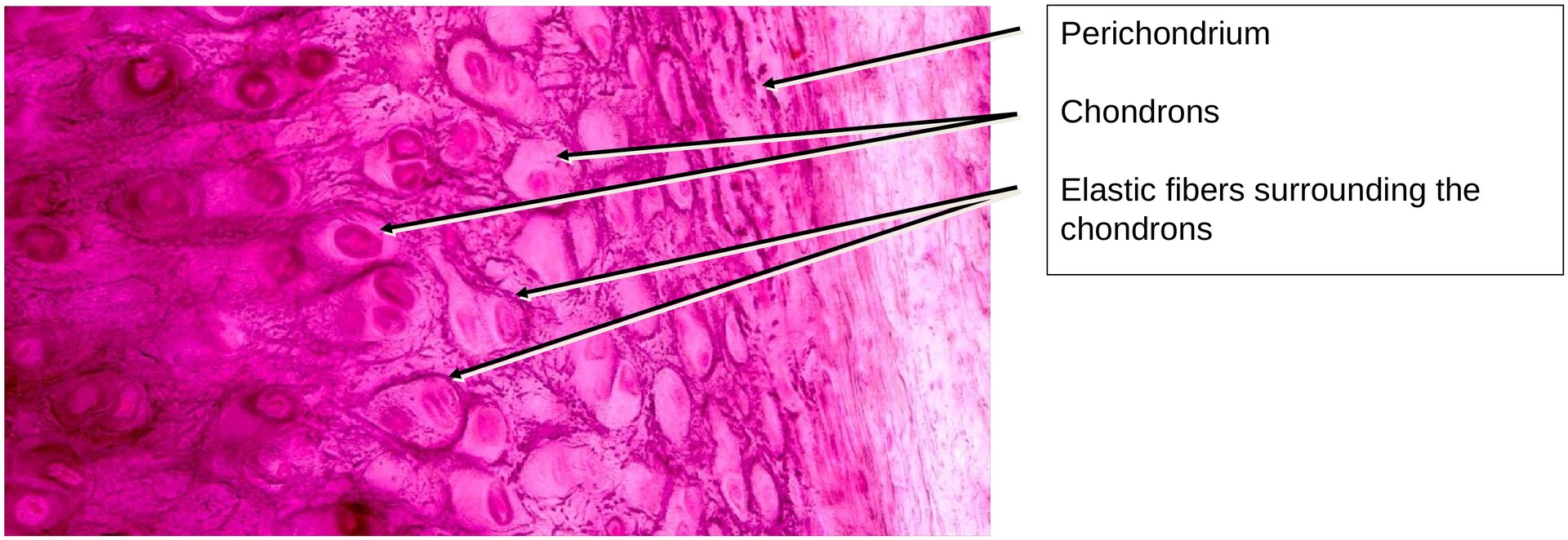
The elastic fibers in the cartilage matrix are clearly visible due to the specific stain used. Each chondron (cartilage cell group) generally contains fewer chondrocytes than those seen in hyaline cartilage.
At medium and high magnification, the course of the elastic fibers becomes apparent. The arrangement of fibers reflects the mechanical stress acting on the tissue:
-
On the convex side, where tensile stress predominates, fibers in the perichondrium and adjacent cartilage layers run tangentially.
-
From this layer, the fibers branch in an arc-like pattern and continue in a radial direction.
There is a clear correlation between the fiber orientation and the shape or long axis of the cartilage cells. The collagen fibers are masked by the ground substance, as in hyaline cartilage, whereas the elastic fibers remain visible due to their distinct refractive index and specific staining. Each chondron is surrounded by a fine network of elastic fibers.
Tasks:
• Examine the entire specimen at low magnification to determine the position and extent of the ear cartilage.
• Identify which side represents the inner and which the outer surface of the ear.
• Assess the size and distribution of chondrons.
• Observe the orientation and branching pattern of the elastic fibres within the matrix.
• Determine on which side of the cartilage the perichondrium is more developed.
License
University of Basel
Downloads