MUSCULATURE (GENERAL HISTOLOGY)
6.3
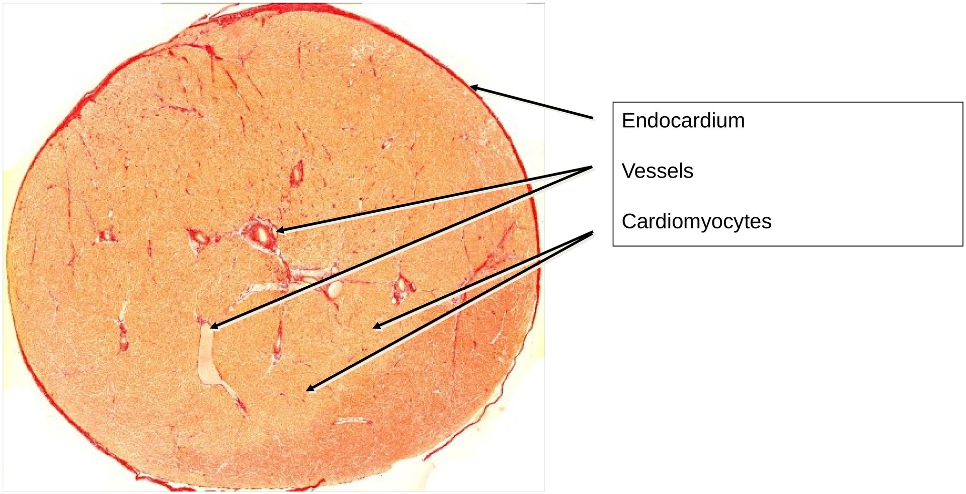
Cardiac muscle, transverse section (papillary muscle)
Specimen Details:
Specimen Details:
Organ: Cardiac muscle
Origin: Human
Staining: Van Gieson
Method and Specimen Description:
Standard histological section stained with Van Gieson, which stains muscle fibers yellow and connective tissue red.
Objective of the Examination:
To understand the characteristic features of cardiac muscle in transverse section and to distinguish it from smooth and skeletal muscle.
Special Features of the Specimen:
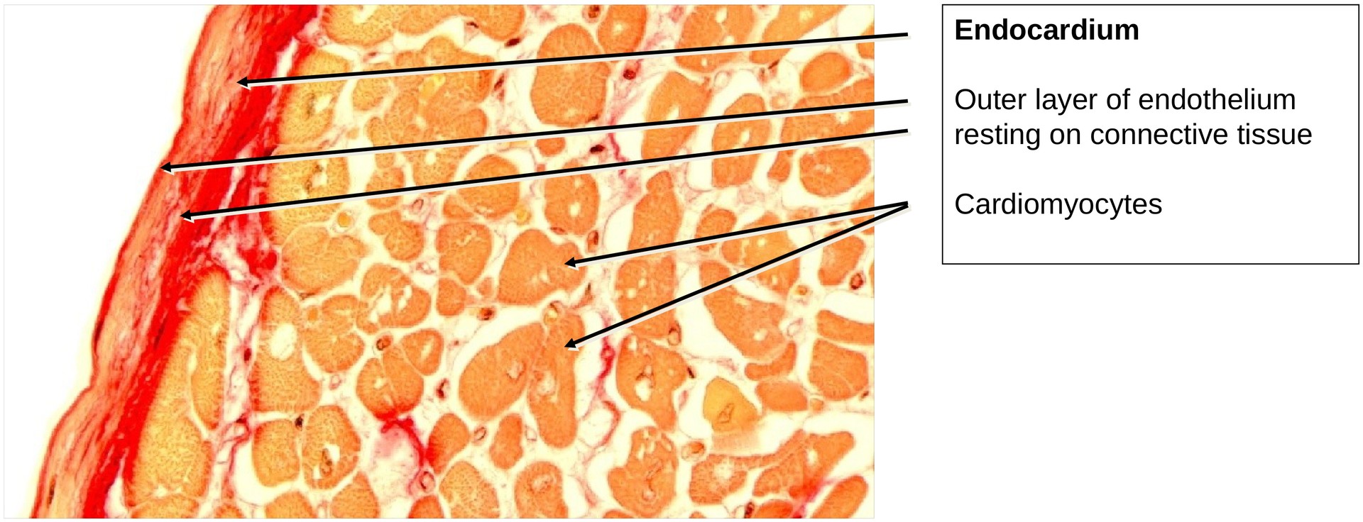
This specimen represents a complete transverse section of a papillary muscle, which can be identified by the fact that it is entirely surrounded by endocardium.
The endocardium consists of a very thin layer of endothelial cells resting upon a layer of connective tissue (stained red in this preparation). At some points, erythrocytes are visible on the outer surface of the endothelium, which in vivo is in direct contact with the blood.
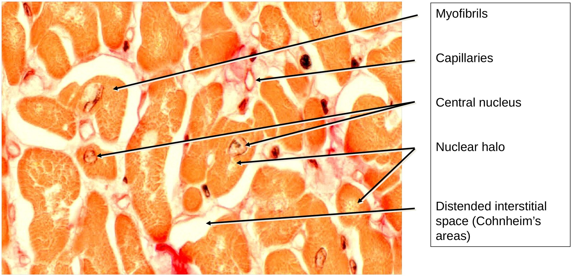
Cross-sections through cardiac muscle cells (high magnification):
Depending on the plane of section, several appearances may be observed:
-
Regions containing only myofibrils.
-
Regions containing a centrally located nucleus.
-
Regions showing a “nuclear halo”, where the nucleus lies above or below the section plane. The section thus passes through the perinuclear, organelle-rich zone, which appears relatively pale or empty under the microscope.
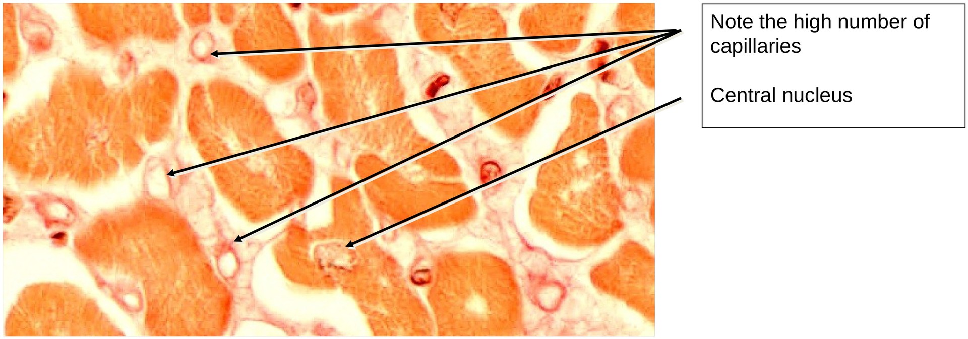
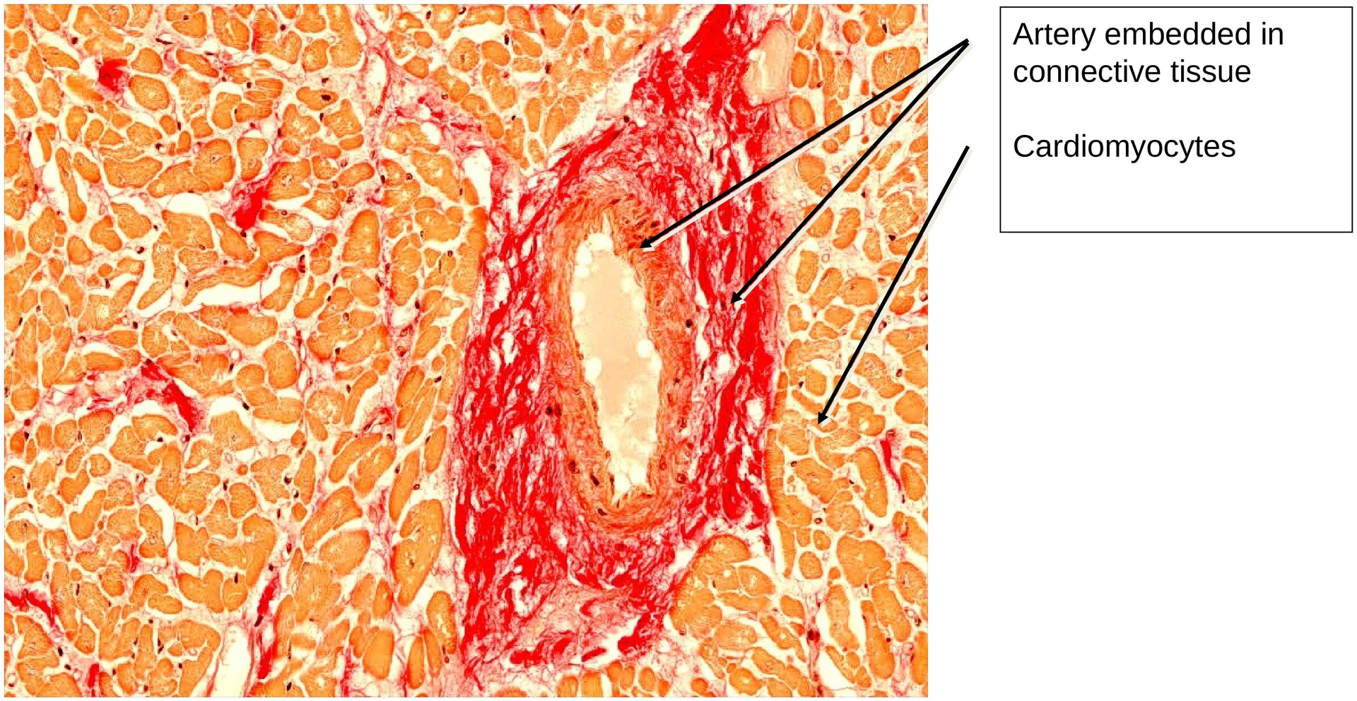
Vascular supply (medium and high magnification):
Smaller and larger blood vessels traverse the connective tissue between cardiac muscle cells, most of which are seen in cross-section. In close proximity to the cardiomyocytes, numerous capillary profiles can be identified.
These abundant capillaries reflect the high oxygen demand of cardiac muscle, which cannot incur an oxygen debt. Each capillary is lined by a single layer of endothelial cells, whose nuclei are clearly visible.
In this specimen, the interstitial spaces appear artificially widened, which is a preparation artefact. In vivo, the cardiomyocytes lie much closer together. Nevertheless, this artefact facilitates the identification of the capillaries and interstitial connective tissue.
Tasks:
• Identify the nuclei of the cardiomyocytes. Where are they located?
• Describe the shape of the cardiomyocytes in cross-section.
• At high magnification, examine the cytoplasm of the cardiomyocytes. Which structures can you identify?
• Trace the outer circumference of the specimen. Which structure encloses the myocardium externally?
• Locate capillaries and observe their relative frequency.
• Compare the diameter of the capillaries with that of the erythrocytes, which appear in some vascular profiles as pale, circular structures.
License
University of Basel
Downloads