CONNECTIVE TISSUE (GENERAL HISTOLOGY)
2.3
Fetal connective tissue (Umbilical Cord)
Specimen:
Specimen Details:
Organ: Umbilical Cord
Origin: Human
Staining: PAS (Periodic acid–Schiff)
Method and Specimen Description:
A standard histological section of the umbilical cord was prepared and stained using the PAS reaction (Periodic acid–Schiff). This staining method highlights glycosaminoglycans (GAGs) and proteoglycans, which are abundant in the gelatinous connective tissue known as Wharton’s jelly — the principal component of the umbilical cord’s stroma.
Objective of the Examination:
• To identify the microscopic structure of gelatinous connective tissue and the overall organisation of the umbilical cord.
• To understand that the amorphous ground substance of Wharton’s jelly has a high capacity for water binding, contributing to its gelatinous consistency and shock-absorbing properties.
Specimen Features:
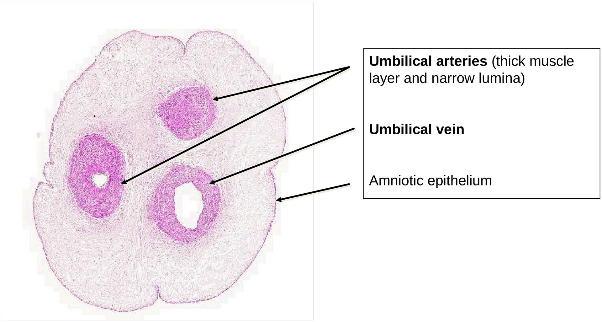
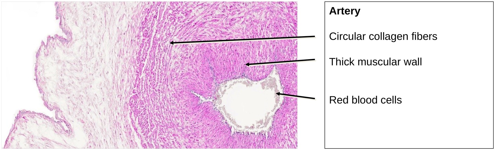
General Features (low magnification): In cross-section, the umbilical cord is surrounded externally by a layer of amniotic epithelium. Within the stroma lie three large blood vessels: two umbilical arteries and one umbilical vein. The arterial lumina appear narrow because of the prominent tunica media, which tends to contract during fixation. The intervening tissue, Wharton’s jelly, is avascular and forms the main bulk of the cord.
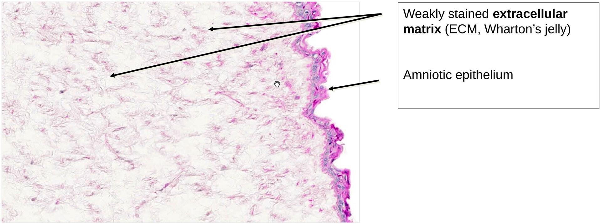
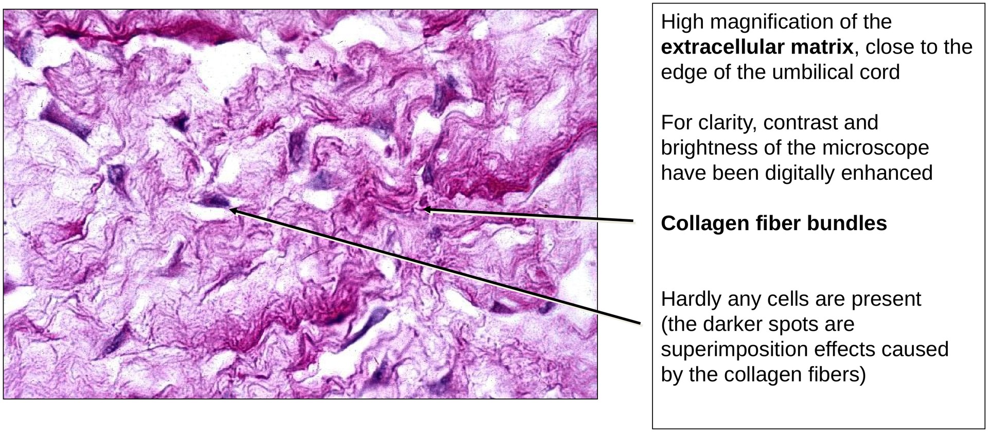
Structure of Wharton’s Jelly (high magnification): The connective tissue cells (primitive fibroblasts or mesenchymal cells) form a three-dimensional network. Their processes frequently make contact with one another, creating a reticular meshwork visible in some section planes. The extracellular matrix (ECM) contains fine collagen fibers and PAS-positive amorphous material, representing proteoglycans and glycosaminoglycans (notably hyaluronic acid). This hydrated ECM confers the gel-like consistency and mechanical resilience characteristic of fetal connective tissue.
Orientation of Connective Tissue Structures (medium to high magnification): The gelatinous connective tissue exhibits regional variation in fiber orientation, reflecting adaptation to mechanical stress. Around the blood vessels and directly beneath the amniotic surface, fibers are arranged concentrically. In other regions, fibers are randomly oriented without any preferred direction, providing flexible support throughout the cord.
Amniotic Epithelium (high magnification): The amniotic epithelium forms the outer covering of the umbilical cord. It is composed of a single layer of cuboidal epithelial cells with distinct cell domes. These cells are strongly PAS-positive due to their glycogen-rich cytoplasm, which reflects their metabolic activity and secretory functions.
Tasks:
-
Examine the amniotic epithelium covering the outer surface of the umbilical cord and note the cuboidal cell morphology and PAS-positive cytoplasm.
-
Identify the three large vessels: distinguish between the single umbilical vein (with a wider lumen and thinner wall) and the two umbilical arteries (with narrower lumina and thicker tunica media).
-
At high magnification, study the interstitium of Wharton’s jelly.
-
Describe the structure of the amorphous ground substance and the distribution of fine collagen fibers.
-
Observe that, even under low mechanical stress, the fibers near the vessel walls are concentrically arranged, optimally resisting pulsatile forces.
-
License
University of Basel
Downloads