EPITHELIUM (GENERAL HISTOLOGY)
1.1
Single-layered epithelium (kidney)
Specimen:
Specimen Details:
Organ: Kidney
Source: Rabbit
Staining: Hematoxylin-Eosin (H&E)
Method and Specimen Description:
This is a sectioned specimen with standard overview staining. The rabbit kidney is unipapillary, meaning it has a single medullary pyramid. The section was taken from the area of the medullary pyramid (papilla).
Objective of the Examination:
To identify different types of simple epithelia—squamous, cuboidal, and columnar—and to understand their structural characteristics.
Specific Features of the Specimen:
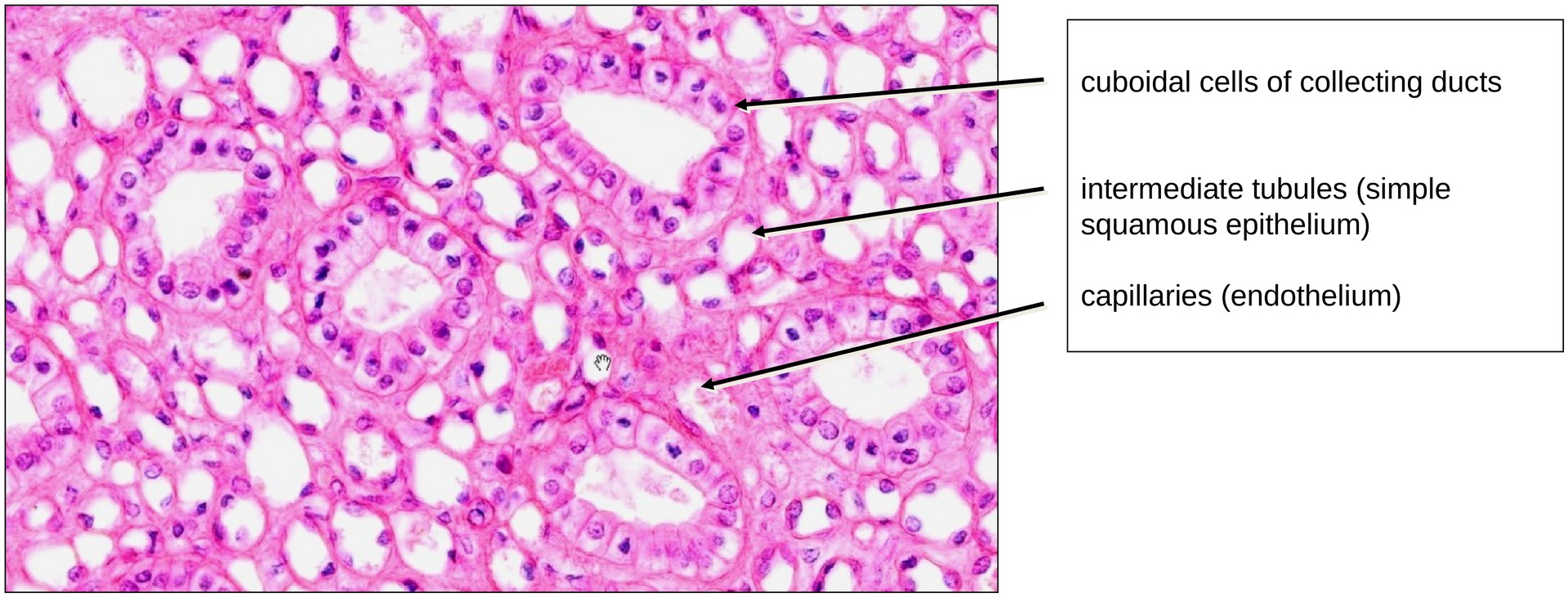
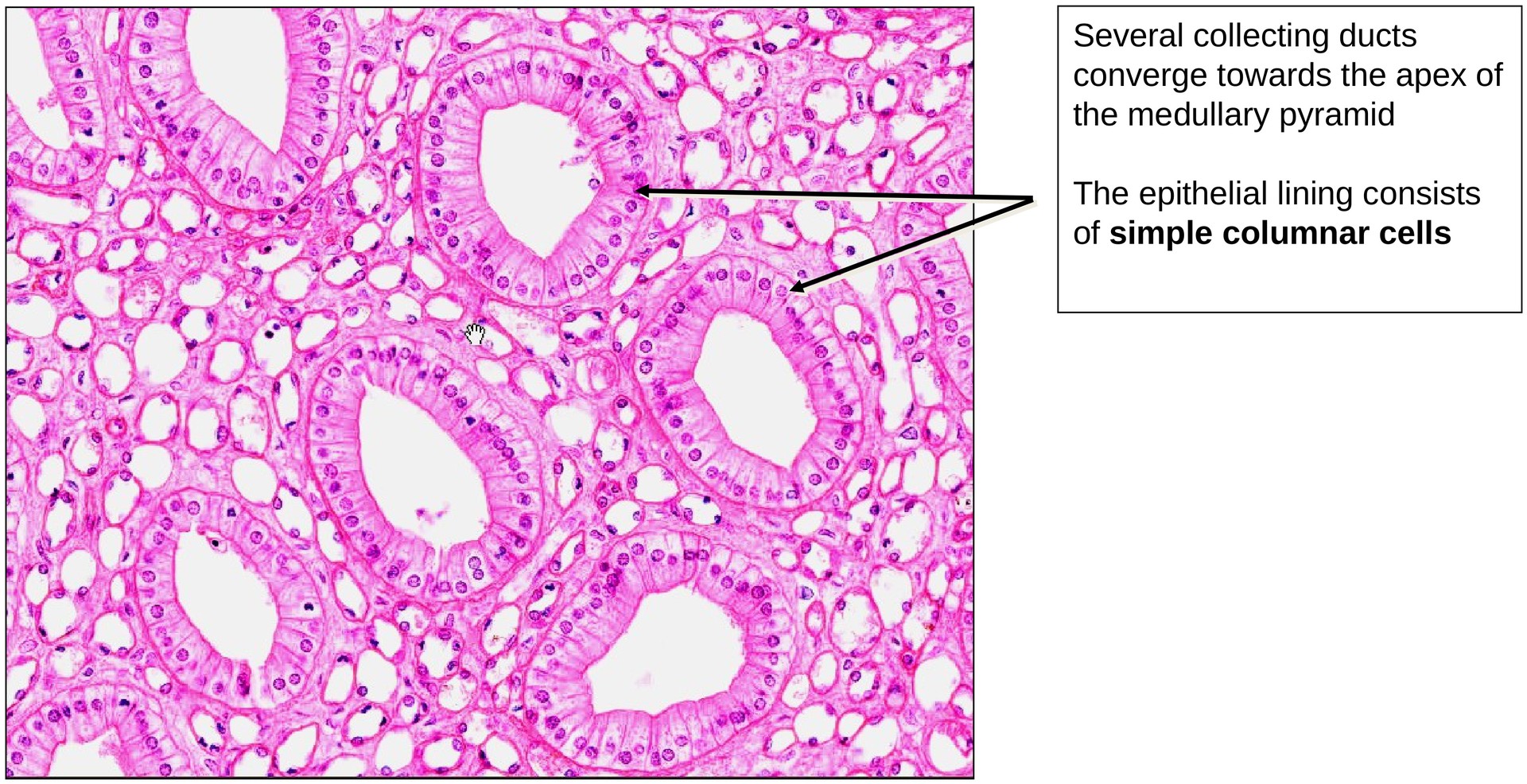
In the rabbit kidney, which is unipapillary, a single medullary pyramid projects as a papilla into the renal pelvis. The papilla is traversed longitudinally by collecting ducts, intermediate tubules, and blood capillaries. Because the section is taken across the medullary pyramid, these tubular systems generally appear in cross-section. The purpose of this specimen is not to analyze individual tubules in detail, but to recognize the different epithelial types present. You can observe:
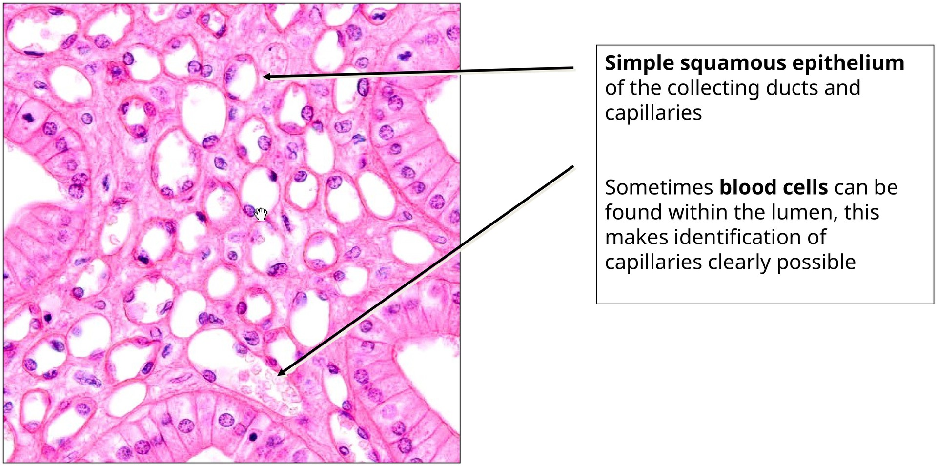
- Simple squamous epithelium — in the intermediate tubules and blood capillaries.
- Simple cuboidal to low columnar epithelium — in the collecting ducts and papillary ducts.
Some of the larger cross-sections (collecting and papillary ducts) may contain detached epithelial cells, which were displaced during tissue sampling, fixation, or processing.
Epithelial Characteristics:
- Simple cuboidal and columnar (prismatic) epithelium (medium to high magnification): Found in the tubules and collecting ducts, which appear in cross or longitudinal section. The epithelial cells rest on a distinct basement membrane.
- Simple squamous epithelium: Forms the lining of smaller-caliber tubules (intermediate tubules) and blood capillaries, located between the collecting and papillary ducts.
- In blood vessels, this lining is termed endothelium.
- Capillaries can be distinguished from tubules when red blood cells (RBCs) are visible in the lumen.
Tasks:
- Identify large cross-sections of the collecting and papillary ducts, and distinguish between cuboidal and columnar epithelial forms.
- Compare these epithelia with the squamous epithelium found in the intermediate tubules and the endothelium of the capillaries.
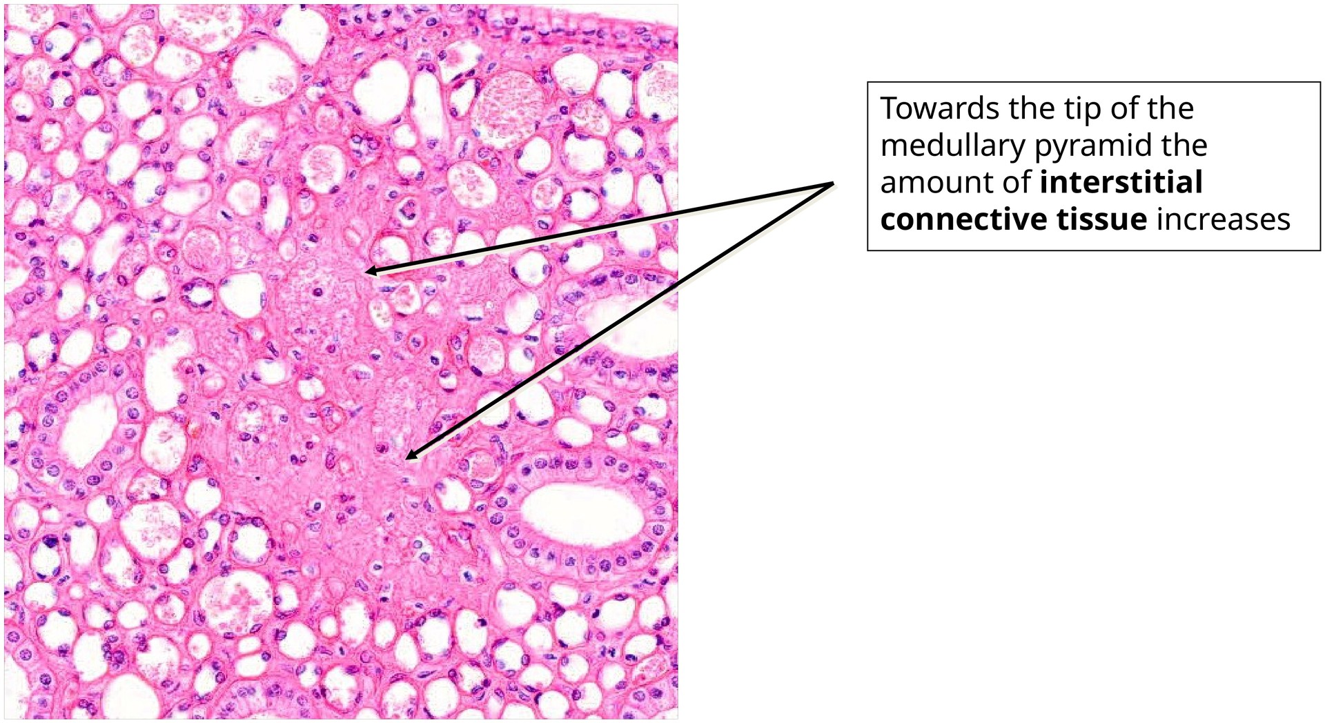
- Locate one or two regions containing connective tissue. Although connective tissue is generally sparse in the kidney, it can be found in certain areas of the medullary zone in this specimen.
License
University of Basel
Downloads