NERVOUS SYSTEM (ANATOMICAL MICROSCOPY)
16.8
Spinal cord, thoracic
Preparation:
PREPARATION DETAILS:
Organ: Spinal cord, thoracic region
Origin: Human
Staining: Luxol Fast Blue/Cresyl Violet
METHOD AND SPECIMEN DESCRIPTION:
Luxol Fast Blue stains myelinated axons blue, allowing clear visualization of myelin and phospholipid-rich structures. Cresyl Violet binds to nucleic acids (RNA and DNA), staining the chromatin, nucleolus, and rough endoplasmic reticulum (Nissl substance) blue-violet.
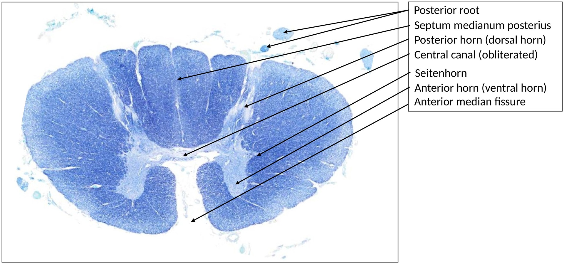
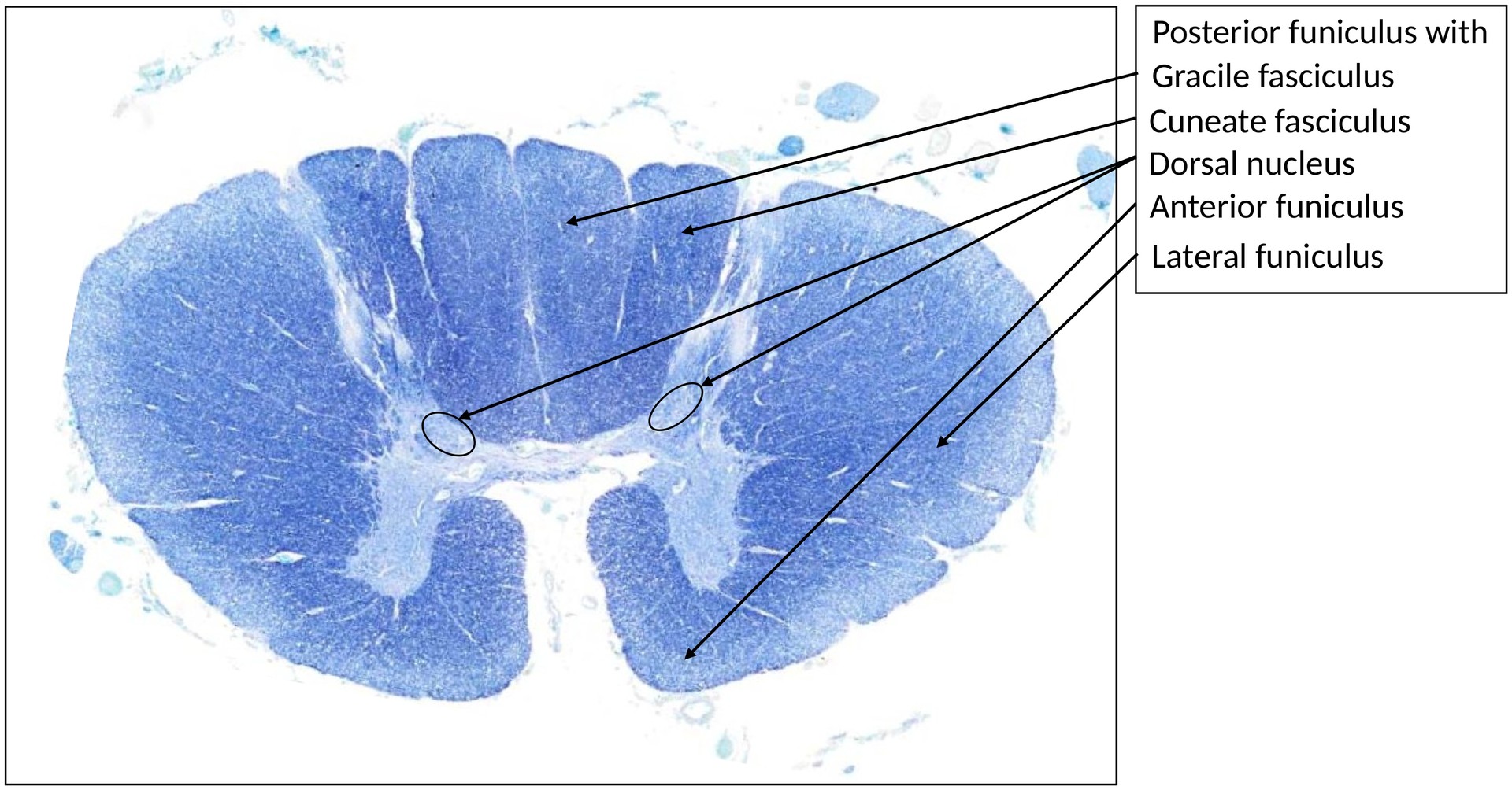
The spinal cord, like the vertebral column, is bilaterally symmetrical, consisting of two halves separated ventrally by the anterior median fissure. In contrast to the cerebrum and cerebellum, the spinal cord exhibits white matter externally and grey matter internally, forming a butterfly-shaped configuration. Motoneurons are located in the anterior (ventral) horn.
OBJECTIVE OF THE EXAMINATION:
To understand the organization of white and grey matter and identify motoneurons in the anterior horn of the thoracic spinal cord.
SPECIAL FEATURES OF THE PREPARATION:
The basic structure corresponds to that of the cervical spinal cord, with:
- Substantia gelatinosa,
- Nucleus proprius, and
- Motoneuron groups in the anterior horn.
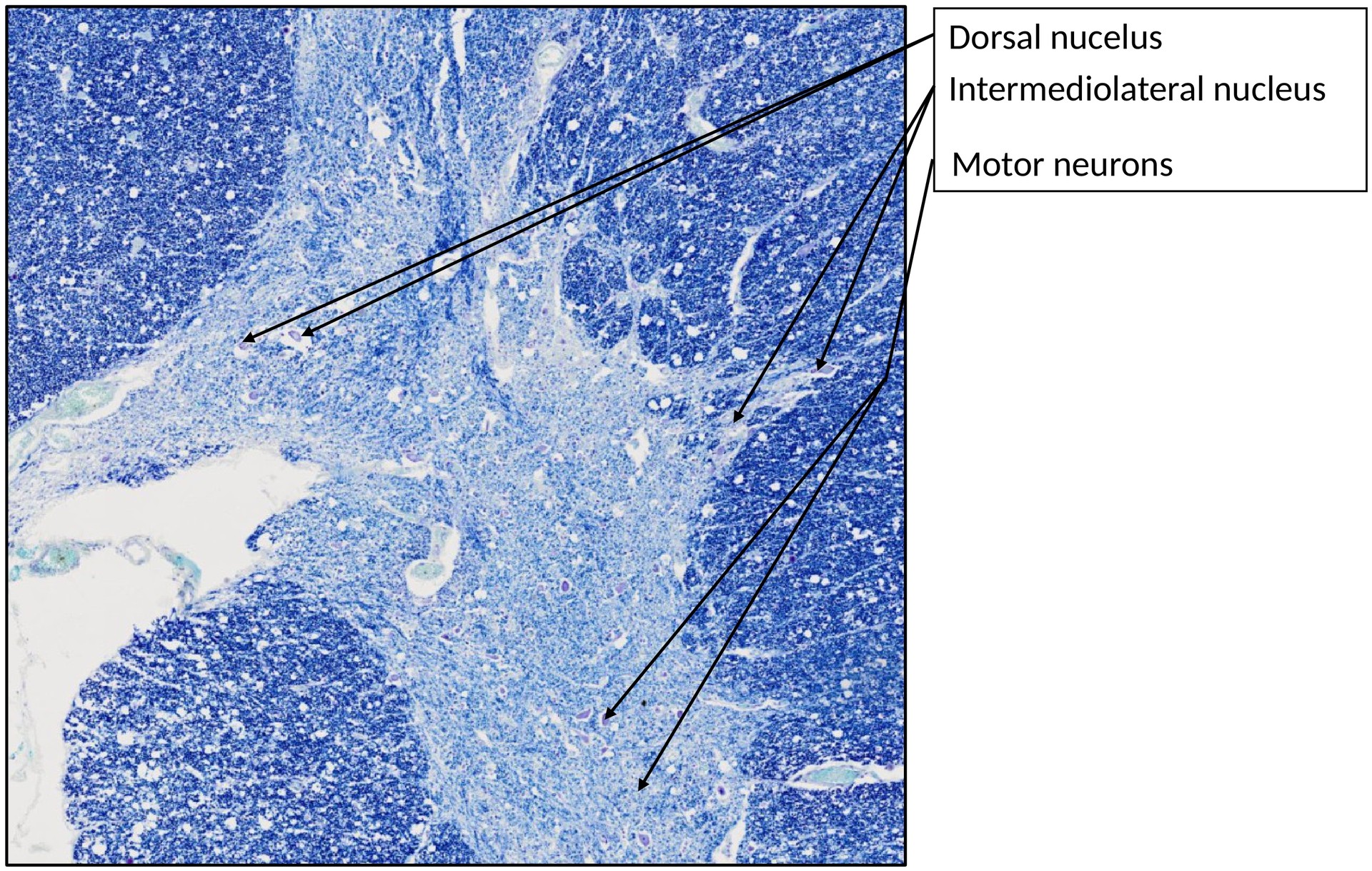
However, the thoracic spinal cord has two additional and characteristic nuclei:
1. Nucleus intermediolateralis (intermediolateral column): - Located in the lateral horn of the grey matter (present only in thoracic and upper lumbar segments). - Contains violet-stained preganglionic sympathetic neurons. 2. Nucleus thoracicus posterior (Clarke’s nucleus): - Situated at the base of the posterior horn (dorsal column). - Comprises larger neurons involved in proprioceptive signal transmission to the cerebellum via the dorsal spinocerebellar tract.
TASKS:
- Identify the following structures:
- Grey matter
- White matter
- Central canal
- Anterior horn
- Posterior horn
- In a section of the anterior horn, identify motoneurons and their components:
- Nucleus
- Dendrites
- Axon
- Glial cells
- Question: Where do the neurons of the posterior thoracic nucleus (Clarke’s nucleus) project to?
License
University of Basel