LYMPHATIC ORGANS (ANATOMICAL MICROSCOPY)
15.1
Ileum (Peyer's Patches)
Specimen Details:
Specimen Details:
Organ: Ileum
Origin: Rabbit
Staining: Haematoxylin - Eosin (H&E)
Method and Specimen Description:
Standard histological section stained with H&E, providing an overview of the small intestinal wall and associated lymphatic structures.
Objective of the Examination:
To study Peyer’s patches, the characteristic lymphoid aggregates of the ileum, including their organization, the dome area, and the high endothelial venules (HEV) responsible for lymphocyte migration.
Specific Features of the Specimen:
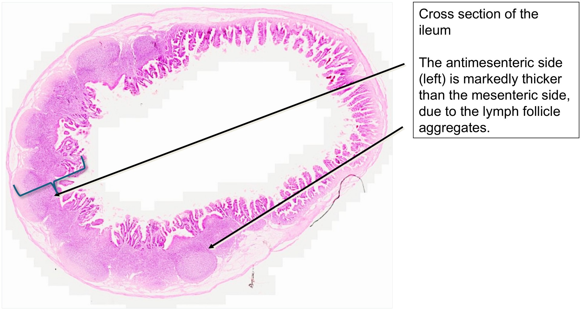
Even at low magnification, an uneven wall thickness of the ileum is visible. The antimesenteric side is markedly thicker than the mesenteric side. This thickening is due to the presence of aggregated lymphoid follicles (Peyer’s patches).
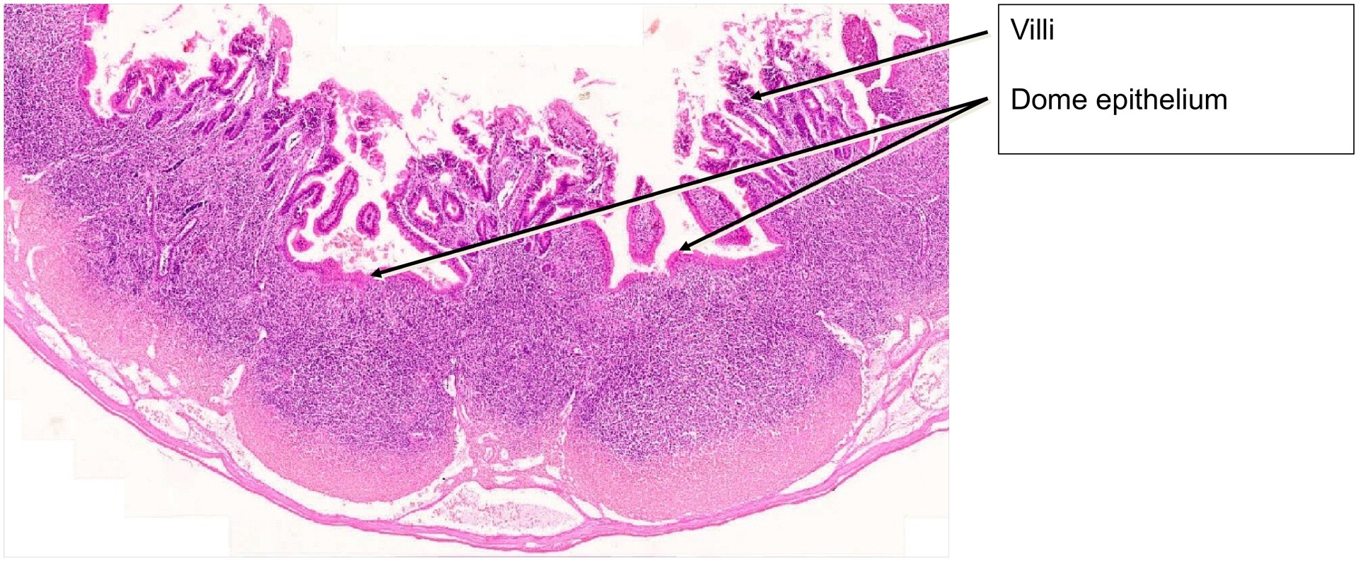
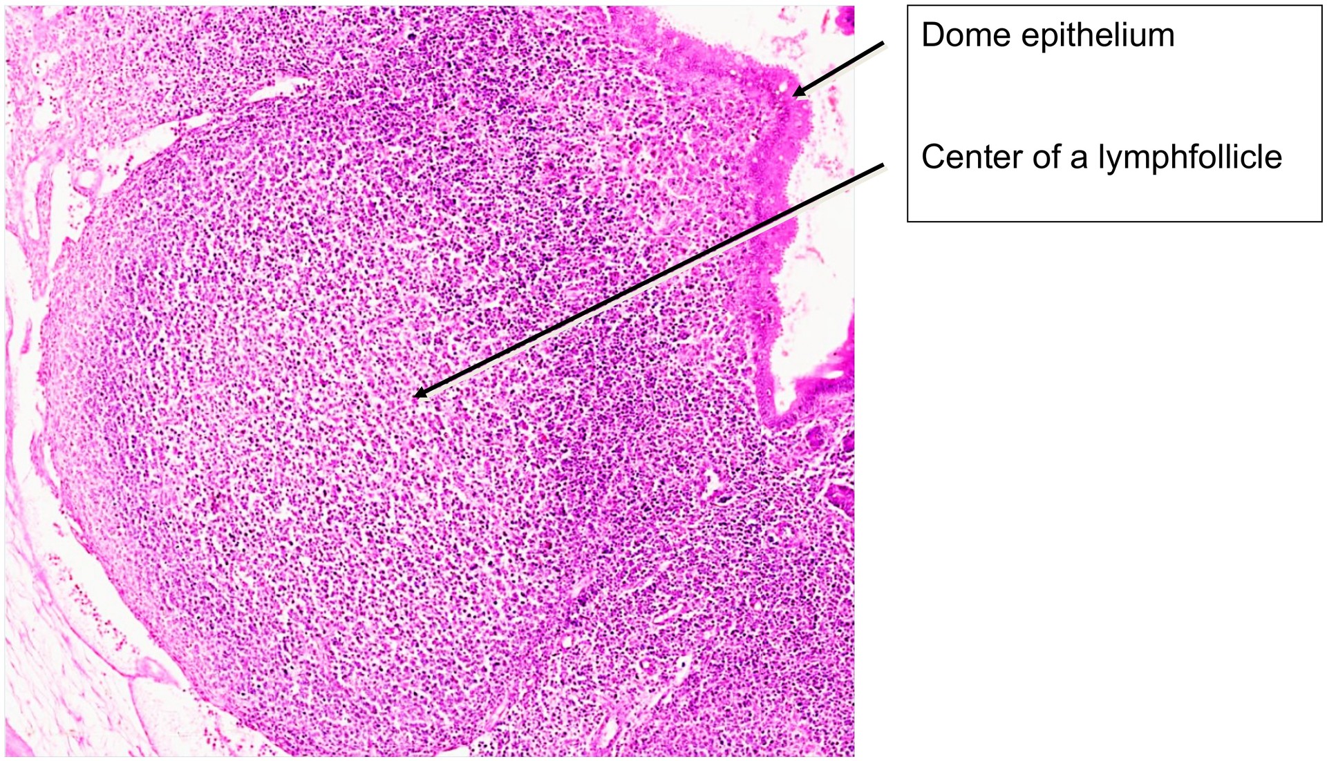
These lymphoid follicles originate primarily within the lamina propria of the mucosa (tunica mucosa) but can extend through the muscularis mucosae into the submucosa. Each follicle is covered towards the lumen by follicle-associated epithelium, forming the dome area.
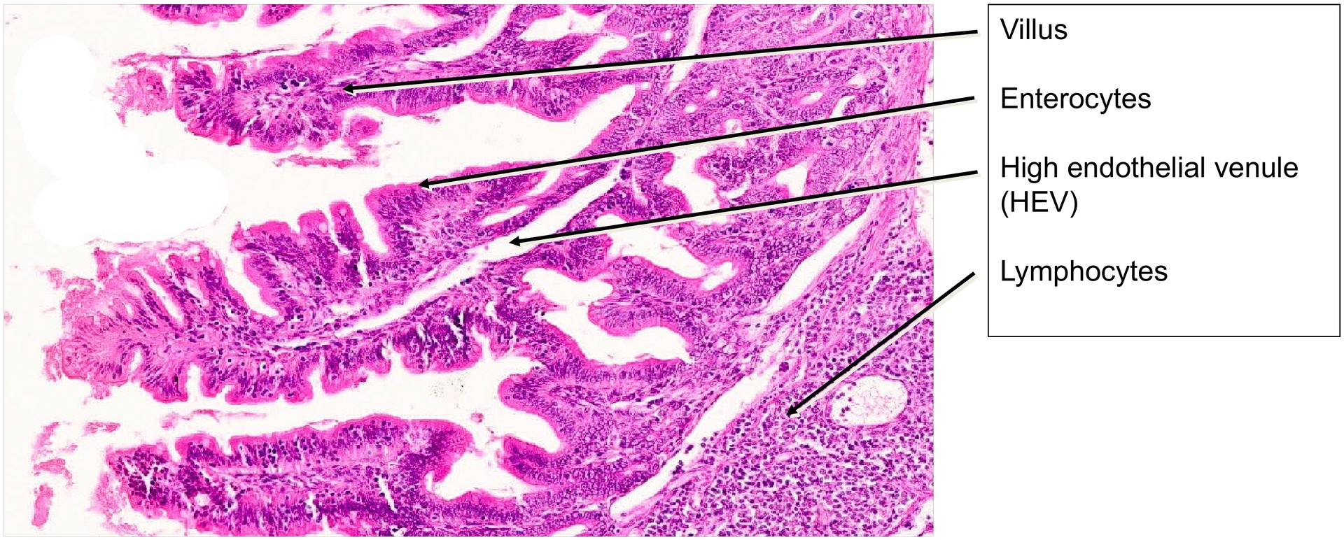
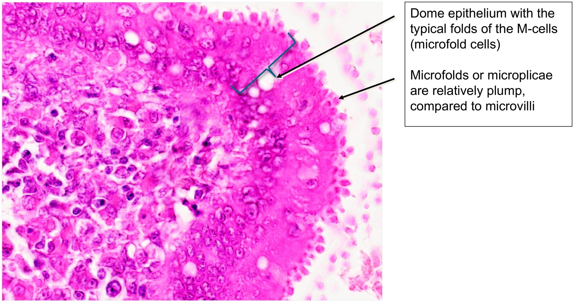
The dome epithelium comprises normal enterocytes and specialized M cells (microfold cells). These M cells possess small surface folds (microplicae) rather than typical microvilli, and are essential for antigen transport from the intestinal lumen to the underlying immunocompetent cells. This gives the dome region its distinctive, uneven appearance.
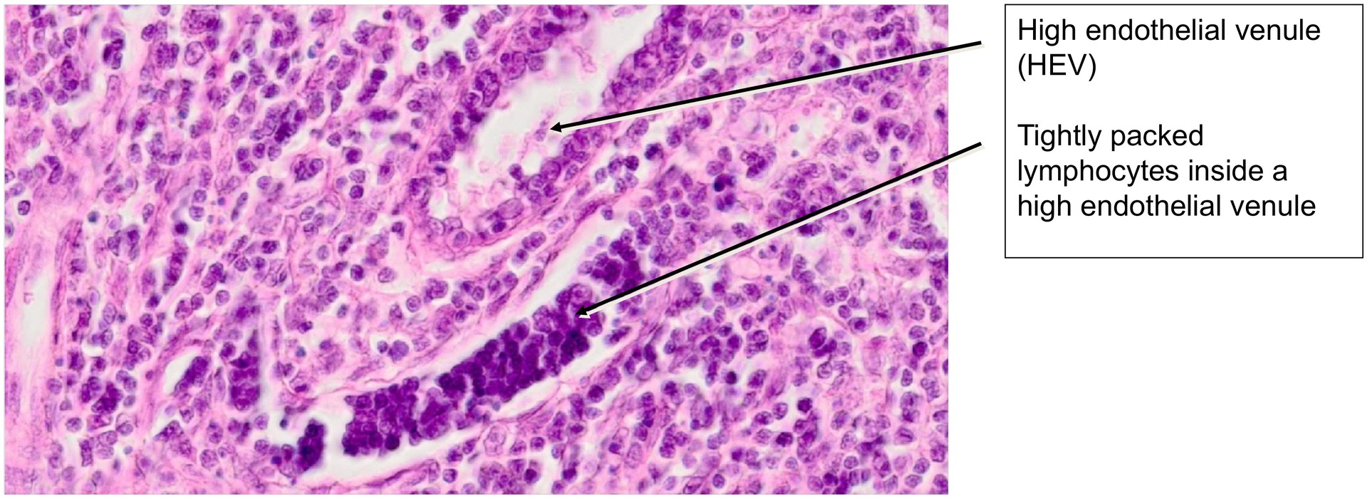
A characteristic feature of lymphoid tissue in the ileum is the presence of high endothelial venules (HEV)—venules lined by cuboidal or prismatic endothelium. These vessels often contain migrating lymphocytes and facilitate their diapedesis into the surrounding lymphoid tissue. HEVs must not be mistaken for crypts of Lieberkühn, which may appear in similar regions depending on the sectioning plane.
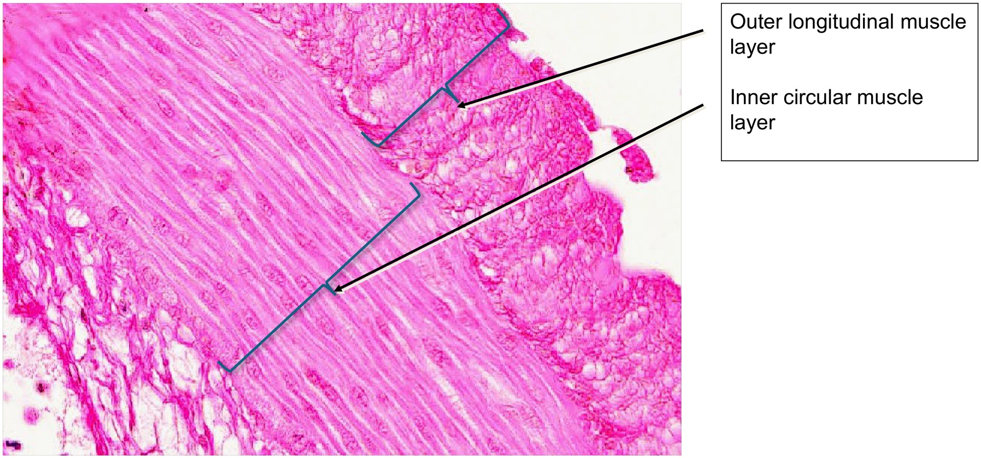
Apart from these immunological specializations, the ileum wall displays the general structure typical of the small intestine, although in this specimen the tunica muscularis (both circular and longitudinal muscle layers) is comparatively thin, especially when compared with other intestinal segments.
Tasks:
- At low magnification, determine which side of the specimen represents the mesenteric and which the antimesenteric aspect.
- Compare the two sides structurally. On which side are the Peyer’s patch aggregates located?
- Observe the relatively low height of the villi and folds in the ileum compared with other intestinal regions.
- Identify the dome epithelium and, under higher magnification, locate the M cells with their characteristic microfolds.
- Search for high endothelial venules (HEV) and note the lymphocytes accumulated within their lumen.
License
University of Basel
Downloads