MALE REPRODUCTIVE ORGANS (ANATOMICAL MICROSCOPY)
11.11
Spermatic cord
Specimen:
Specimen Details:
Organ: Spermatic cord
Origin: Human
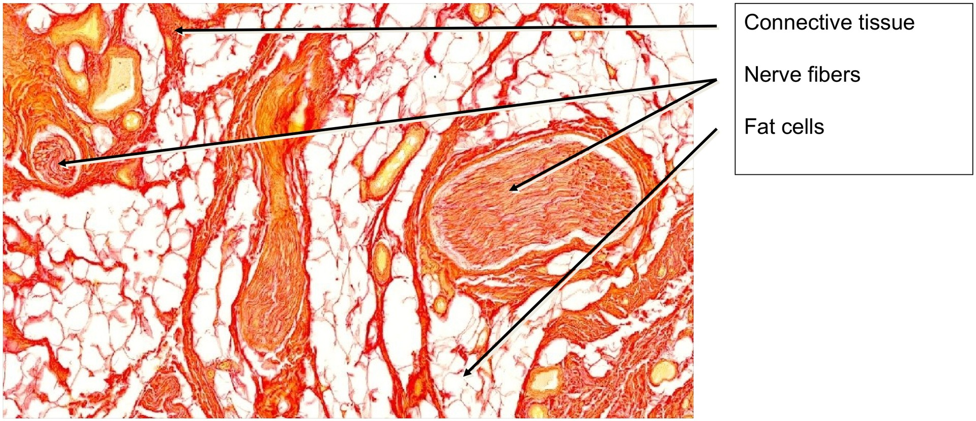
Staining: Van Gieson
Method and Specimen Description:
Normal histological section stained with Van Gieson, which colors muscle cells and fibers yellow, while epithelium and connective tissue appear red.
Objective of the Examination:
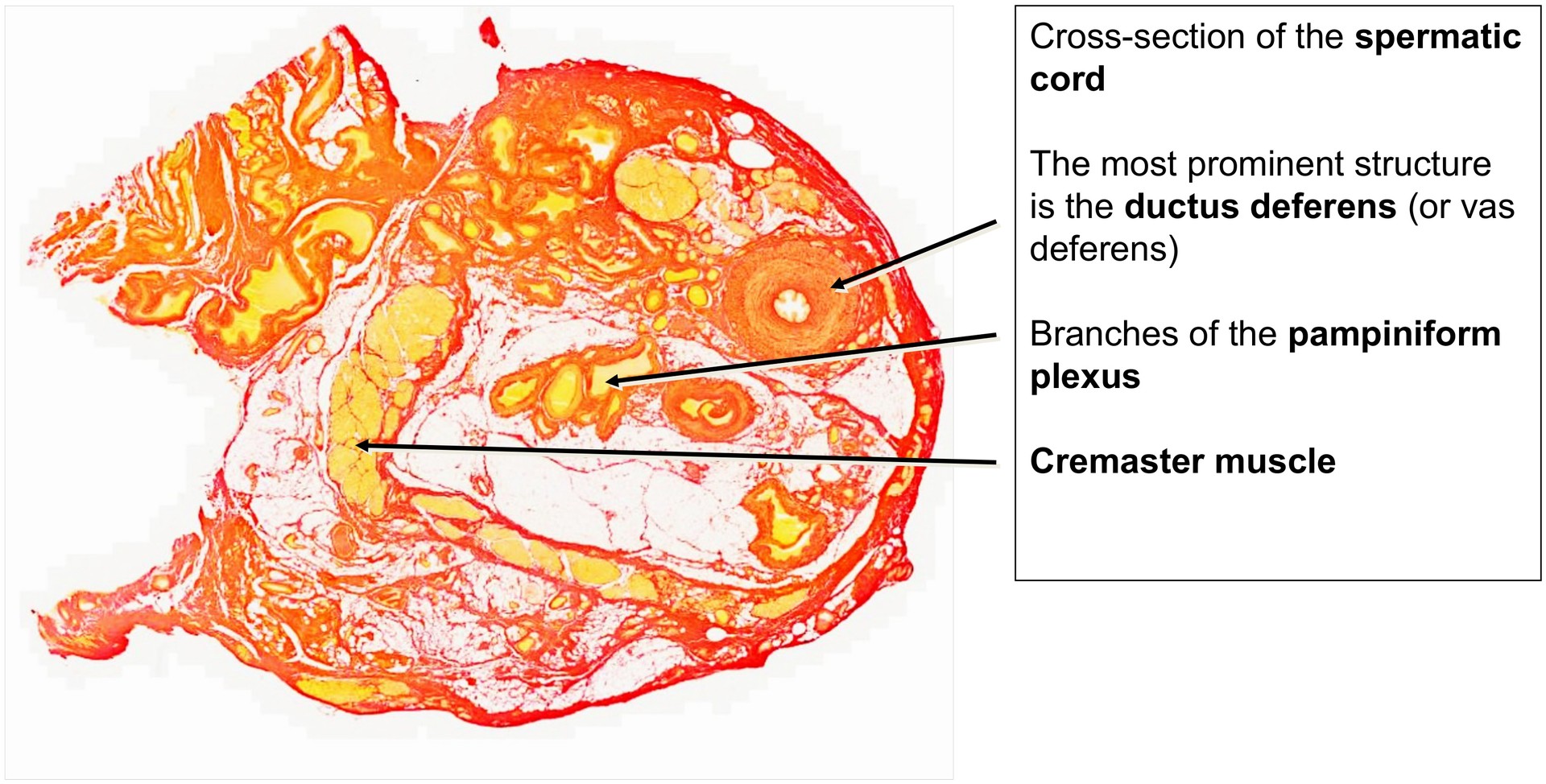
To study the ductus deferens and its three-layered musculature, the distinctive wall structure of the veins of the pampiniform plexus, and to identify muscles and nerves within the spermatic cord.
Special Features of the Specimen:
The spermatic cord contains several clearly recognizable structures, including the ductus deferens (vas deferens), and branches of the artery to the ductus deferens, the cremasteric artery, and the testicular artery. These arterial branches are present in the specimen, but cannot all be specifically identified.
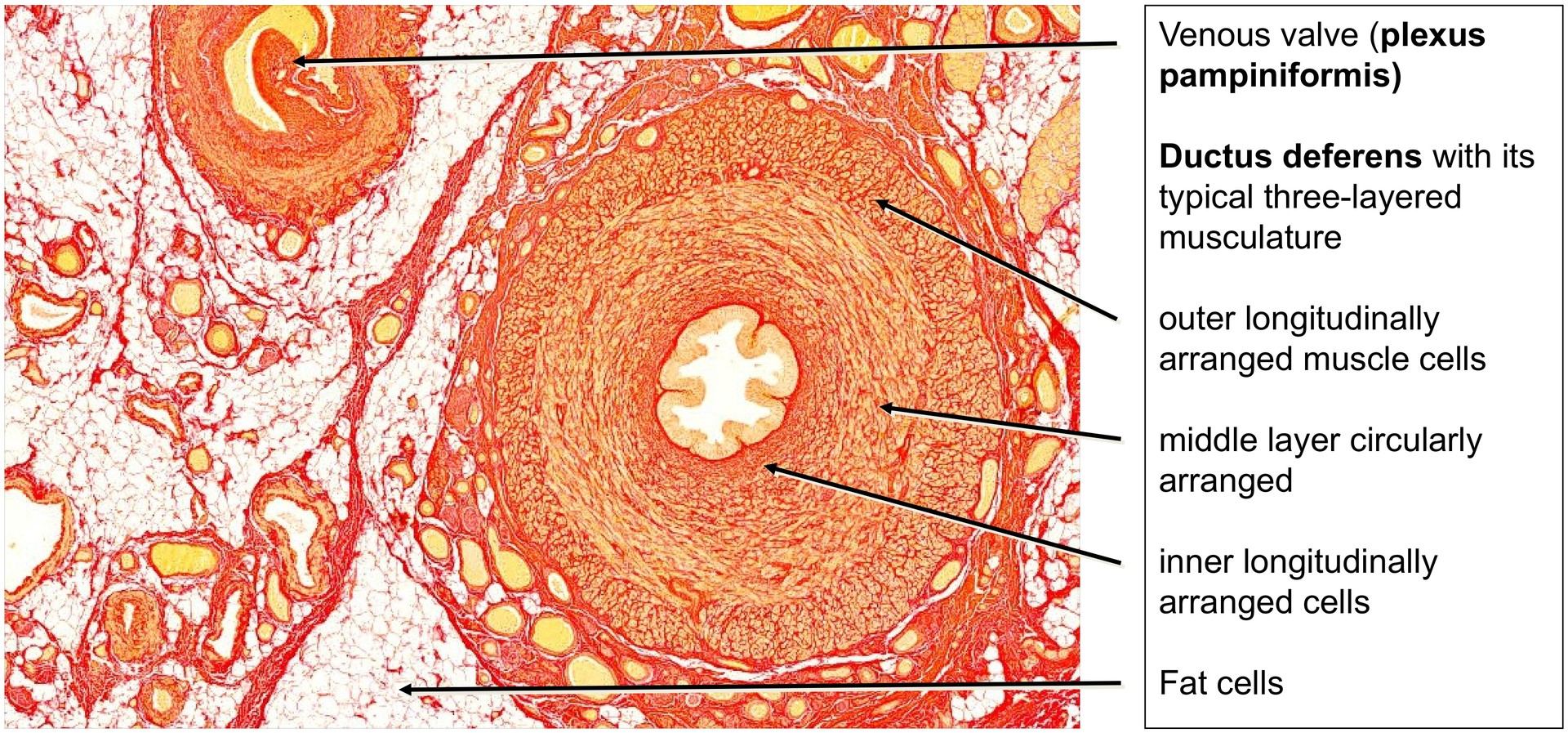
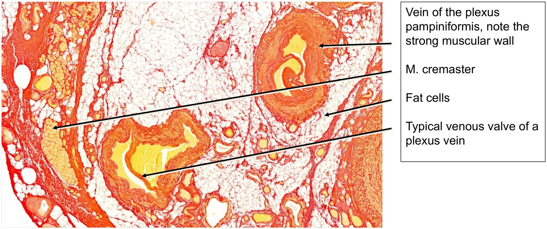
The veins of the pampiniform plexus are notably muscular, in some cases containing more smooth muscle within their walls than the arteries of the same region. They can be identified as veins by the presence of venous valves, which are absent in arteries.
The cremaster muscle fibers are strongly highlighted by the yellow Van Gieson stain and are visible even at low magnification. In addition, nerve fibers, including branches of the genital branch of the genitofemoral nerve, are present and contribute to innervation of the cremaster muscle. Adipose tissue is interspersed among the various structures of the spermatic cord.
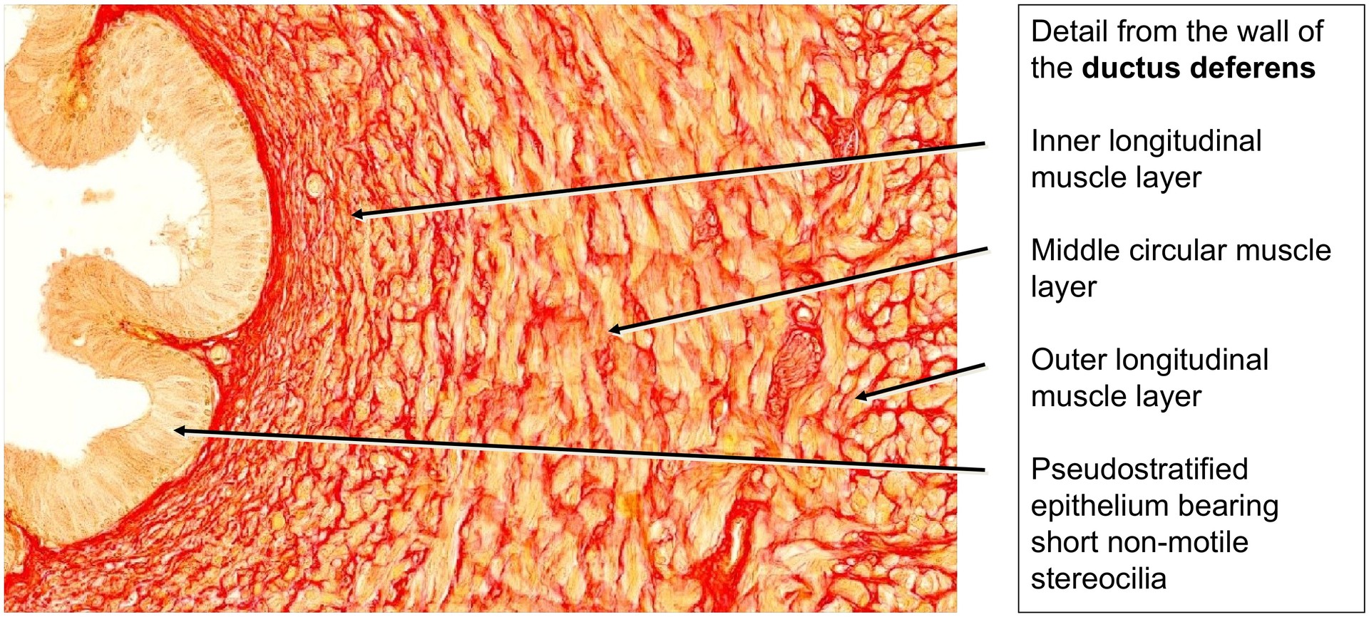
The ductus deferens exhibits a characteristic three-layered musculature, giving it a firm, cord-like consistency easily palpable in situ. The inner and outer layers consist of longitudinally arranged smooth muscle cells, while the middle layer is circularly arranged. Multiple nerve fiber sections lie close to the ductus deferens, mediating motor control of the muscular wall and peristaltic contractions during ejaculation.
The mucosa of the ductus deferens is lined by a two-row (pseudostratified) epithelium, composed of basal cells and principal cells. The latter bear a surface covering of short stereocilia (non-motile), particularly evident in the proximal vas deferens. The lamina propria is narrow and directly contiguous with the inner longitudinal muscle layer.
Tasks:
- Gain an overview at low magnification and identify the ductus deferens.
- Examine the three-layered musculature and determine the orientation of the muscle cells (circular versus longitudinal).
- Locate veins of the pampiniform plexus and assess the structure of their tunica media. At two points, identify how these vessels can still be recognized as veins.
- Identify the fibers of the cremaster muscle.
- Search for and identify nerve fibers, both in the vicinity of the ductus deferens and elsewhere in the specimen.
License
University of Basel
Downloads