MALE REPRODUCTIVE ORGANS (ANATOMICAL MICROSCOPY)
11.8
Penis, child
Specimen Details:
Specimen Details:
Organ: Penis
Origin: Human (Child)
Staining: Haematoxylin - Eosin (H&E)
Method and Specimen Description:
Normal histological section stained with H&E as a general overview stain.
Objective of the Examination:
To study the histological structure of the human penis, particularly the corpora cavernosa, the corpus spongiosum, and the glans penis, with special attention to the skin features and the absence of subcutaneous tissue (subcutis).
Special Features of the Specimen:
The penis contains specialized erectile tissue within its corpora cavernosa and corpus spongiosum, enabling its dual functions of urine excretion and sexual intercourse. During arousal, these tissues fill with blood, producing erection; during micturition, they remain flaccid, allowing the urethra to conduct urine.
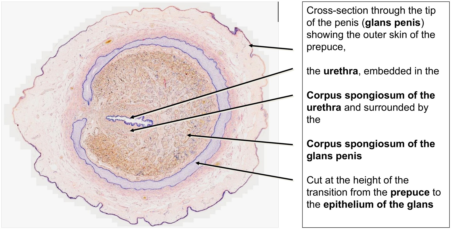
In this specimen, the section is taken through the tip of the penis (glans penis), specifically at the transition between the penile shaft and the glans.
Accordingly, part of the corpus cavernosum is visible peripherally, while the corpus spongiosum occupies the central region.
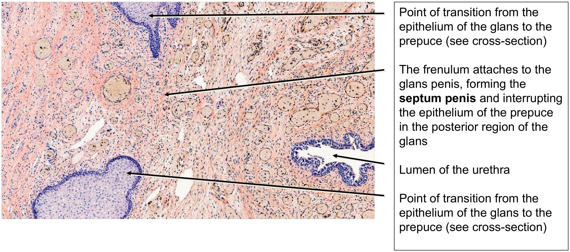
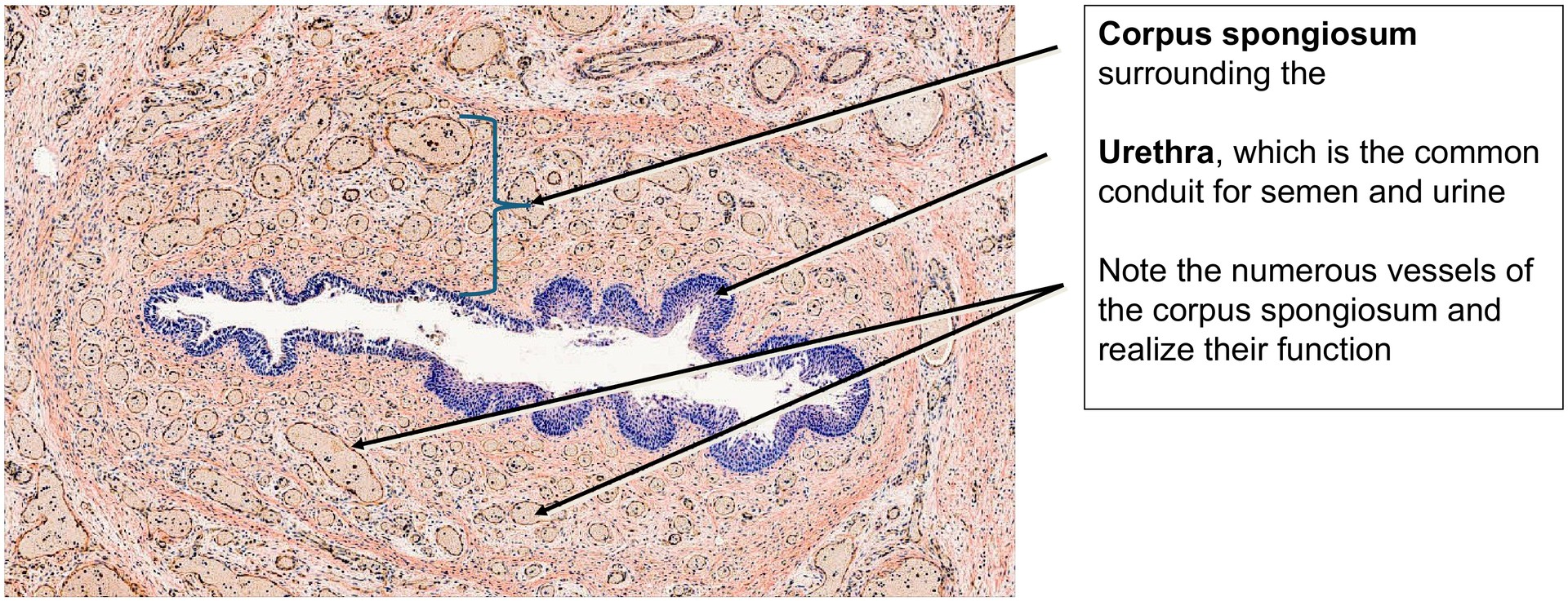
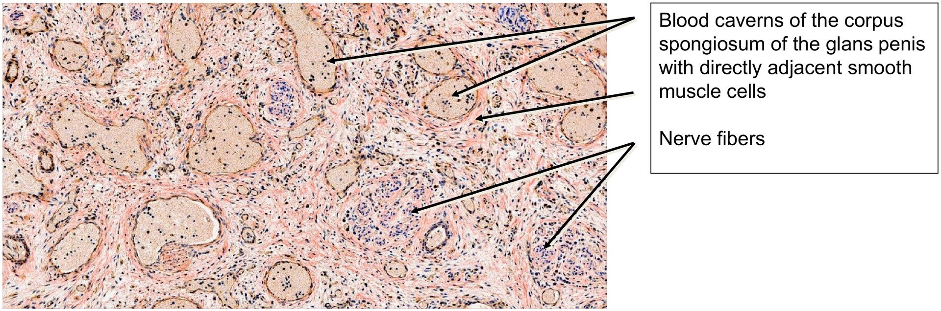
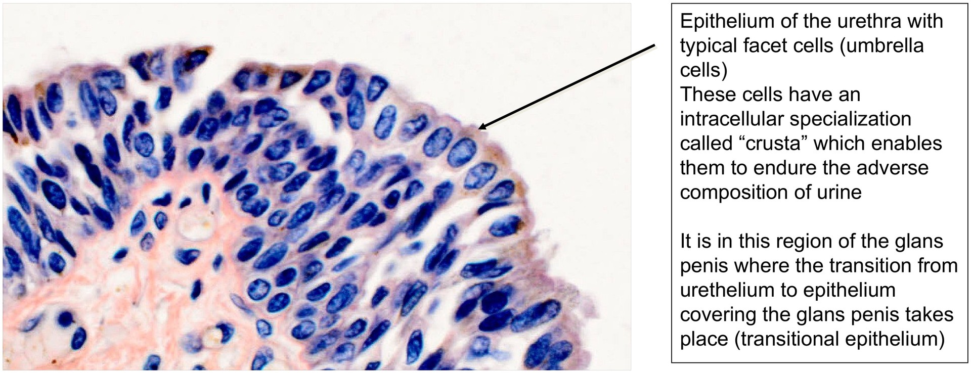
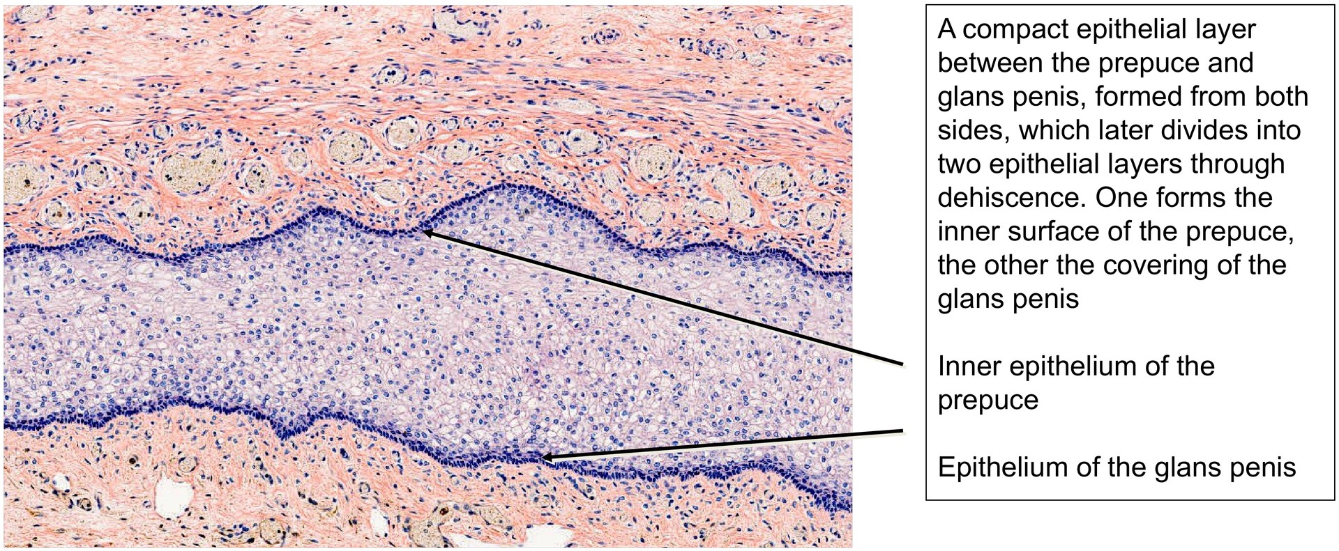
The corpus spongiosum displays numerous vascular spaces (blood caverns), which during sexual arousal become engorged with blood. The surrounding epithelium, lightly basophilic in H&E staining, is sectioned across the transition from the inner surface of the prepuce (foreskin) to the epithelium of the glans. A narrow potential space between the glans penis and the foreskin can already be identified at two points.
At the frenulum—the ventral fold connecting glans and prepuce—the epithelial ring is interrupted by connective tissue of the septum penis.
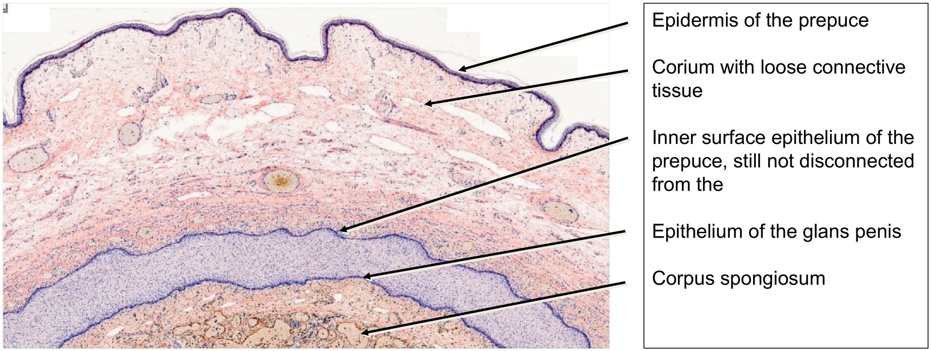
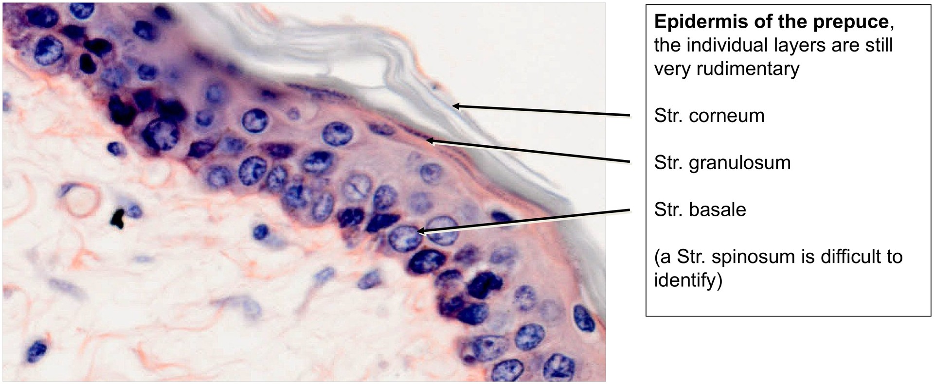
Externally, the glans penis is covered by the foreskin (prepuce), whose structure differs from that of general skin. The subcutis (subcutaneous fat) is absent, and the foreskin represents a double fold of epidermis with a loosely arranged dermis (corium).
A slight cornification of the outer epithelium can already be seen, even in this juvenile specimen.
The vascular spaces of the corpus spongiosum form a venous plexus, whose walls contain smooth muscle cells—still only weakly developed in a child. These smooth muscle fibres, together with the fibroelastic connective tissue, regulate blood flow within the erectile tissue.
Tasks:
• Orient yourself on the specimen and identify the urethra. Why does it have this double name (urethra and pars spongiosa)?
• Examine the inner epithelium of the prepuce and glans penis. Why might it appear as a single epithelial layer but with two strata basalia?
• Study the skin of the prepuce and list its layers. Which layer, normally present below the corium in other skin types, is absent here?
• Locate the numerous nerve fibers present in this region.
• Identify and describe the structure of the blood caverns within the glans penis and corpus spongiosum.
License
University of Basel
Downloads