EPITHELIUM (GENERAL HISTOLOGY)
1.6
Cell boundaries (mesothelium)
Specimen:
Specimen Details:
Organ: Mesentery
Origin: Rat
Staining: Silver nitrate (AgNO₃)
Method and Specimen Description:
This specimen has been treated with silver nitrate, which reacts with strongly reducing substances in the extracellular matrix (ECM) to form metallic silver precipitates. In this preparation, the small amount of ECM present in the intercellular spaces between epithelial cells becomes visible through this reaction, effectively highlighting cell boundaries.
Objective of the Examination:
To observe the cell boundaries of a simple squamous epithelium (mesothelium), to understand the complex interdigitations between neighboring cells, and to appreciate that the intercellular space is extremely narrow.
Special Features of the Specimen:
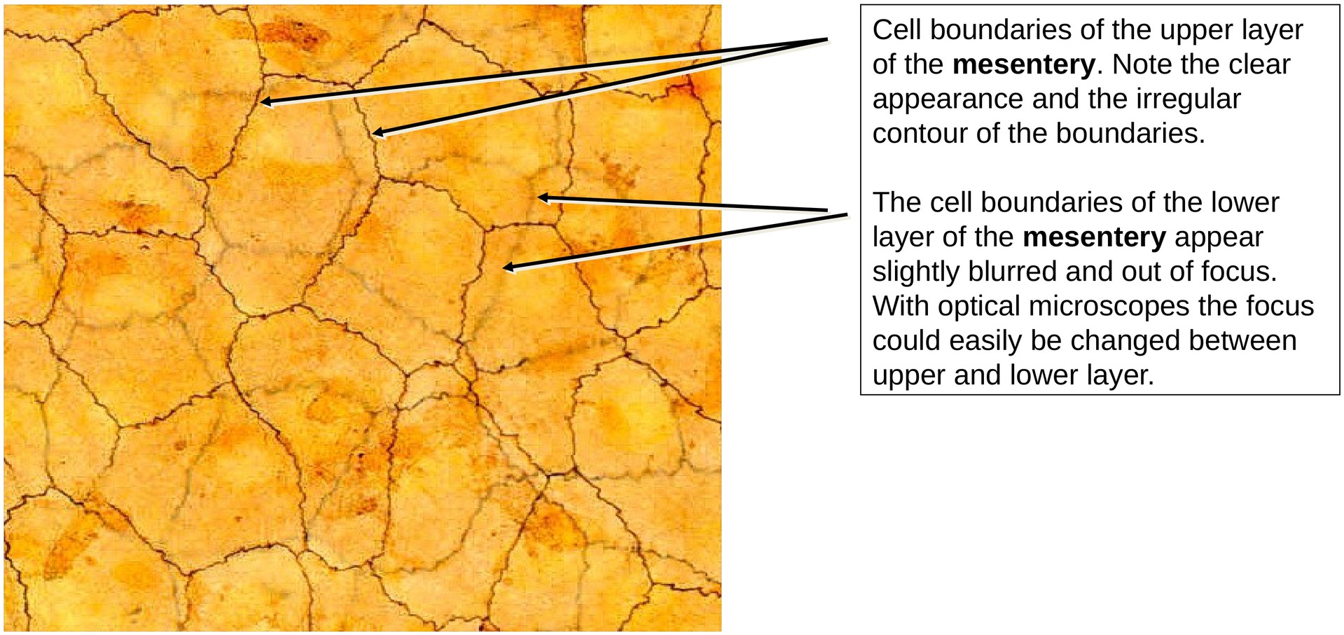
General: The mesentery consists of a double layer of peritoneum. Between these two layers lies connective tissue rich in blood vessels and nerves. This is a whole-mount preparation, meaning that only the two epithelial (mesothelial) layers — the upper and lower — are visible in the overview. The intermediate connective tissue and intracellular components are not stained.
Structure of Cell Boundaries (low to medium magnification): Two planes of focus can be distinguished: one corresponding to the upper mesothelial surface, and the other to the lower surface. Because the microscope can only focus on one plane at a time, cell boundaries in one layer appear sharp and distinct, whereas those in the opposite layer appear blurred. The cell borders are irregular and exhibit interdigitations, reflecting the interlocking arrangement of neighboring mesothelial cells. The intercellular space is extremely narrow — effectively virtual.
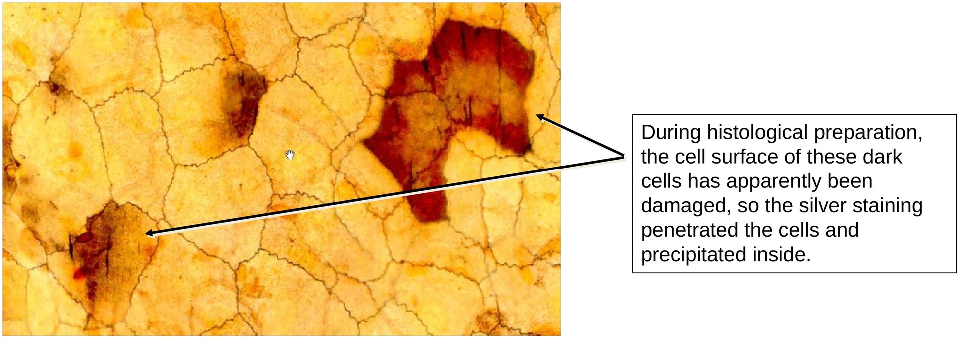
Additional Notes: Intracellular structures are not visible with this staining method. Small surface particles may take up stain and appear dark. Cells that were damaged during dissection or fixation also appear darker, due to silver deposition within the injured cytoplasm.
Tasks:
• Identify a region in the specimen where both epithelial layers of the mesentery can be clearly seen. Depending on the separation between these layers, the opposite layer may appear faintly or blurred in the background.
• Examine the course of the cell boundaries and observe how they interdigitate with one another.
• Recognize that the apparent intercellular spaces are extremely narrow and largely potential spaces, with adjacent cell membranes lying in close contact.
License
University of Basel
Downloads