DIGESTIVE ORGANS: GASTROINTESTINAL TRACT (ANATOMICAL MICROSCOPY)
19.7
Jejunum 1
Specimen:
Specimen Details:
Organ: Jejunum
Origin: Human
Staining: Periodic acid–Schiff (PAS)
Method and Specimen Description:
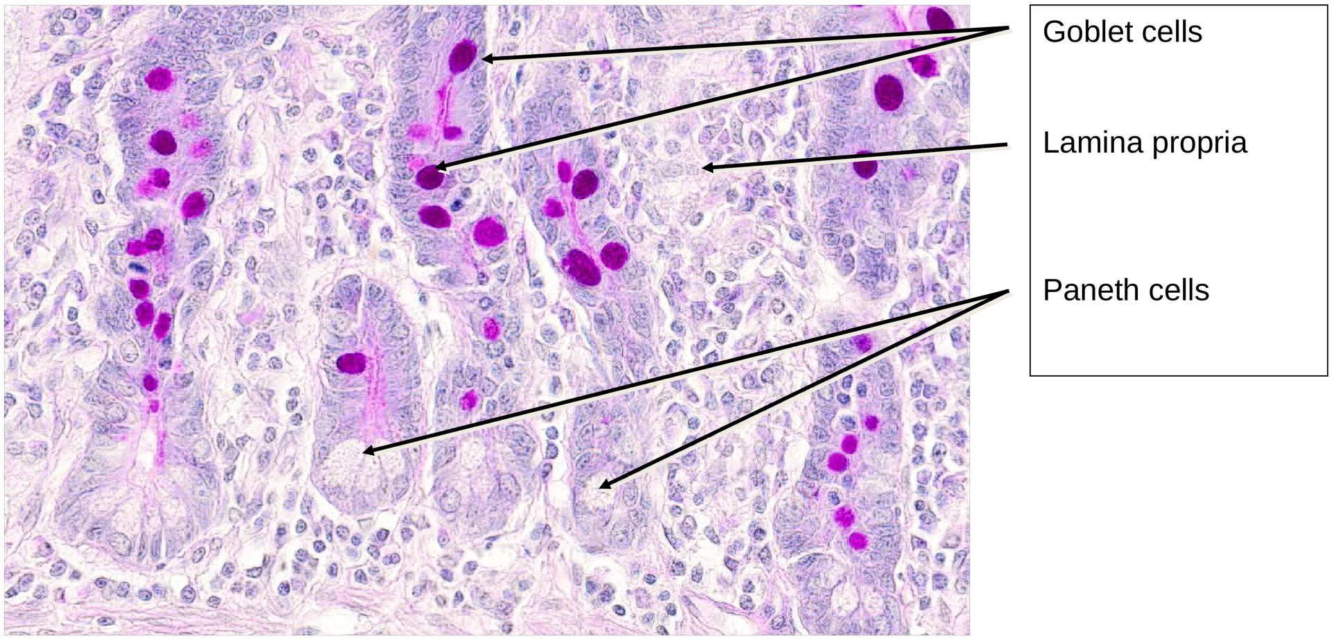
Normal histological section stained with PAS (Periodic acid–Schiff reagent), which primarily stains glycogen, glycoproteins, and glycosaminoglycans, as well as mucus within goblet cells.\ In this specimen, Paneth cell granules in the crypts are poorly stained and therefore appear almost empty.
Objective of the Examination:
To study the structure of the jejunum, focusing on its mucosal architecture and the distribution of goblet cells within the brush border epithelium (enterocytes).
Specimen Specifics:
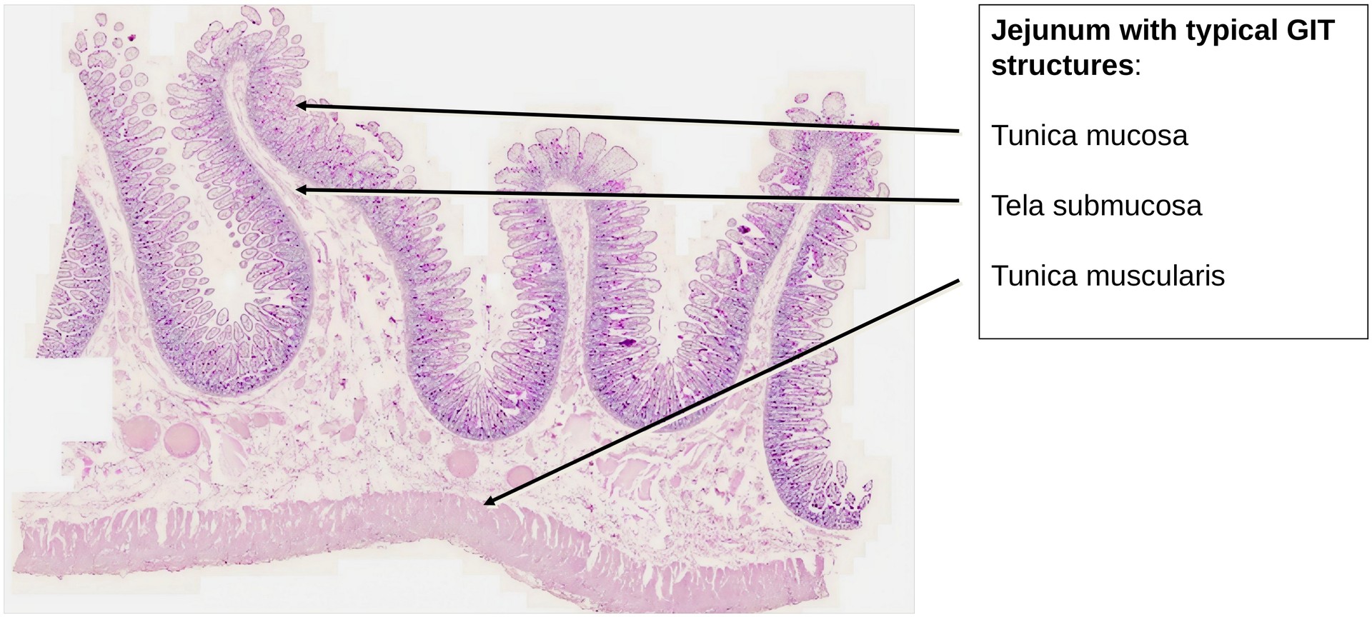
The jejunum shows the typical layered organization of the gastrointestinal tract (GIT):
- Tunica mucosa (comprising the lamina epithelialis, lamina propria, and lamina muscularis mucosae)
- Tela submucosa (containing the submucosal plexus, or Meissner’s plexus)
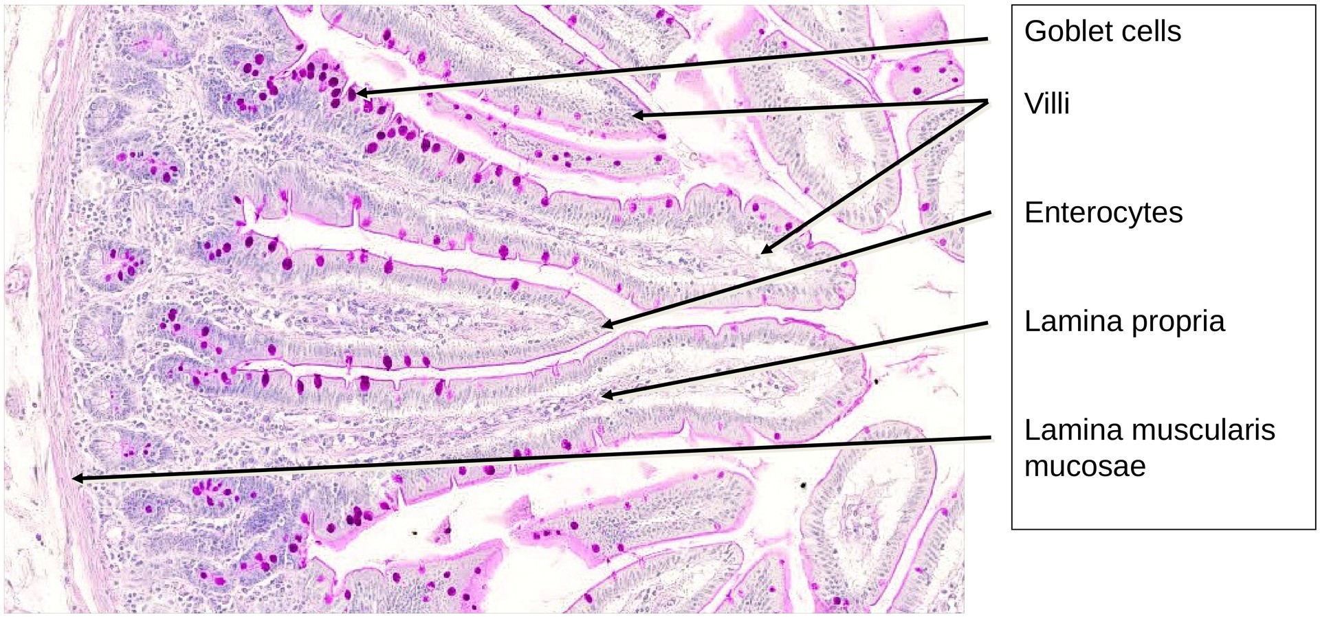
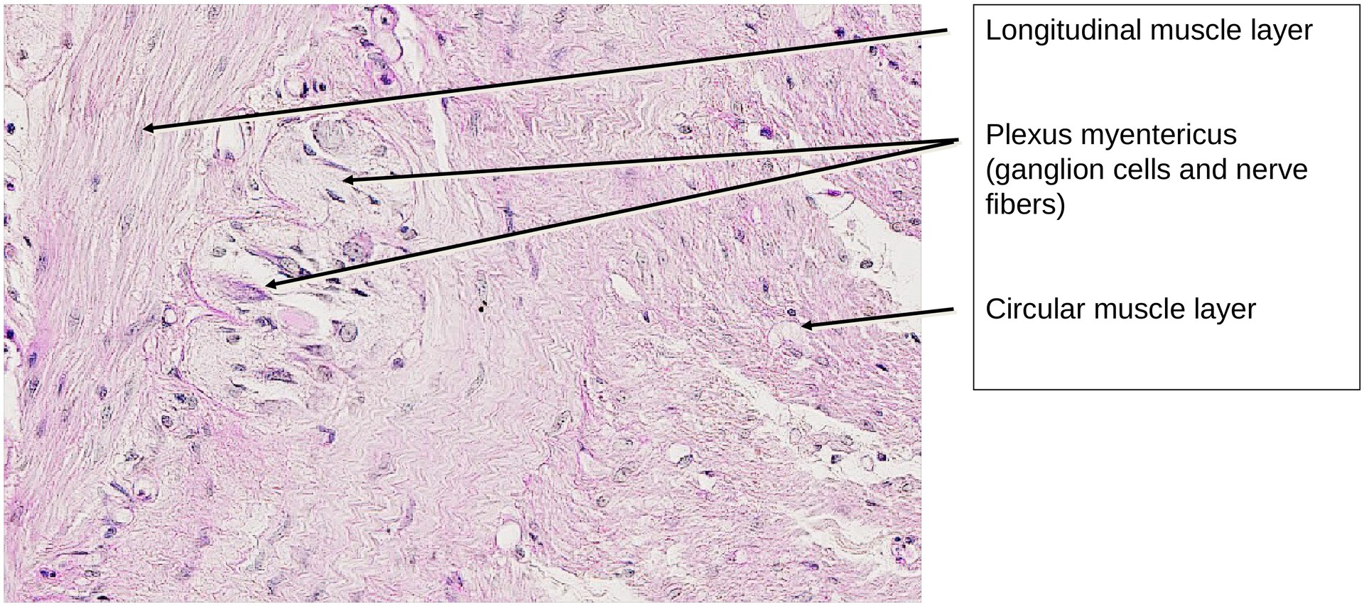
- Tunica muscularis (composed of an inner circular and an outer longitudinal smooth muscle layer) The epithelium consists of two main cell types:
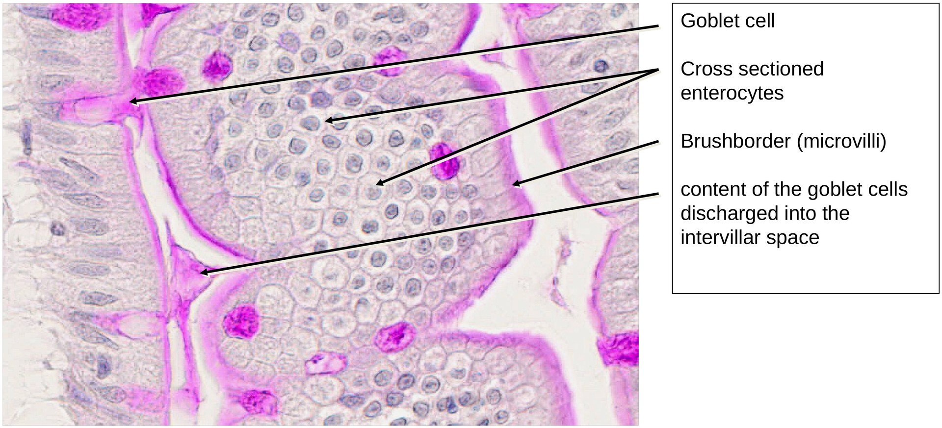
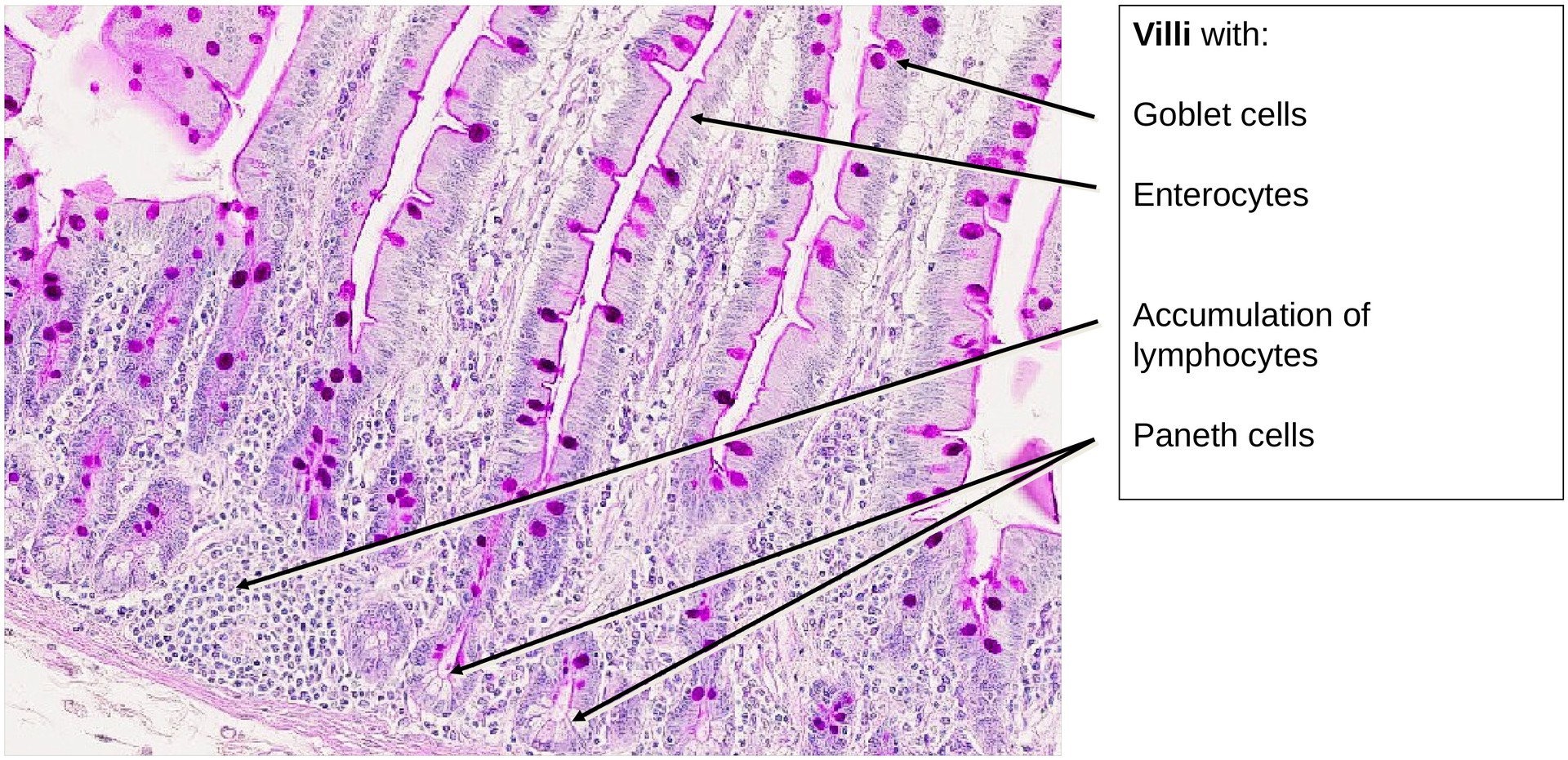
- Enterocytes (brush border cells), and
- Goblet cells, which are distinctly PAS-positive.
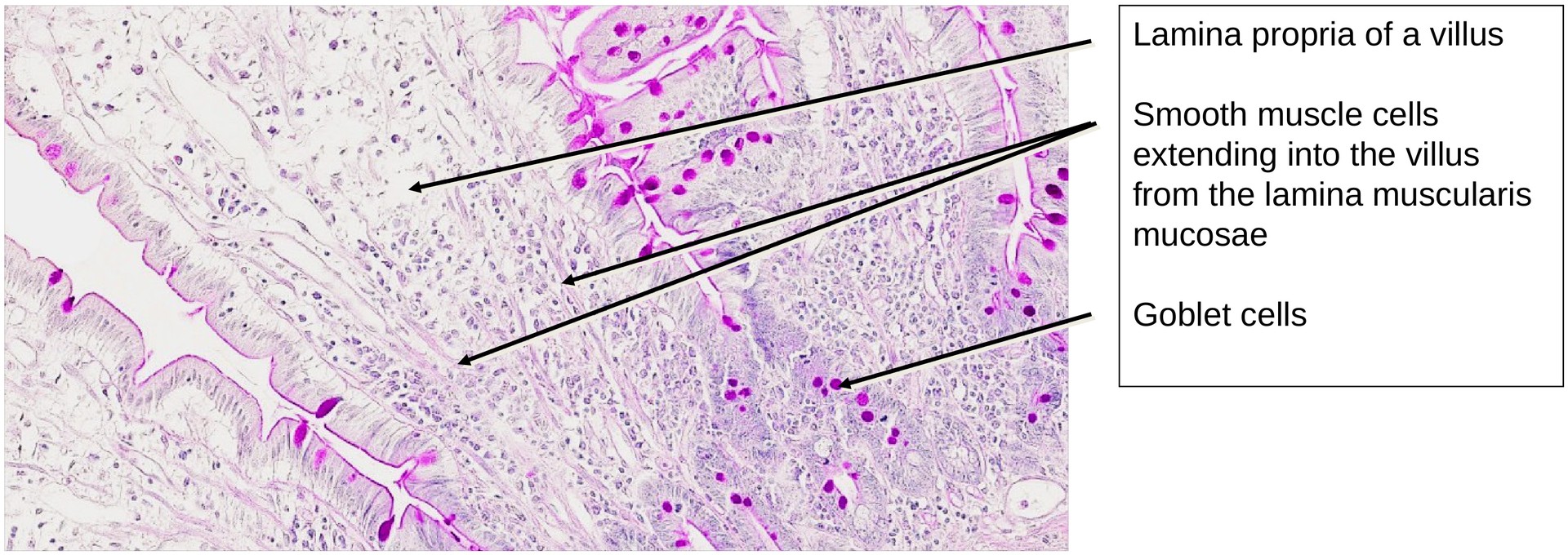
The mucosa is folded into plicae circulares (folds), upon which finger-shaped villi project. At the base of the villi lie the openings of the intestinal crypts (crypts of Lieberkühn).
In PAS staining, goblet cells are intensely highlighted, and in some areas, mucus secretion can be seen as goblet cells release their contents.
The lamina propria forms a relatively thin layer beneath the epithelium. The lamina muscularis mucosae is well developed; its smooth muscle fibers extend into the mucosal folds, and occasionally into individual villi.
The submucosal plexus (Meissner’s plexus) is present within the submucosa but not as distinct as the myenteric plexus (Auerbach’s plexus), which lies between the circular and longitudinal muscle layers and contains ganglion cells and nerve fibers.
Within the crypts, Paneth cells are visible. Their apical granules are not stained by PAS, leaving the apical cytoplasm bright and clear, distinguishing them from surrounding crypt cells.
Tasks:
- Identify the layers of the gastrointestinal tract (GIT) in low magnification:
- Tunica mucosa (lamina epithelialis, lamina propria, lamina muscularis mucosae)
- Tela submucosa
- Tunica muscularis (inner circular and outer longitudinal muscle)
- Describe the difference between folds and villi. Which components of the typical GIT structure are found in both?
- Determine where most goblet cells are located in this specimen.
- Name the second cell type of the lamina epithelialis (besides goblet cells).
- Locate and identify the submucosal (Meissner’s) plexus and the myenteric (Auerbach’s) plexus. Where must you look to find each?
- Identify the Paneth cell granules. In which structures are they found, and what distinguishes them histologically?
License
University of Basel
Downloads