MUSCULATURE (GENERAL HISTOLOGY)
6.4
Cardiac muscle, longitudinal section (intercalated disks)
Preparation:
Preparation Details:
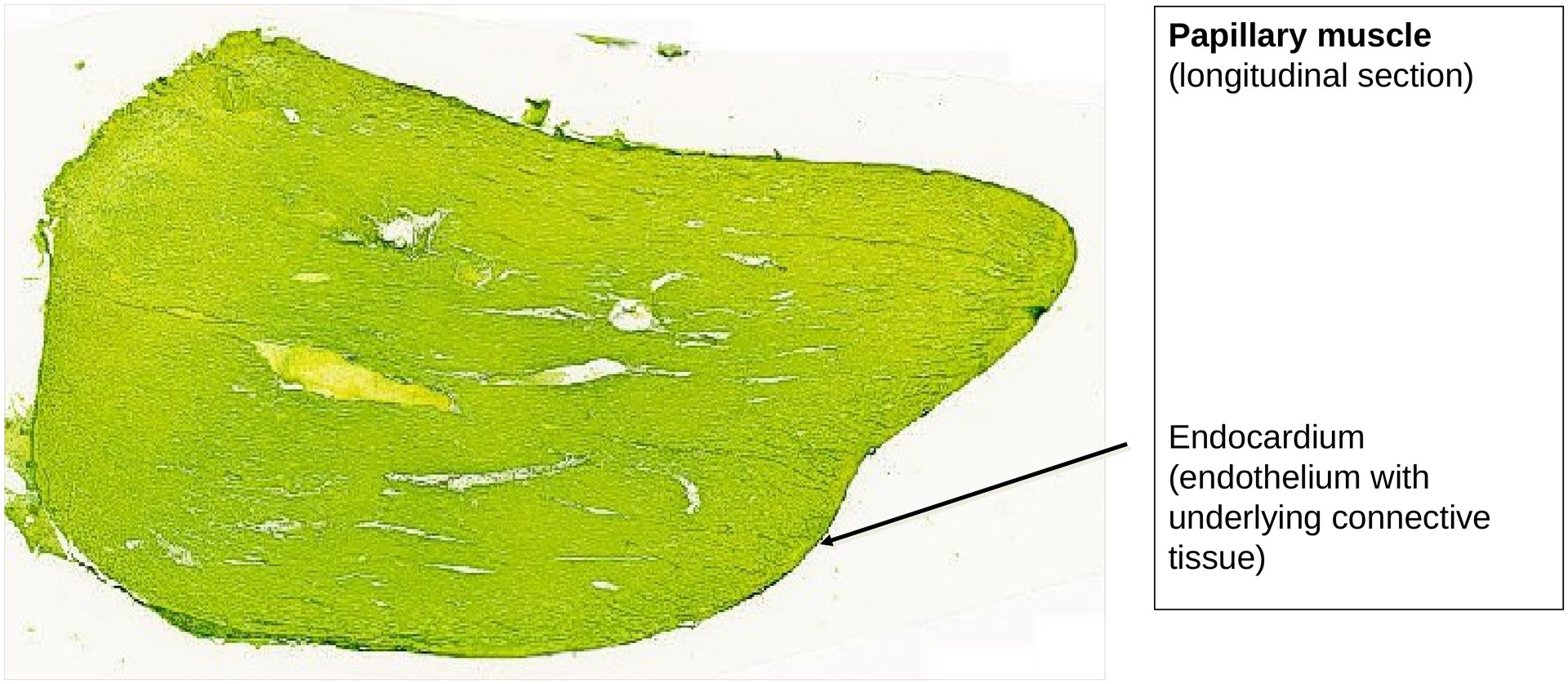
Organ: Papillary muscle
Origin: Human
Staining: Mikado yellow-toluidine blue
Method and Specimen Description:
Papillary muscles are particularly suitable for examining cardiac muscle, as the muscle fibers in these regions run almost parallel to one another. In contrast, the remaining myocardium forms a network of interwoven muscle strands.
Staining with Mikado yellow and toluidine blue makes the striations and intercalated discs especially distinct. The mixture of dyes produces an intense greenish hue in the preparation.
Objective of the Examination:
To study the microscopic structure of cardiac muscle and to differentiate it from skeletal muscle.
Special Features of the Preparation:
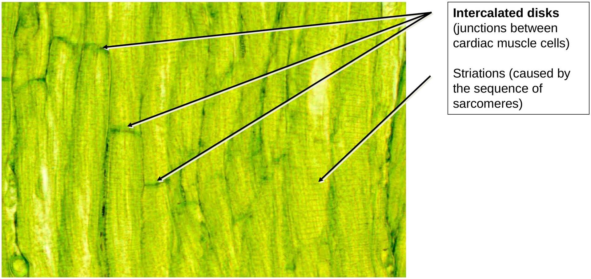
Even at low magnification, the longitudinal cell boundaries of the cardiac muscle cells can be clearly observed. In certain regions, transverse boundaries are also visible — these are the intercalated discs. Such areas are particularly suitable for examination under higher magnification.
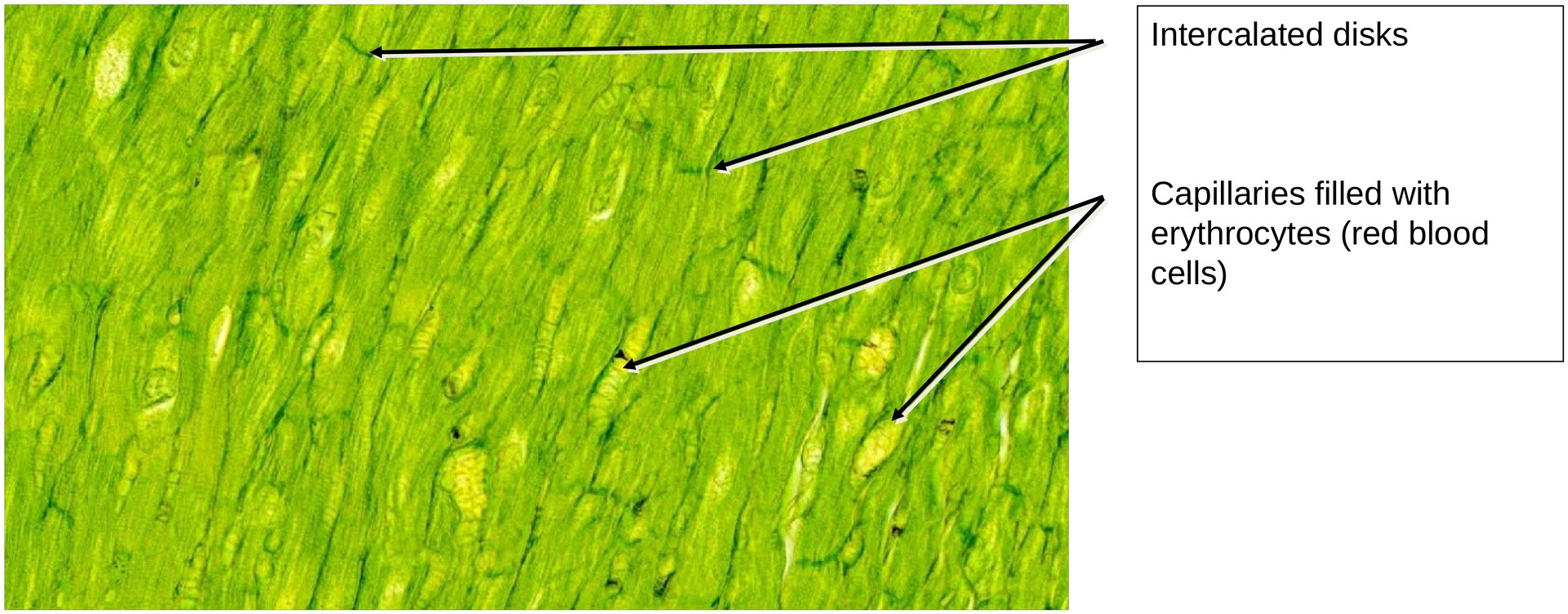
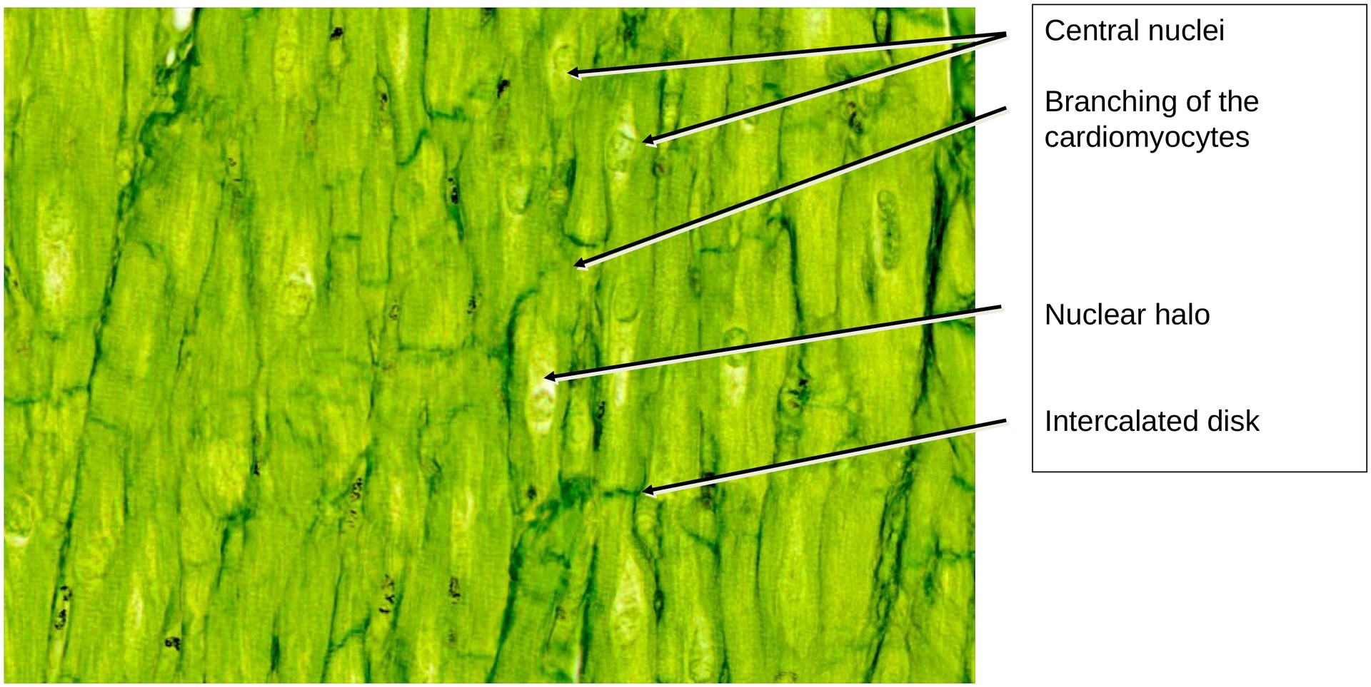
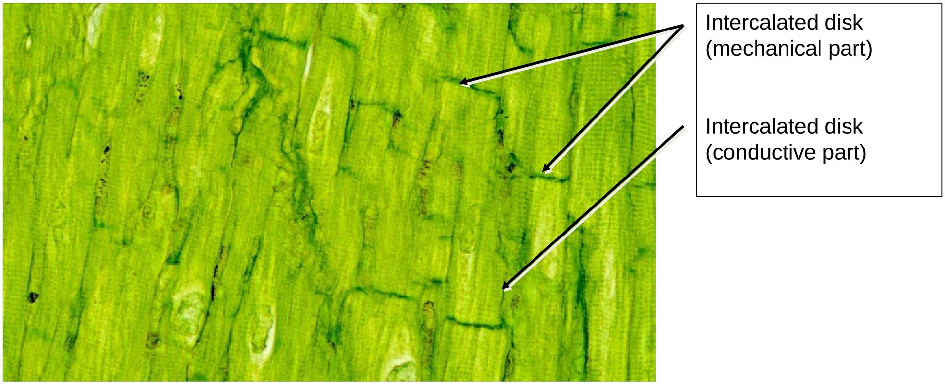
At medium and high magnification, the intercalated discs can be recognized as the junctions between adjacent cardiac muscle cells. They often appear branched or step-like and are composed of desmosomes, adherens junctions (fasciae adhaerentes), and gap junctions — the latter being visible only under electron microscopy.
The nuclei of the cardiac muscle cells are centrally located and are often surrounded by perinuclear halos, which contain organelles and glycogen. Generally, only the A and I bands of the sarcomeres are visible, and the striations are less distinct than those seen in skeletal muscle.
Erythrocytes, which are relatively easy to identify, can be used to locate capillaries and other vascular profiles. These form a dense capillary network between the cardiac muscle fibers. The capillary walls are not always visible, but the presence of erythrocytes indicates their location.
Tasks:
-
First, obtain an overview of the section and examine its periphery. Note that this region — even if not perfectly focused in some areas due to scanning artefacts — represents the natural boundary of the tissue, the endocardium.
-
Locate and examine the intercalated discs.
-
Identify the position of the cell nuclei.
-
Observe that cardiac muscle cells are branched.
-
Locate blood vessels (mainly capillaries) and comment on their frequency, explaining the reason for this abundant vascularization.
License
University of Basel
Downloads