FEMALE REPRODUCTIVE ORGANS (ANATOMICAL MICROSCOPY)
10.6
Decidua
Specimen Details:
Specimen Details:
Organ: Decidua
Origin: Human
Staining: Azan
Method and Specimen Description:
Routine histological section of human decidua, stained with Azan. In this stain, cell nuclei of epithelial cells and erythrocytes appear red, while connective tissue fibers and fibrinoid material at the uteroplacental border zone are stained blue.
Objective of the Examination:
To understand the decidua as a key component of the uteroplacental interface, the region where the implanting blastocyst interacts with, and must be tolerated by, the maternal tissue. This site represents a dynamic balance between invasion and immune regulation.
Special Features of the Specimen:
True decidua cells differentiate from predecidual cells of the late secretory phase following successful implantation. These cells are most prominent in the decidua basalis — the region where the chorionic villi, and later the anchoring villi of the placenta, attach.
The decidua therefore represents the maternal component of the placenta, while the chorion forms the fetal component. The developing embryo tends to implant as deeply as possible, while the maternal tissue must limit this invasion to prevent overpenetration. Disruption of this delicate balance can result in conditions such as placenta increta or, in severe cases with uterine wall rupture, placenta percreta.
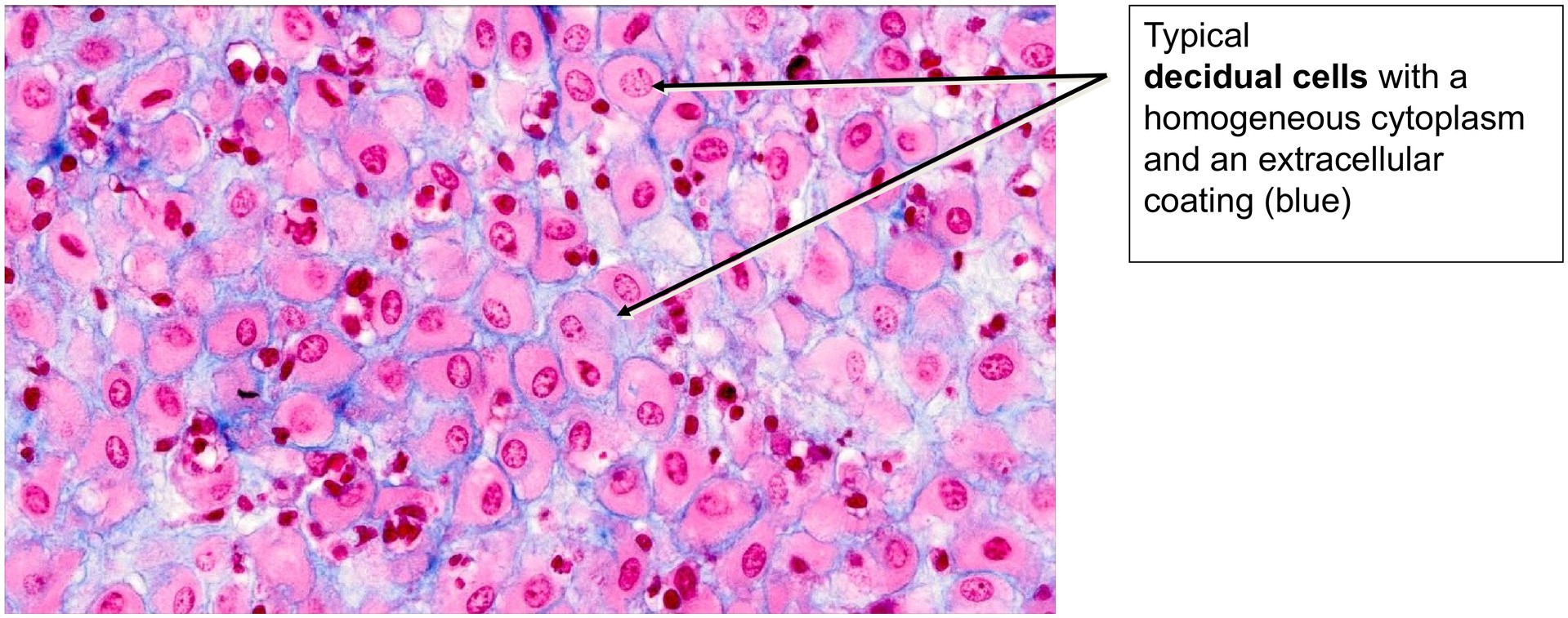
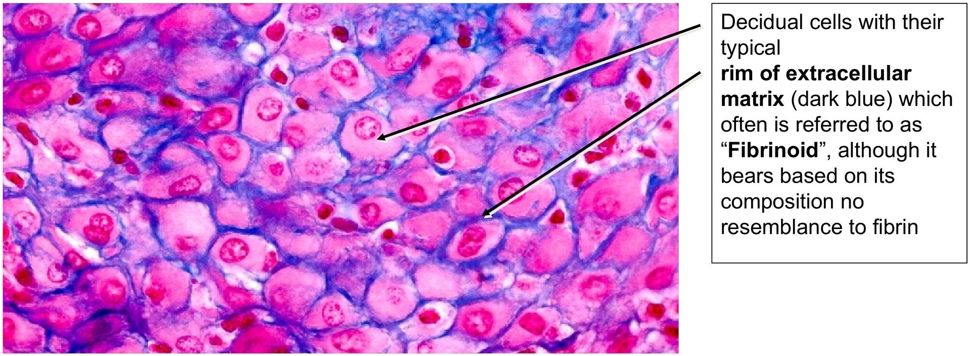
Decidua cells play a central regulatory role in this process. Compared with their precursors, the endometrial stromal fibroblasts, they are significantly enlarged, rich in glycogen and often lipid inclusions, and display a pale cytoplasm. These cells secrete extracellular matrix material, partly by apocrine secretion, which includes collagen and forms a bluish rim around each cell in Azan-stained sections.
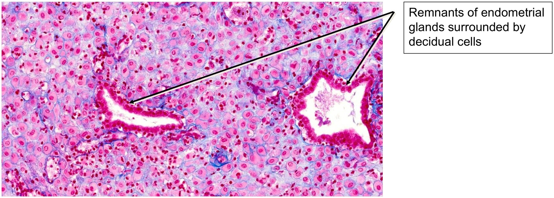
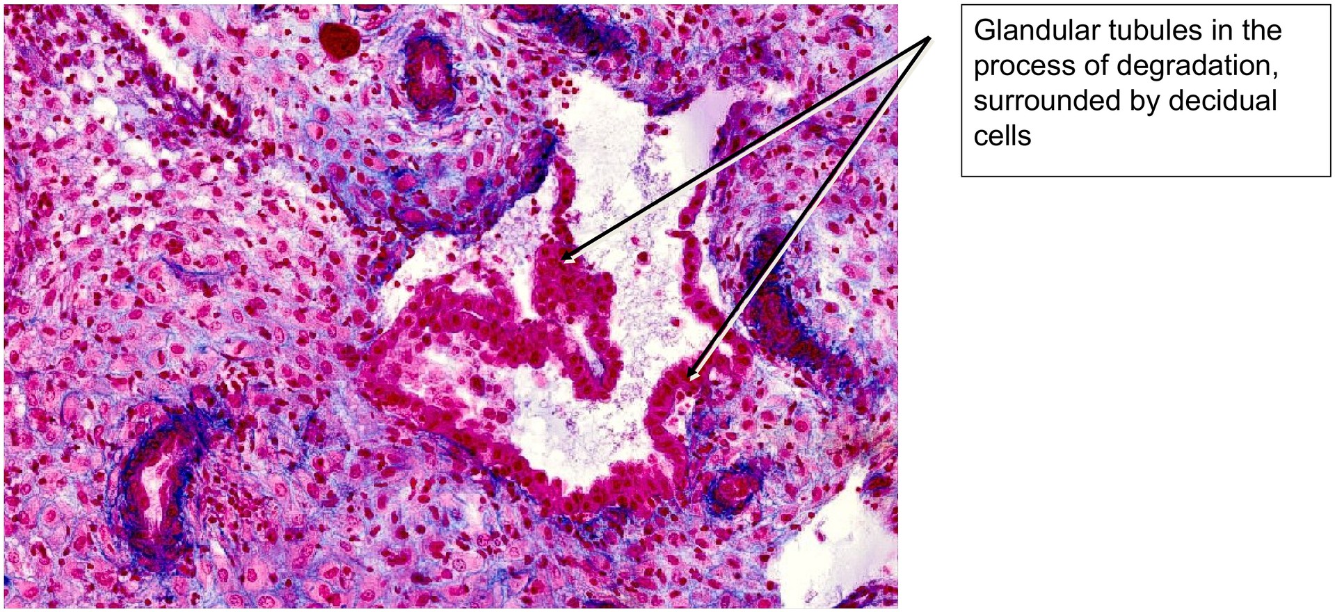
Remnants of endometrial glandular tubules can often be identified between the decidua cells; some are undergoing degeneration and may lie in direct contact with the decidual tissue. Blood vessels can also be recognized within the connective tissue framework.
Tasks:
• Obtain an overview of the specimen and identify the general arrangement of the tissue.
• Locate remnants of glandular tubules within the decidua.
• Identify the large decidua cells and note their pale cytoplasm and prominent nuclei.
• Observe the blue-stained rim surrounding the decidua cells (representing extracellular matrix and fibrinoid material).
• Search for blood vessels within the connective tissue of the decidua.
License
University of Basel
Downloads