URINARY ORGANS (ANATOMICAL MICROSCOPY)
12.3
Kidney, Human 1
Specimen:
SPECIMEN DETAILS:
Organ: Kidney
Origin: Human
Staining: Azan
METHOD AND SPECIMEN DESCRIPTION:
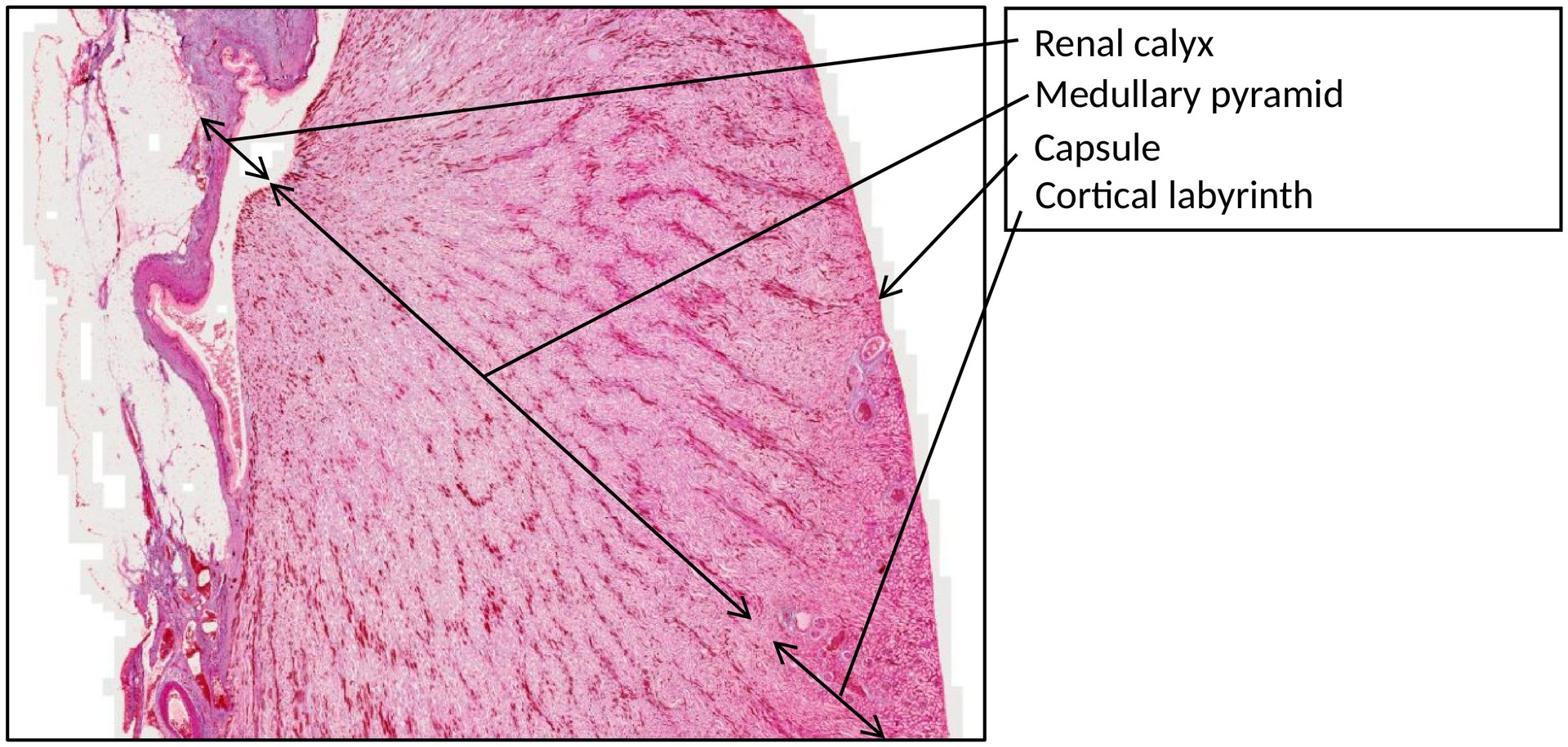
Normal histological section of the human kidney stained with Azan, which distinctly colors connective tissue (collagen) blue and the cytoplasm and muscle fibers red.## OBJECTIVE OF THE EXAMINATION:
To study the structure of the human multipapillary kidney and its functional units, and to compare this specimen with the kidney of the rabbit.
Special Features of the Specimen:
The paired kidneys excrete water-soluble metabolic end products and foreign substances as urine-producing organs. They play an essential role in maintaining water, electrolyte, and acid–base balance.
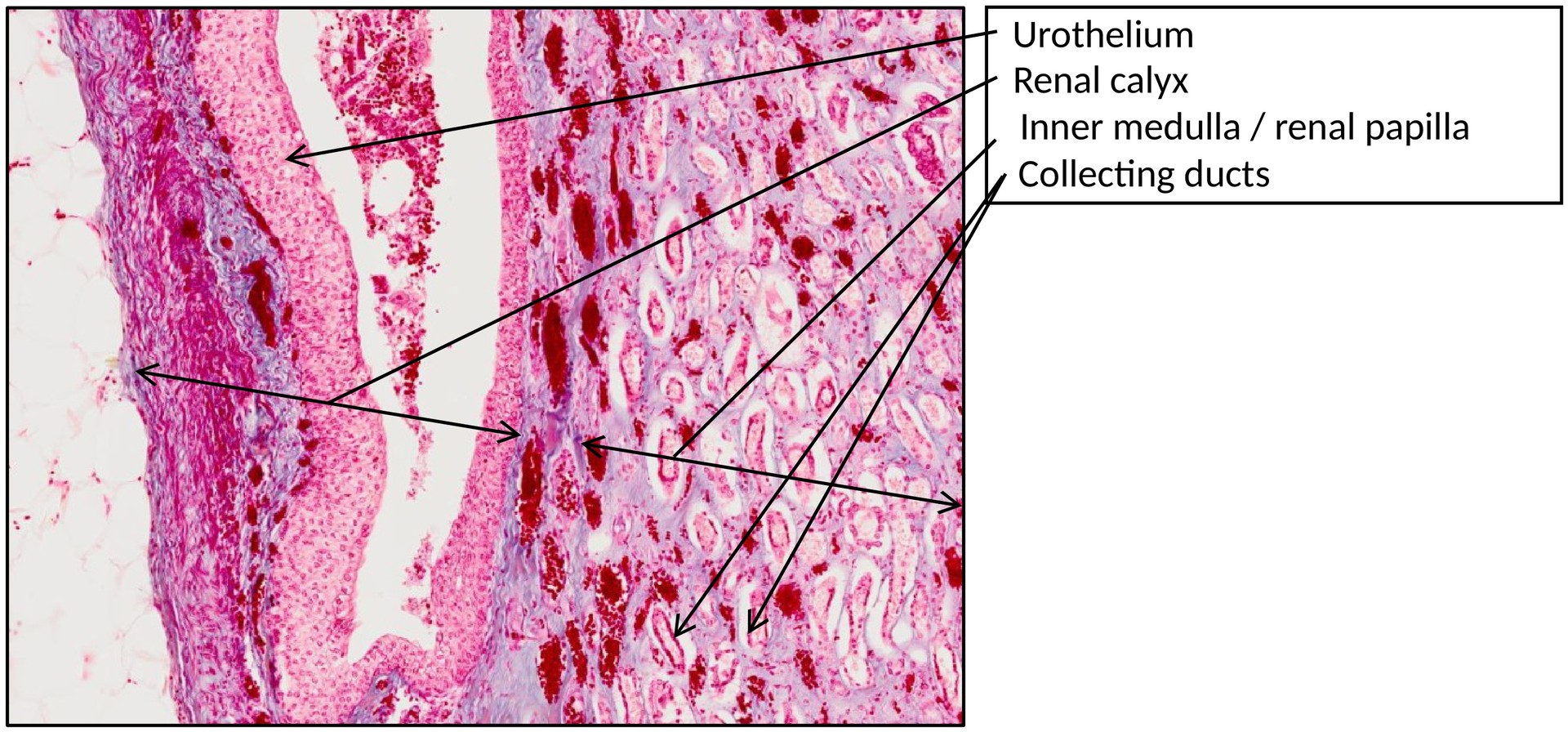
The smallest functional unit of the kidney is the nephron, consisting of a renal corpuscle and a draining unbranched tubule, which connects to a collecting duct via a connecting tubule. Approximately ten to twelve nephrons drain into a single collecting duct, which merges with others to form the papillary ducts, opening into the renal calyces and subsequently into the renal pelvis.
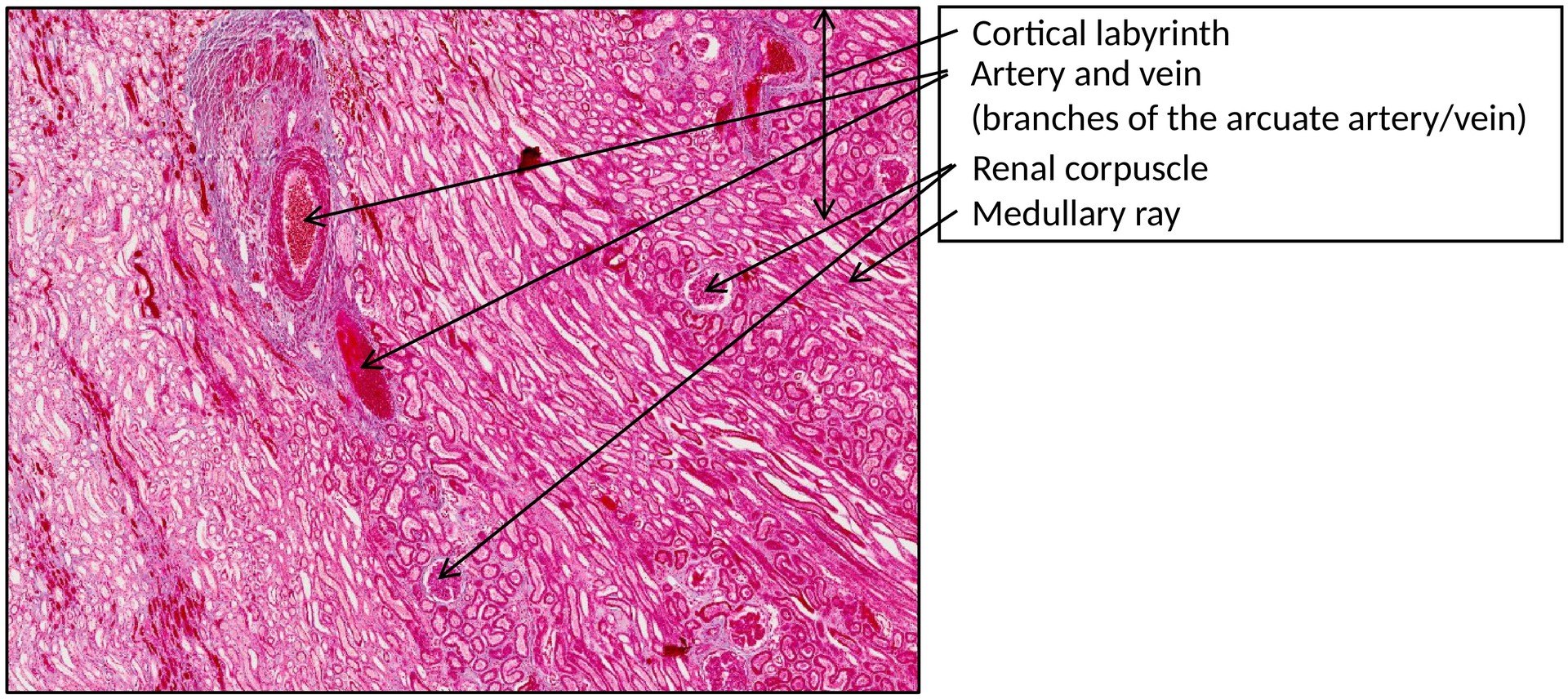
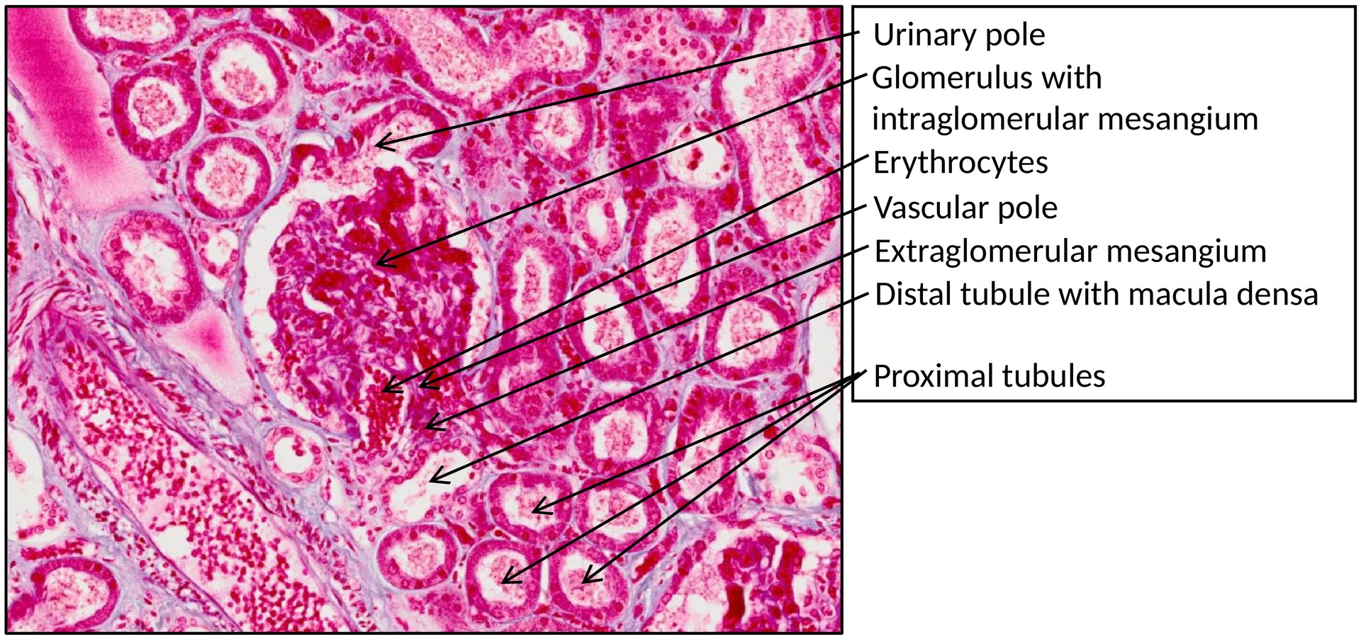
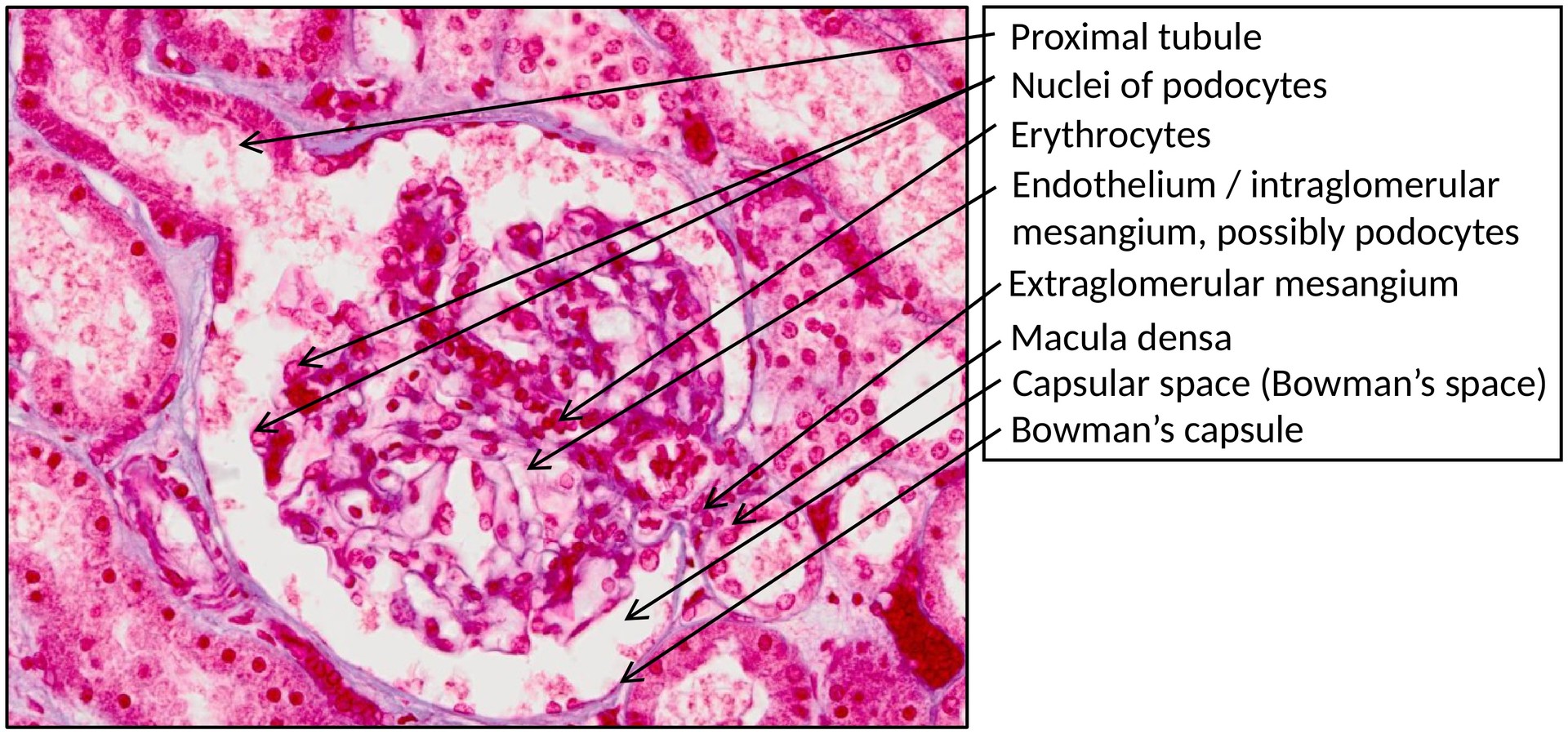
Within the renal corpuscle—composed of a capillary tuft (glomerulus) enclosed by a double-layered Bowman’s capsule—primary urine is produced as a blood ultrafiltrate. This filtrate flows through the tubular system, where the final urine is formed by reabsorption and secretion processes before being excreted via the collecting ducts.
The kidney also functions as an endocrine organ, secreting renin, erythropoietin, and calcitriol into the bloodstream.
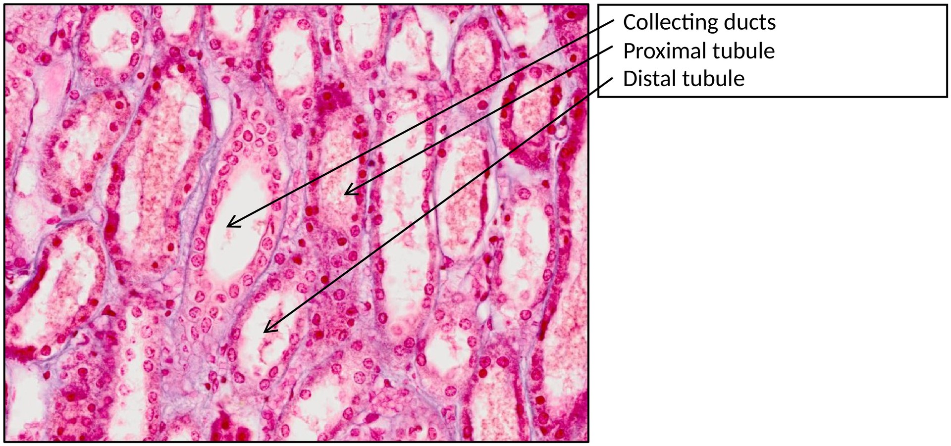
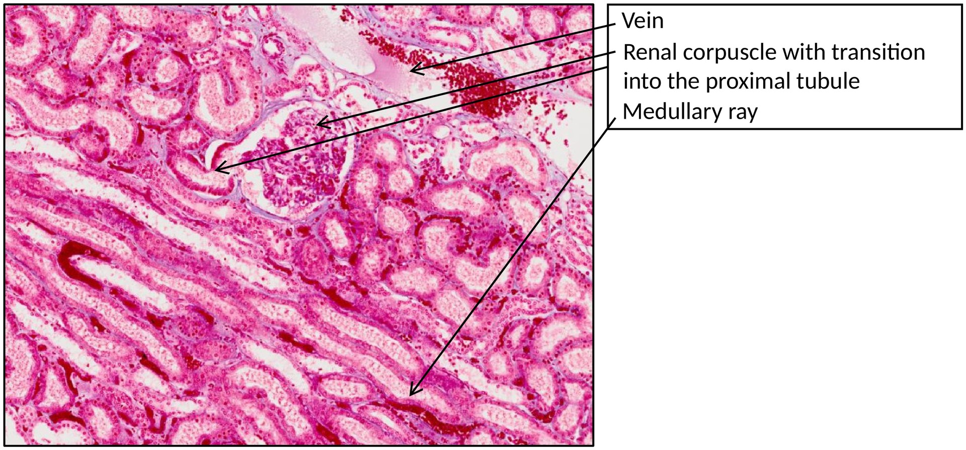
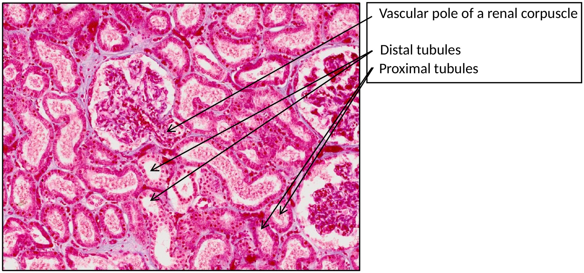
The renal cortex contains renal corpuscles, proximal and distal tubules:
- The proximal tubule is characterized by a brush border (microvilli) giving the apical cell pole a frayed appearance. Its epithelium is cuboidal to columnar, with a strongly stained cytoplasm. Basal striations are sometimes visible.
- The distal tubule has clearer cell boundaries, a flatter cuboidal epithelium, and lacks a brush border. Distal tubule profiles are seen less frequently in section.
- Intermediate (thin) tubules are narrow, lined by a simple squamous epithelium.
In the renal medulla and medullary rays, numerous collecting ducts can be identified by their well-defined cell borders. These ducts contain principal cells (pale cytoplasm, basally striated) and intercalated or dark cells (darker cytoplasm, with apical microplicae).
TASKS:
- Gain an overview of the kidney’s gross architecture, identifying the capsule, cortex, and medulla.
- Examine the renal corpuscles within the cortex, noting the vascular and urinary poles.
- Identify proximal and distal tubule segments, and collecting ducts within the medulla.
- In the region of a renal corpuscle, search for the macula densa, mesangium, and juxtaglomerular cells.
- Identify the visceral and parietal layers of Bowman’s capsule.
License
University of Basel