RESPIRATORY ORGANS (ANATOMICAL MICROSCOPY)
8.1
Lung, adult
Specimen:
Specimen Details:
Organ: Lung
Origin: Human
Staining: Haematoxylin - Eosin (H&E)
Method and Specimen Description:
A normal histological specimen stained with H&E, providing an overview of the structural organization of the lung.
Objective of the Examination:
To understand the structure of the lung, including its gas-conducting and gas-exchanging regions.
Specimen Specifics:
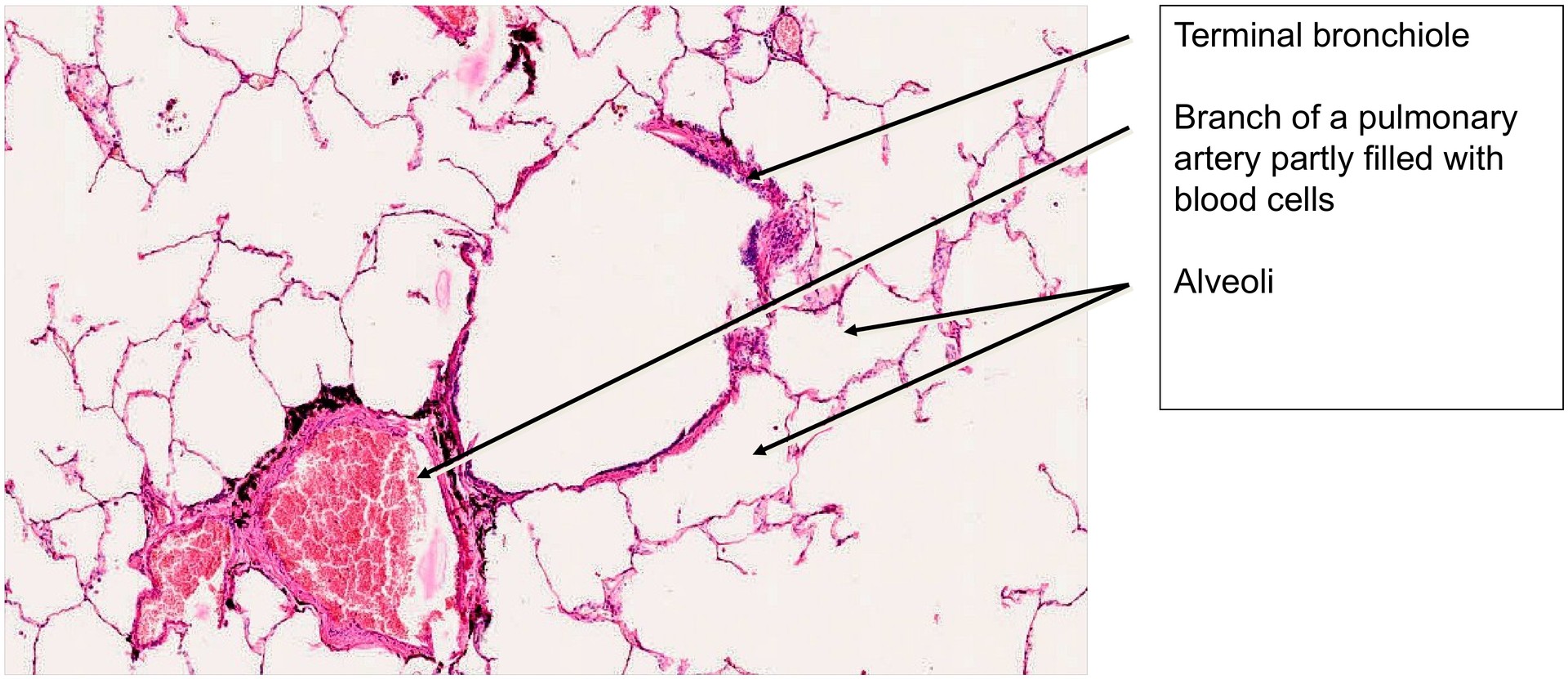
The adult lung, unlike the fetal lung, functions in gas exchange and therefore contains only minimal interstitial connective tissue, mainly around the branches of the bronchial tree and the accompanying blood vessels. Most of the lung tissue consists of a delicate network of alveoli, alveolar ducts, and alveolar sacs. Capillaries within the alveolar walls are difficult to identify in this specimen but are more apparent in the “injected lung” preparation.
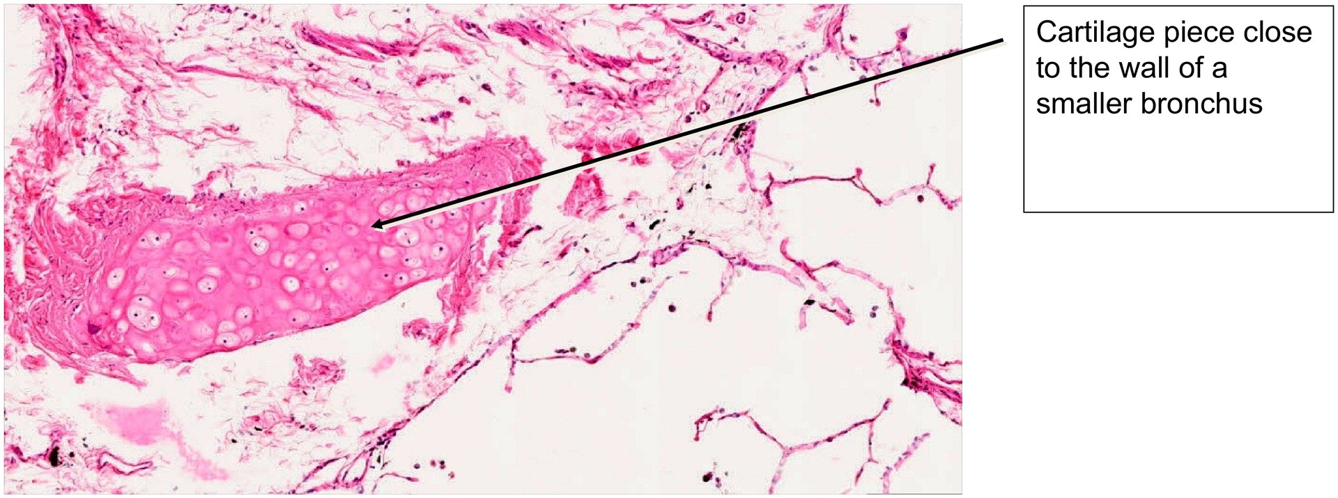
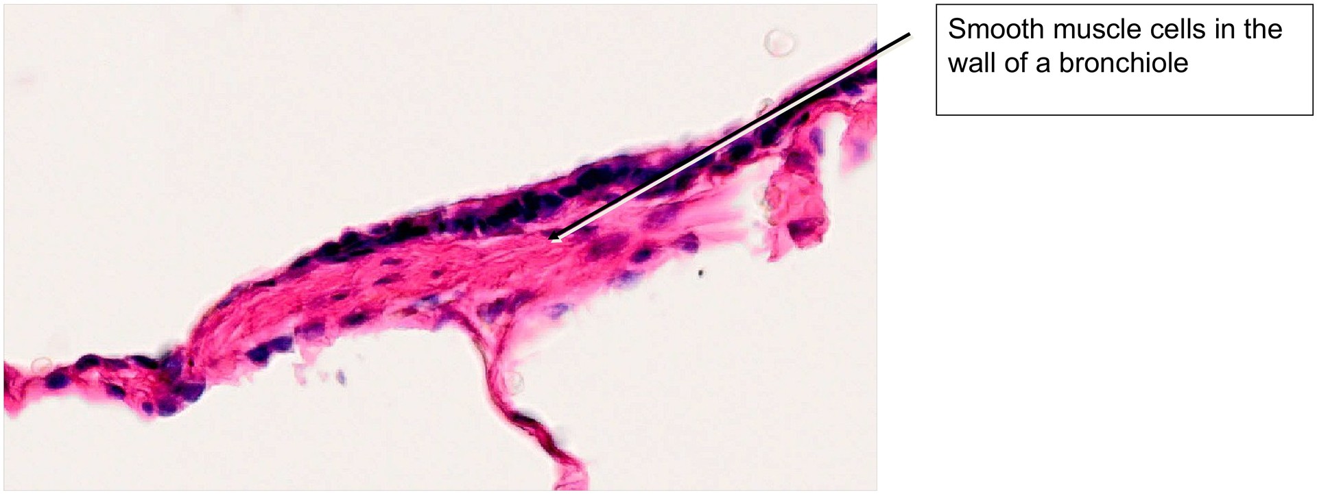
The larger bronchial branches are kept open by cartilaginous plates and, nearer the trachea, by cartilaginous rings. In this specimen, a small piece of cartilage can be seen near one of the smaller bronchi. In the lung, branches of the bronchial tree and the pulmonary artery usually run together, while the pulmonary veins course independently. Cartilage and bronchial glands are found in the walls of bronchi but not in bronchioles; the small bronchus present here lacks bronchial glands.
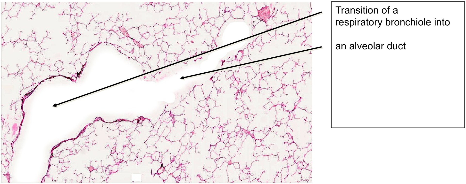
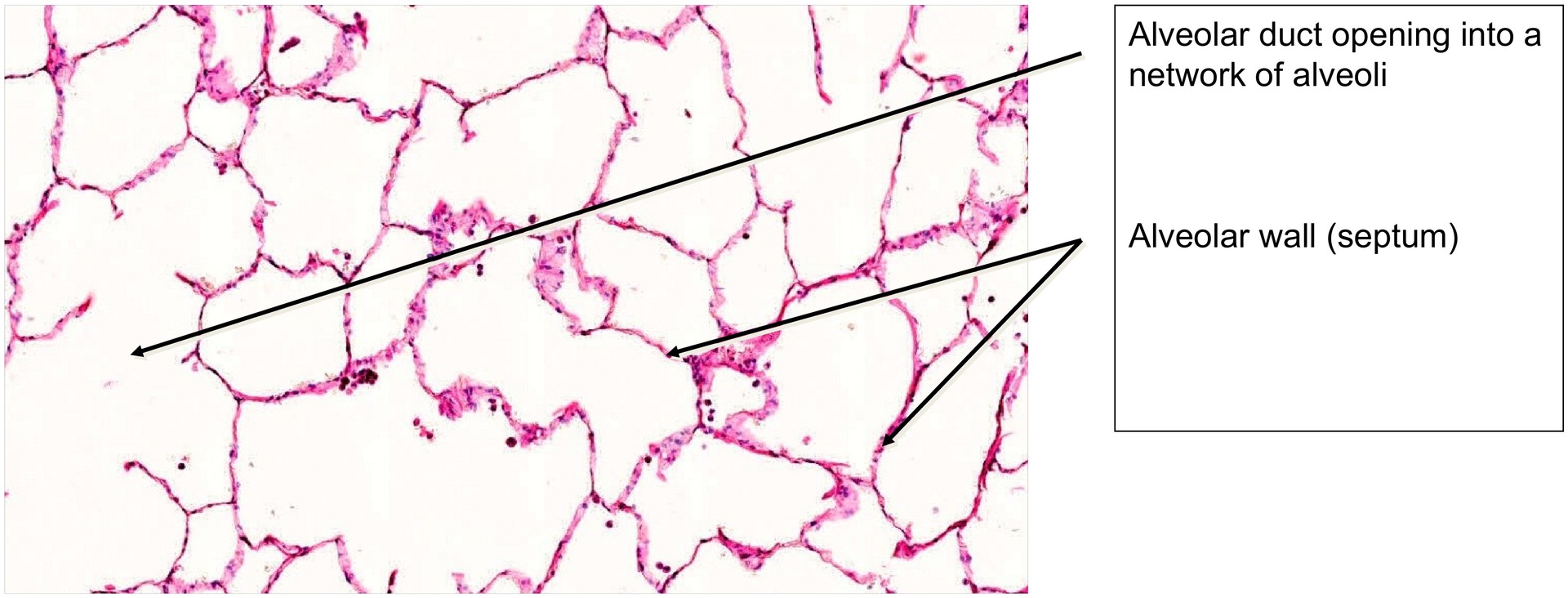
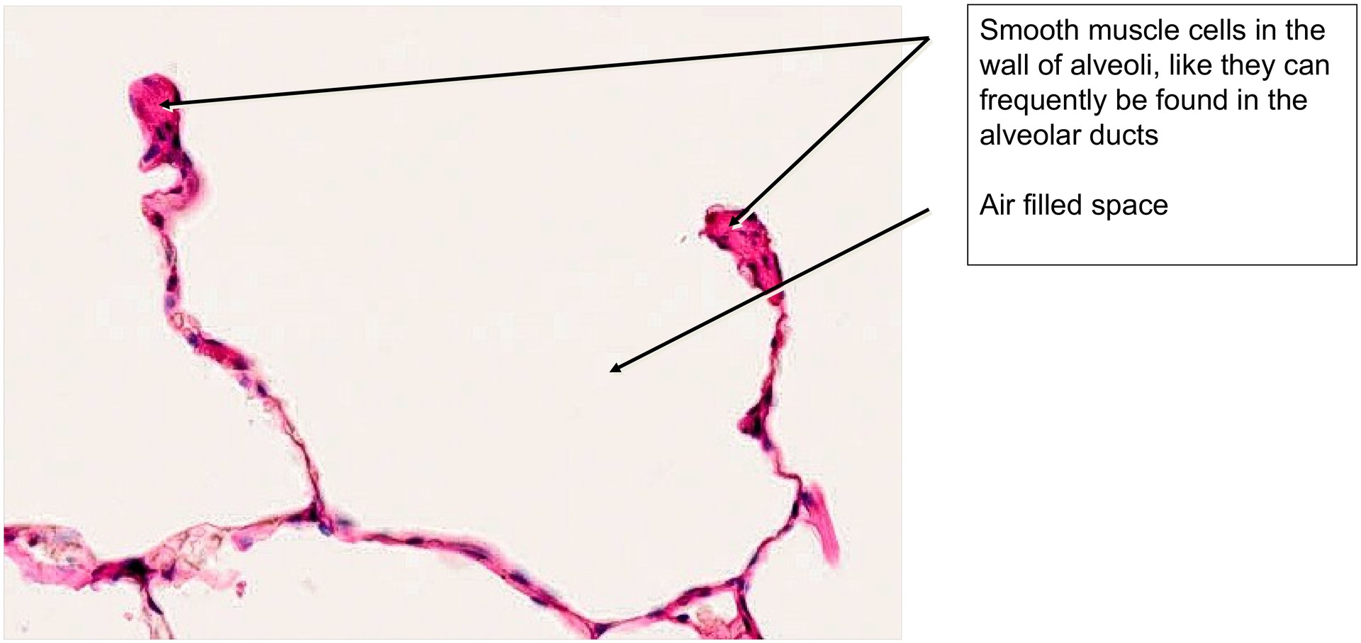
A larger air-filled space bounded by alveoli is an alveolar duct, and several alveoli that open into a single duct form an alveolar sac. Occasionally, the transition from a respiratory bronchiole to an alveolar duct can be observed.
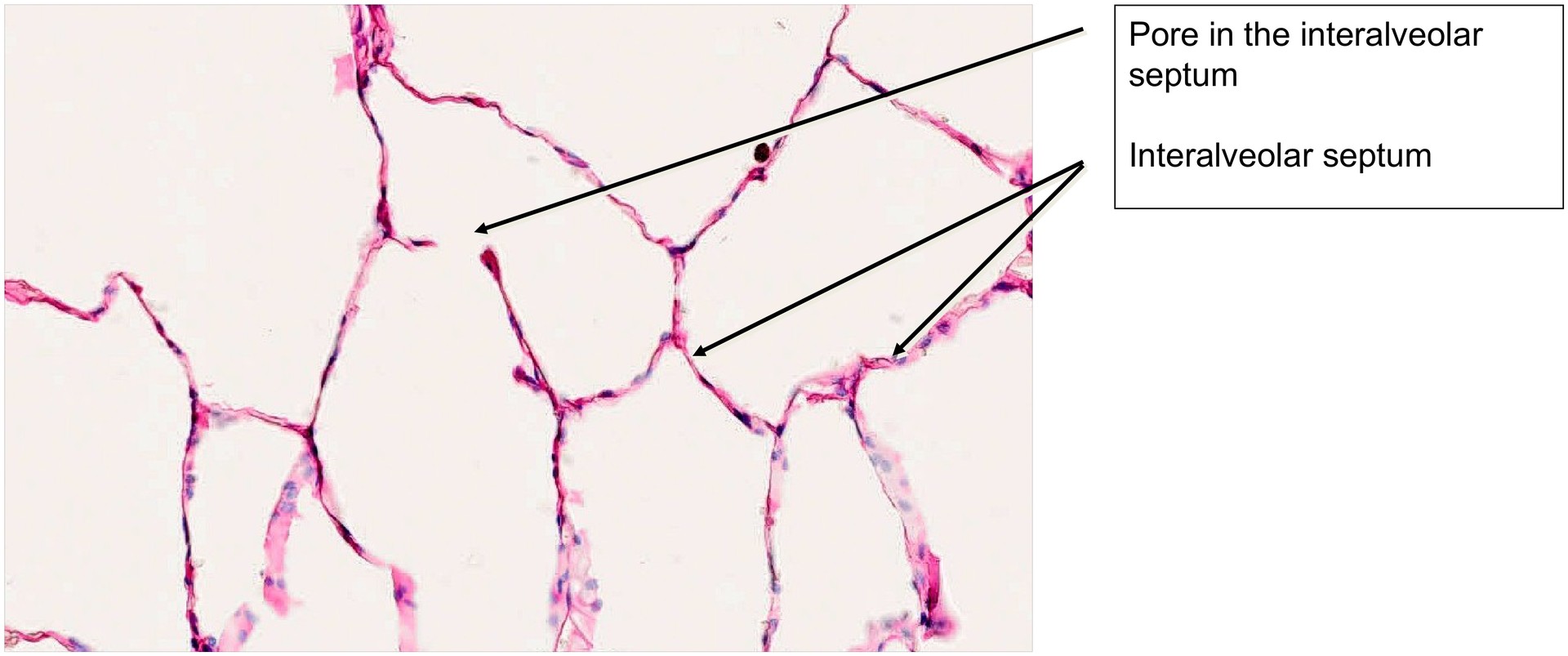
Pores between neighboring alveoli (pores of Kohn) can sometimes be seen, allowing for collateral air circulation. The walls of the alveoli are composed mainly of type I pneumocytes, with a smaller number of type II pneumocytes (responsible for surfactant secretion), and very few club cells. In this preparation, distinguishing these individual cell types or alveolar macrophages is practically impossible due to the limitations of the staining technique.
Tasks:
• Locate several bronchioles and examine their structure.
• Identify the smooth muscle within the bronchiolar wall.
• Locate the cartilaginous fragment near a bronchus.
• Identify the branches of the pulmonary artery.
• Examine the alveolar walls and compare their thickness to the size of the lining cells.
• Identify an alveolar duct and an alveolar sac.
• Locate the transition from a bronchiole to an alveolar duct.
• Identify an alveolar pore between two adjacent alveoli.

License
University of Basel
Downloads