MALE REPRODUCTIVE ORGANS (ANATOMICAL MICROSCOPY)
11.6
Testis, cat
Specimen:
Specimen Details:
Organ: Testis
Origin: Cat
Staining: Azan
Method and Specimen Description:
Normal histological section stained with Azan, in which connective tissue (collagen) appears blue, while epithelial cells, blood, and muscle cells are red.
Objective of the Examination:
To study the structure and organization of the cat testis and the various cell types involved in spermatogenesis, including Sertoli and Leydig cells.
Special Features of the Specimen:
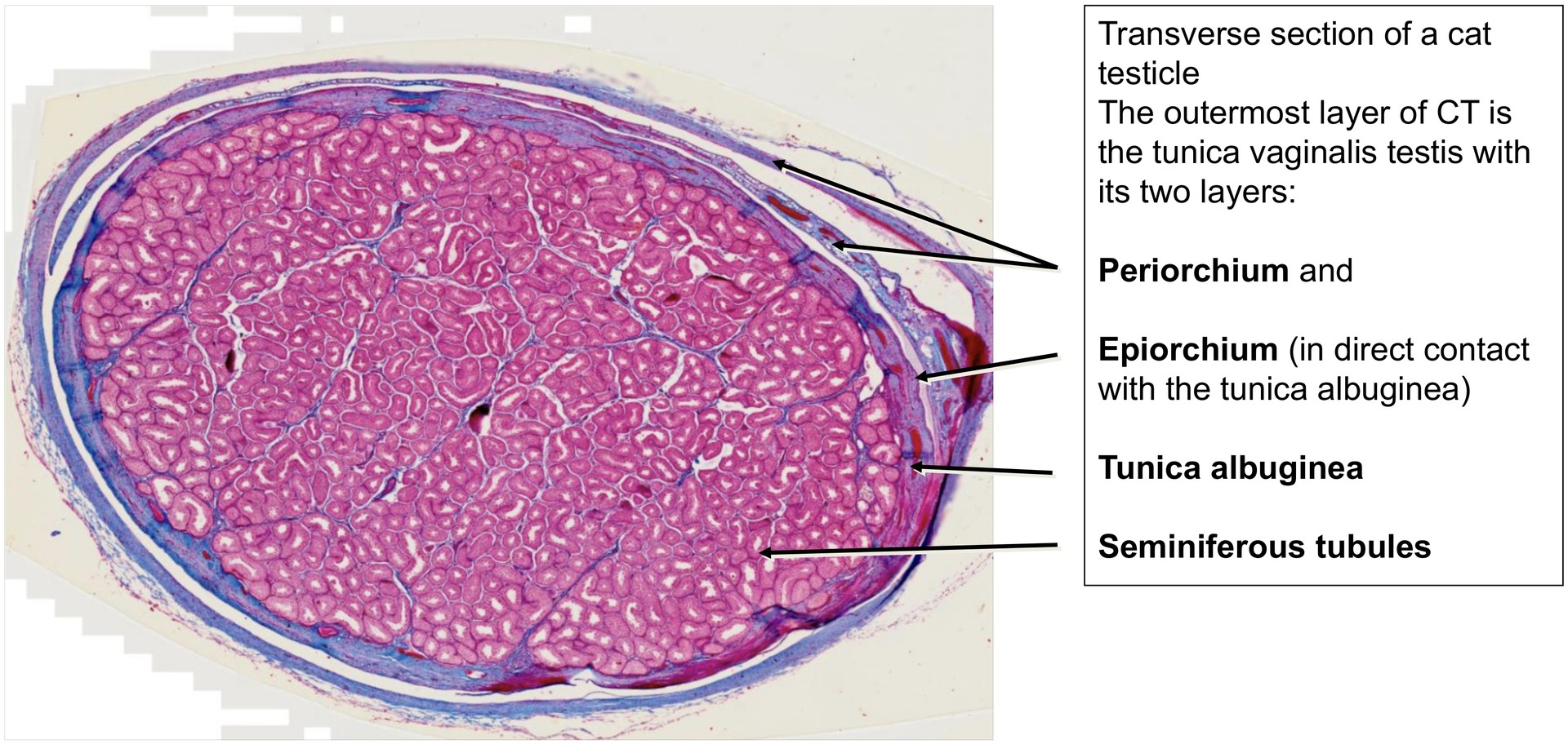
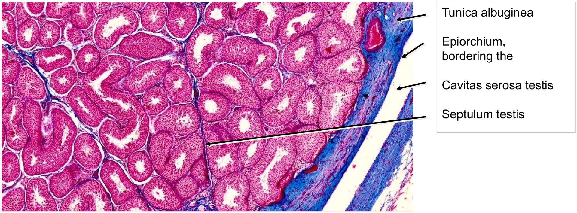
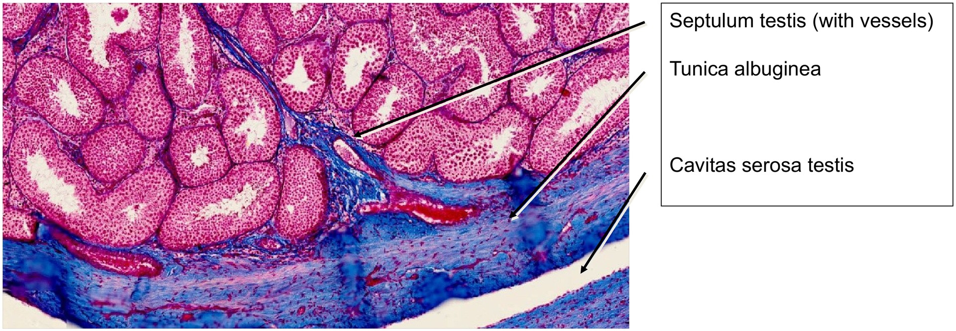
In this specimen, the tunica albuginea is clearly visible and well-defined in Azan staining. It gives rise to connective tissue septa (septula testis) that subdivide the testis into lobules. The testis is externally enveloped by the tunica vaginalis testis, which consists of two layers: the epiorchium, directly covering the tunica albuginea, and the periorchium, which defines the outer boundary of the cavitas serosa testis, allowing free movement of the testis within the scrotum.
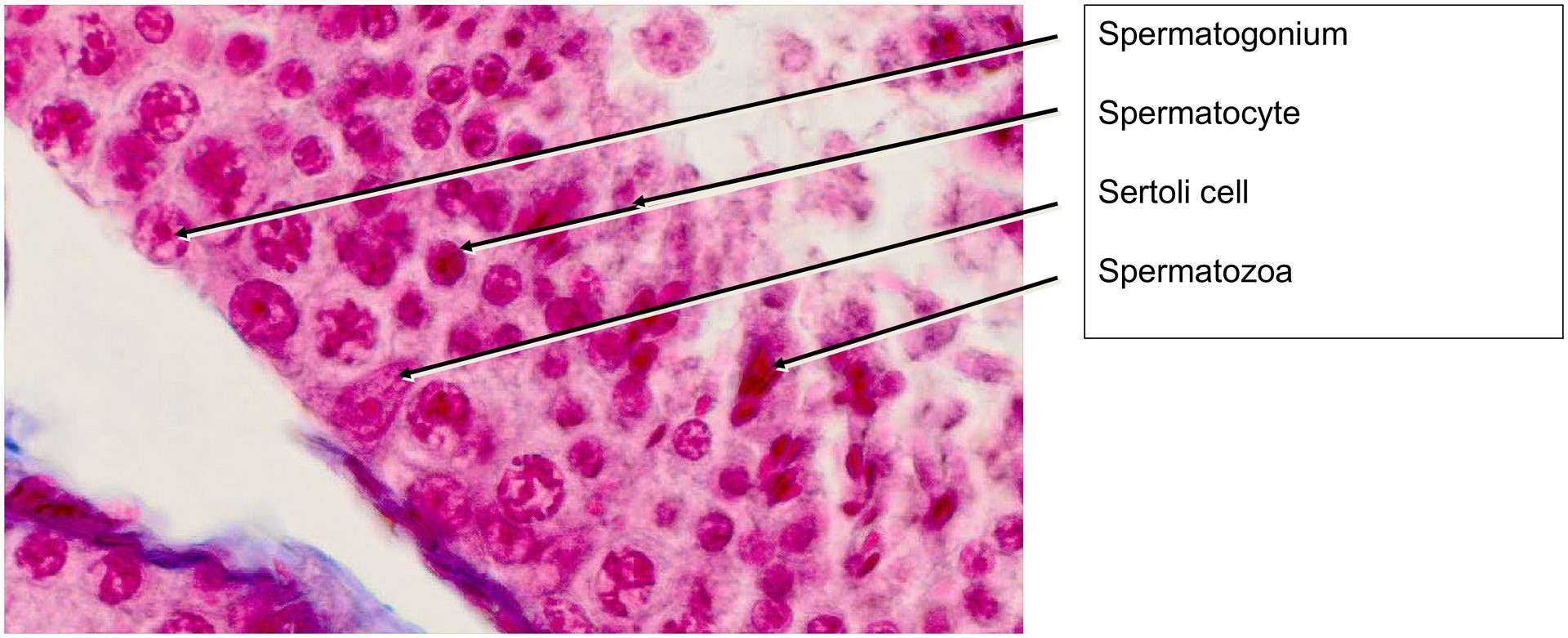
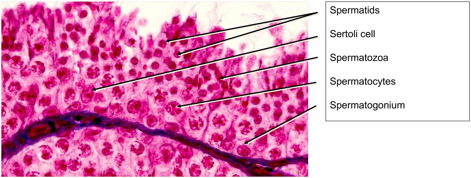
The seminiferous tubules are highly active, as evidenced by the abundance of mature spermatozoa in the adluminal region of the epithelium. The sperm heads, filled with condensed chromatin, stain bright red, making them easily identifiable. Within the epithelium, all major stages of spermatogenesis are visible — from spermatogonia (basal layer) through primary spermatocytes, spermatids, and finally spermatozoa. Because of their short lifespan and resemblance to primary spermatocytes, secondary spermatocytes are rarely distinguishable.
Sertoli cells, the supporting (nurse) cells of spermatogenesis, are well represented. Their oval to pear-shaped nuclei are situated roughly between the first and second thirds of the epithelial height (from the basal membrane). The spermatogonia rest on the basal lamina, exhibiting round nuclei. The primary spermatocytes are located in the middle third, with loosely structured chromatin, while the spermatids, smaller and more condensed, lie adluminally in small groups.
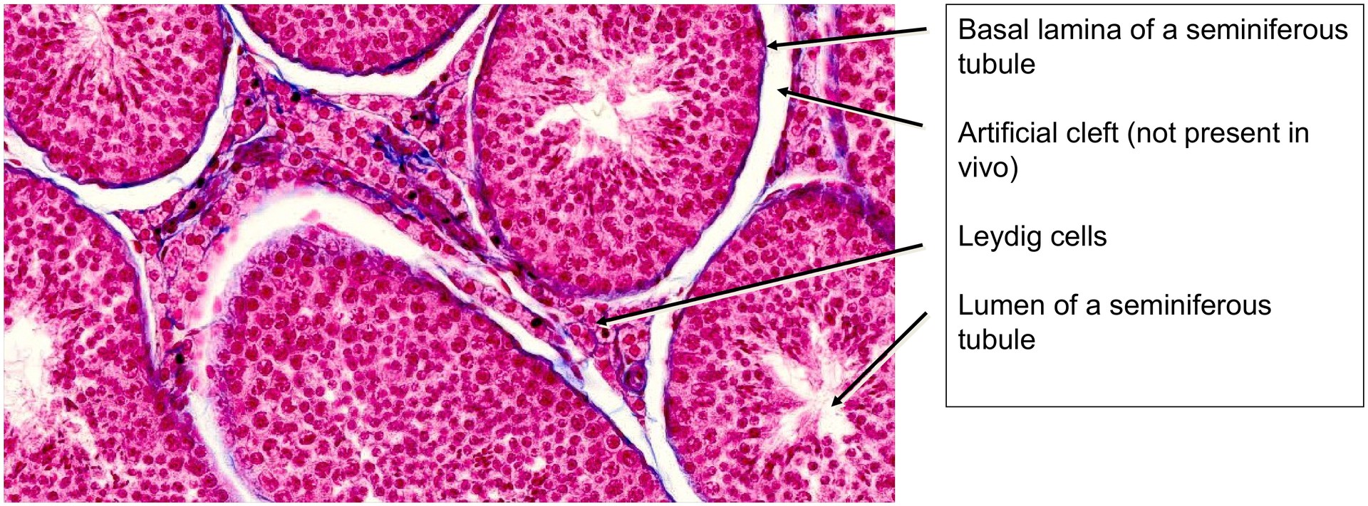
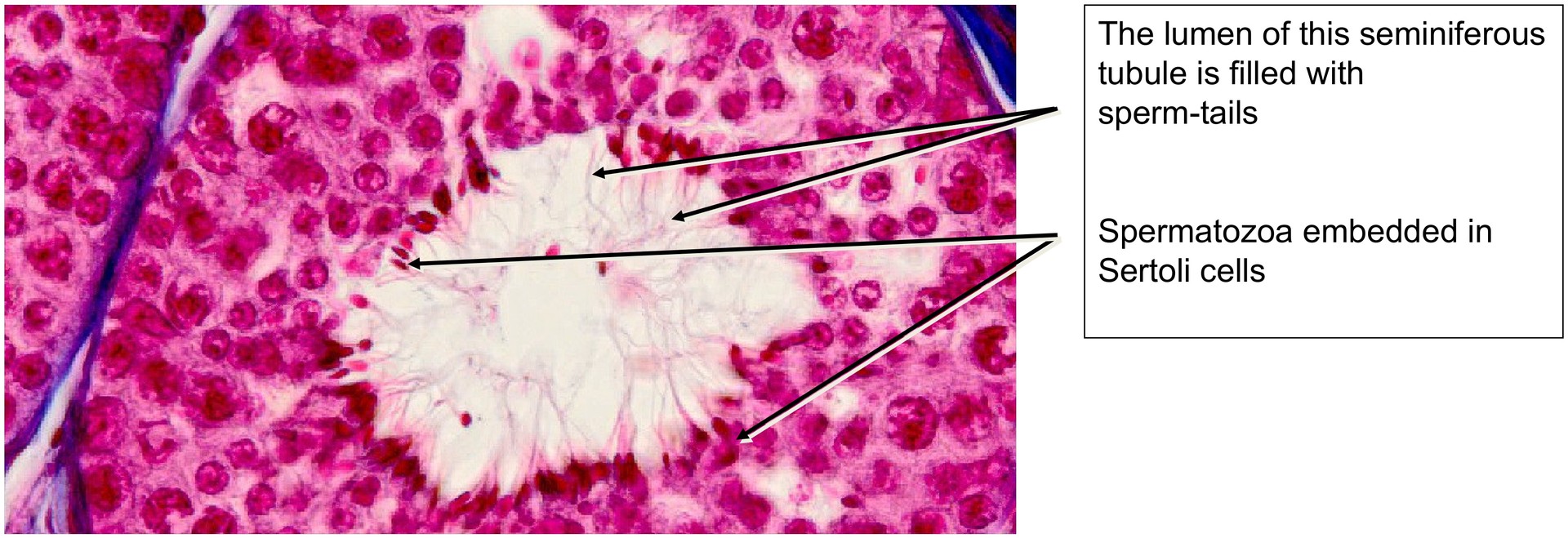
The spermatozoa are often seen with their heads still embedded in Sertoli cells, while their tails project into the lumen of the seminiferous tubules. The interstitial tissue between tubules contains numerous Leydig cells, easily recognized as polygonal cells with pale cytoplasm, responsible for testosterone production. The basement membrane of the seminiferous tubules, rich in collagen fibers, stains intensely blue, giving the tubules a distinct outline.
Tasks:
-
Obtain an overview at low magnification and delineate the layers of the tunica vaginalis testis (epiorchium and periorchium) relative to the tunica albuginea.
-
Identify Leydig cells in the interstitial tissue. What is their function?
-
Locate the septula testis (incomplete partitions between lobules) and describe their composition.
-
Identify Sertoli cells within the seminiferous tubules. What are their roles in spermatogenesis?
-
Recognise spermatogonia based on their basal position and nuclear morphology.
-
Identify primary spermatocytes and spermatids, and compare their nuclear appearance.
-
Discuss the chromosome number at each stage of spermatogenesis:
-
Spermatogonia: diploid (2n)
-
Primary spermatocytes: diploid (2n, after DNA replication 4c)
-
Secondary spermatocytes: haploid (n, 2c)
-
Spermatids and spermatozoa: haploid (n, 1c)
-
License
University of Basel
Downloads