EPITHELIUM (GENERAL HISTOLOGY)
1.2
Non-keratinized stratified squamous epithelium (esophagus)
Specimen:
Specimen Details:
Organ: Esophagus
Origin: Human (Child)
Staining: Hematoxylin - Eosin (H&E)
Method and Specimen Description:
This is a standard histological section of a child’s esophagus stained with hematoxylin and eosin (H&E). Because of its smaller size, the entire circumference of the child’s esophagus can be accommodated on a single slide, unlike that of an adult, which would be too large for complete sectioning.
Objective of the Examination:
To study the structure of stratified, non-keratinized squamous epithelium and to identify portions of the esophageal glands (tubulo-acinar secretory end-pieces and their excretory ducts).
Special Features of the Specimen:
Structure of the Epithelium (medium and high magnification)
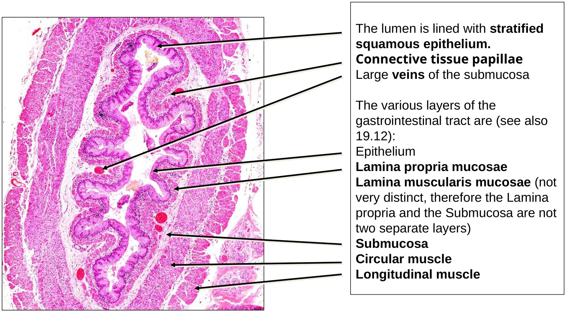
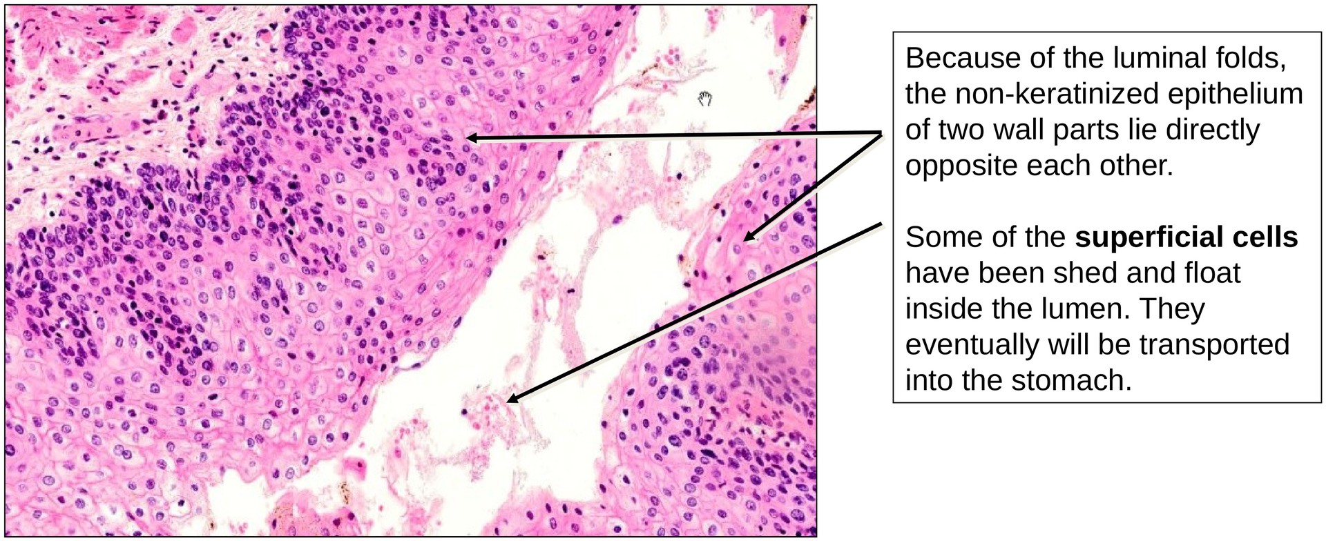
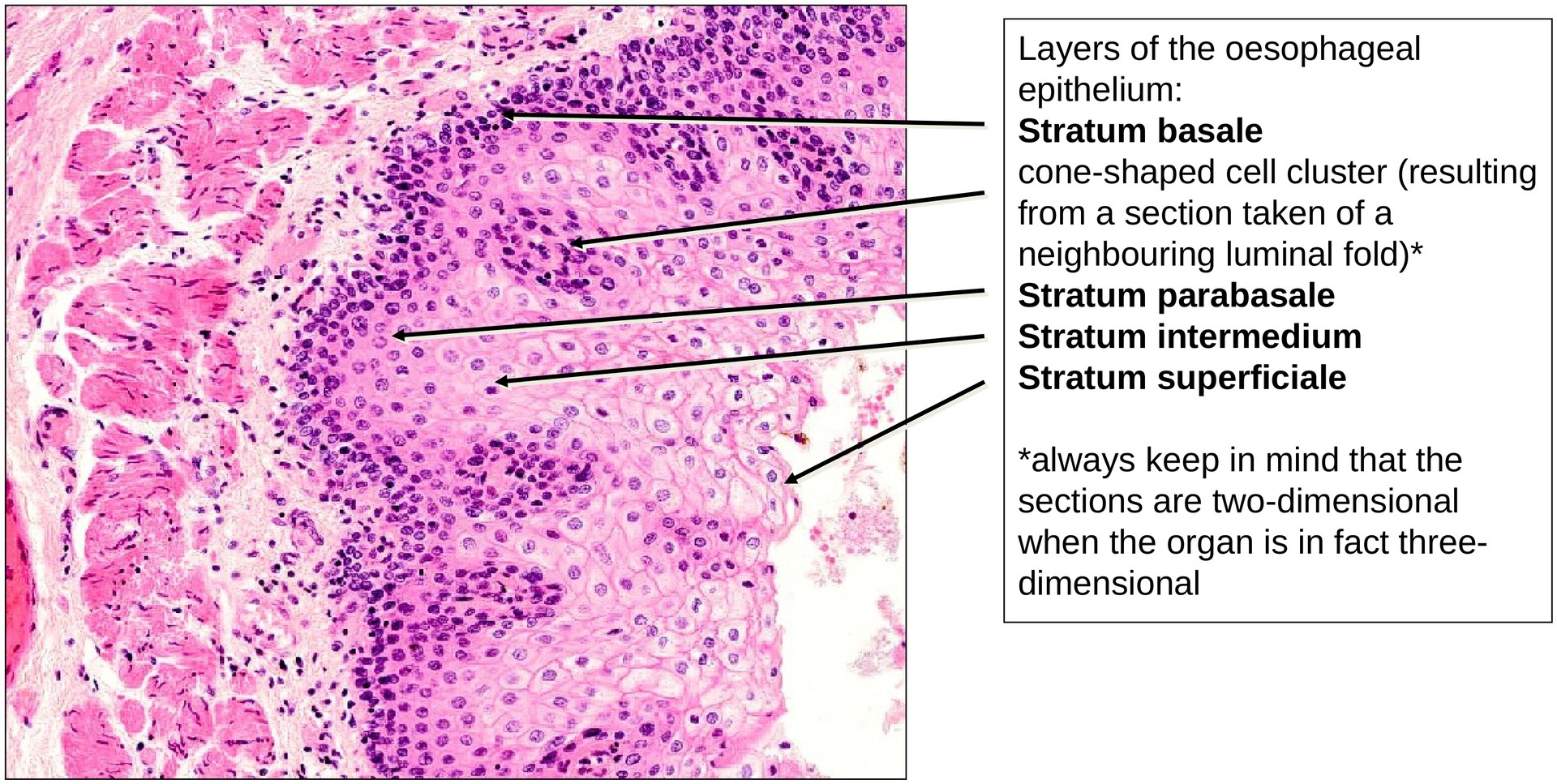
The mucosa of the esophagus is lined by stratified squamous non-keratinized epithelium, in which the most superficial cells retain their nuclei. From the basement membrane to the lumen, the following layers can be distinguished:
- Stratum basale
- Stratum parabasale
- Stratum intermedium
- Stratum superficiale
Cells of the stratum basale and stratum parabasale show strong basophilia, reflecting high levels of protein synthesis necessary for constant cell renewal, as superficial cells are shed during swallowing.
In the stratum intermedium, the cells progressively lose cytoplasmic organelles and become more eosinophilic. Nuclear condensation (pyknosis) may also be observed.
The stratum superficiale consists of flattened, squamous cells which still contain visible nuclei — a defining feature distinguishing this epithelium from keratinised squamous epithelium.
Within the submucosa, tubulo-acinar glands can be observed. Their mucous components stain bluish and secrete mucus that lubricates the esophageal lumen, facilitating the passage of food. The ducts of these glands are rarely captured in the plane of section.
Epithelium–Connective Tissue Junction:
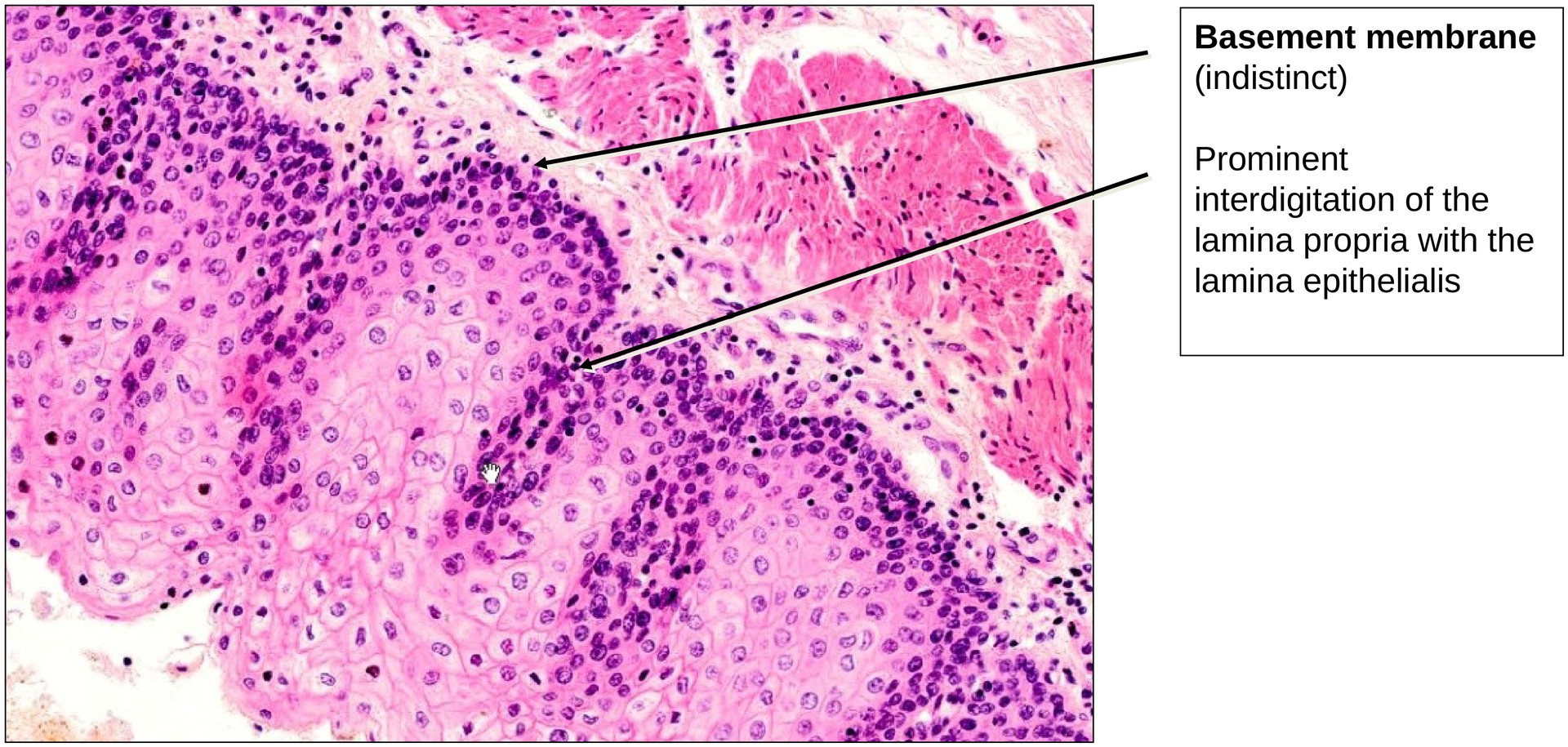
As in all epithelia subjected to significant mechanical stress, the junction between the epithelium and the underlying lamina propria is highly interdigitated. Finger-like connective tissue papillae project into the lower epithelial layers, strengthening the mechanical attachment between tissue layers.
Because of oblique sectioning, some papillae may appear as isolated islands within the epithelium; these are sectioning artefacts, not true histological features.
Tasks:
- Identify the type of epithelium and describe its layers.
- Observe the gradual loss of cytoplasmic stainability towards the lumen and interpret its cause.
- Confirm the presence of cell nuclei in the most superficial cell layers.
- Recognise sectioning artefacts, such as the apparent isolated connective tissue papillae.
- Examine the interdigitation between the epithelium and the underlying lamina propria.
License
University of Basel
Downloads