DIGESTIVE ORGANS: GASTROINTESTINAL TRACT (ANATOMICAL MICROSCOPY)
19.8
Jejunum 2
Präparat:
Specimen Details:
Organ: Jejunum
Origin: Human
Staining: Hematoxylin - Eosin (H&E)
Method and Specimen Description:
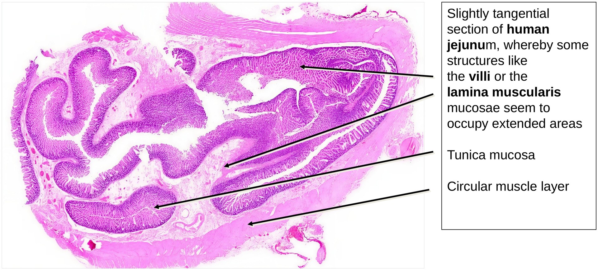
Normal histological specimen stained with H&E, a general overview stain that clearly highlights Paneth cell granules in the crypts. The section is tangential in parts, which makes certain layers—such as the lamina muscularis mucosae—appear planar in places.
Objective of the Examination:
To study the structure of the jejunum, particularly the mucosa and the typical layered organization of the gastrointestinal tract (GIT).
Special Features of the Specimen:
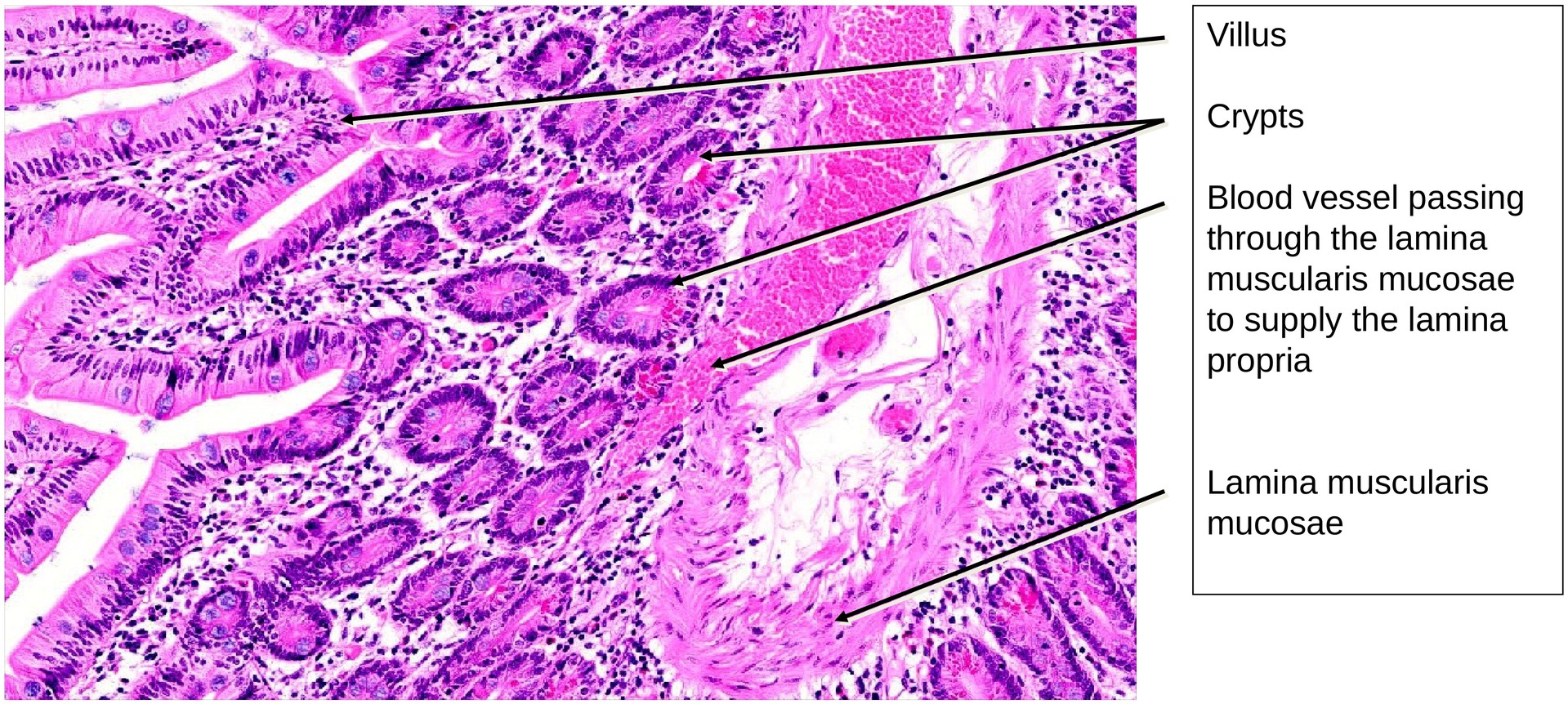
The jejunum shows the characteristic structure of the GIT, consisting of the following layers: - Tunica mucosa (with the lamina epithelialis, lamina propria, and lamina muscularis mucosae) - Tela submucosa (containing the submucosal plexus) - Tunica muscularis (composed of inner circular and outer longitudinal muscle layers)
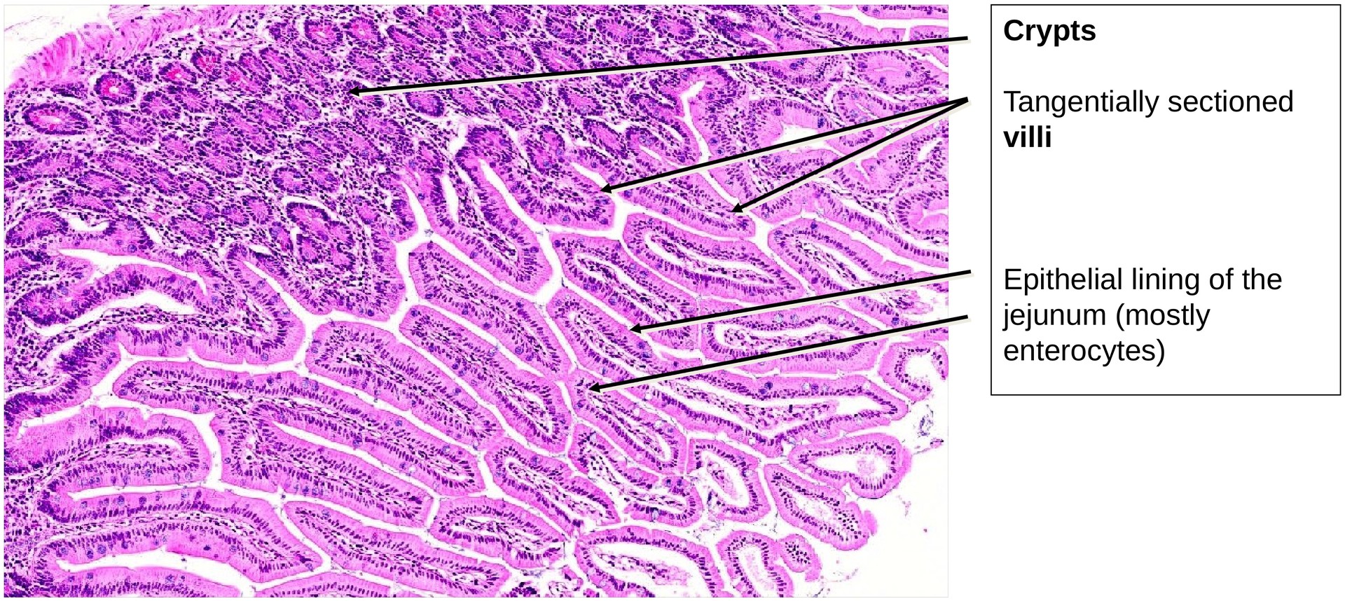
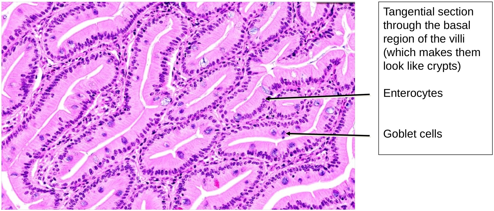
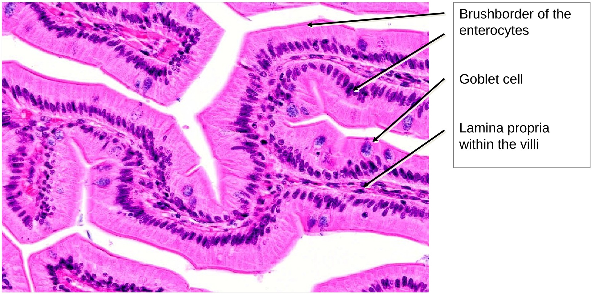
The epithelium is composed of enterocytes (brush-border cells) and goblet cells. The mucosa forms plicae circulares (folds), bearing finger-like villi on their surface. At the base of the intervillar spaces lie the openings of the crypts (crypts of Lieberkühn).
The goblet cells are easily visible even without special staining; in some areas, they appear to have been fixed during mucus secretion.
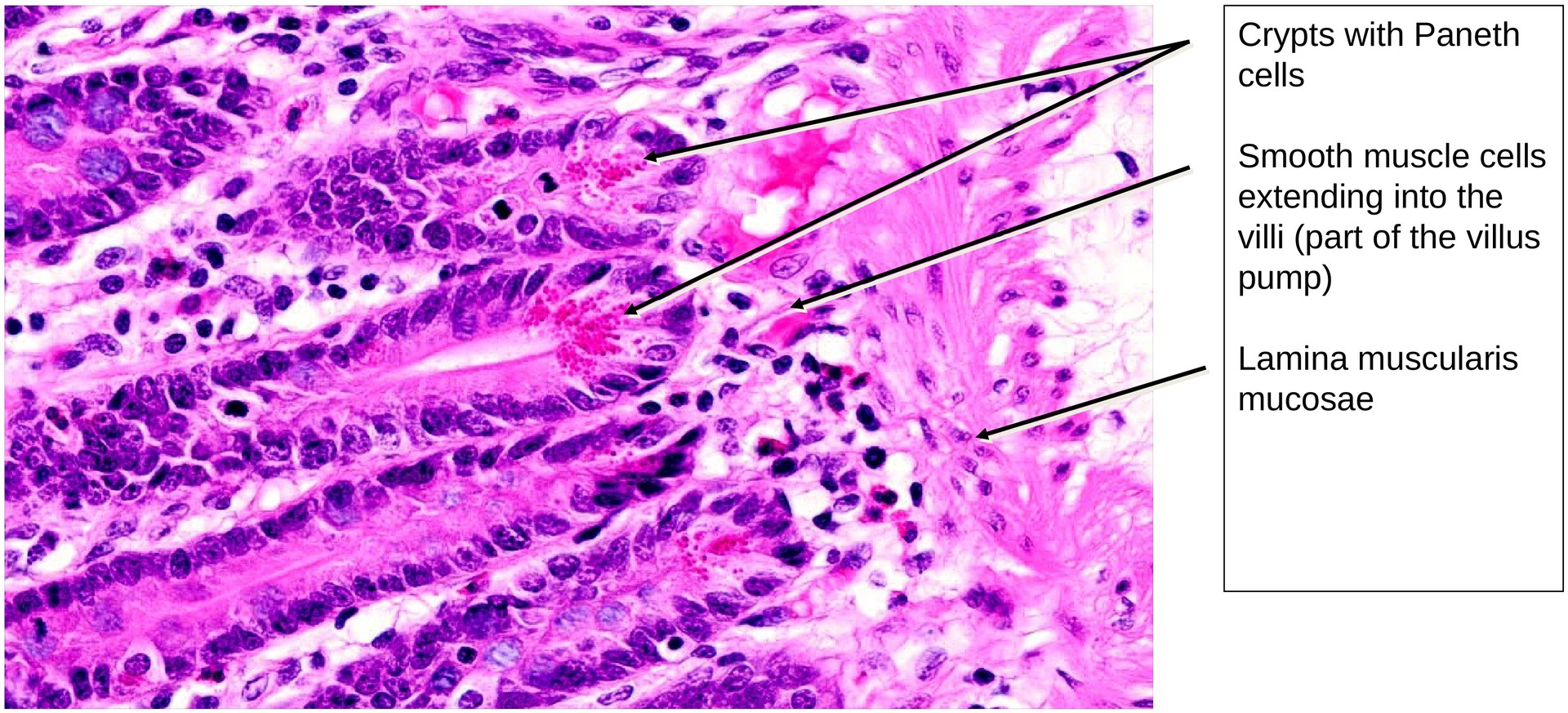
The lamina propria forms a relatively thin layer beneath the epithelium. The lamina muscularis mucosae is clearly developed; due to the tangential orientation of the section, it appears planar in several areas. Individual smooth muscle cells can be seen extending into the villi, forming the basis of the villus pump mechanism.
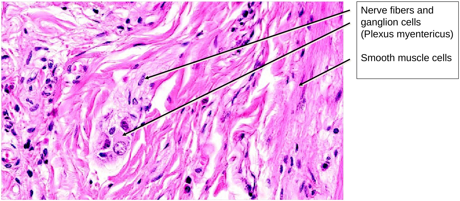
The submucosal plexus (Meissner’s plexus) in the tela submucosa is not as distinct as the myenteric plexus (Auerbach’s plexus), which lies between the circular and longitudinal muscle layers and contains ganglion cells at various points.
Within the crypts, Paneth cells are clearly visible; their apical granules are strongly eosinophilic, giving them a characteristic appearance in H&E staining.
Tasks:
- Identify the layers of the gastrointestinal tract (GIT) at low magnification:
- Tunica mucosa (lamina epithelialis, lamina propria, lamina muscularis mucosae)
- Tela submucosa
- Tunica muscularis (inner circular and outer longitudinal muscle layers)
- Trace the course of the muscle cells in the lamina muscularis mucosae where they appear planar.
- Describe the difference between folds and villi. Which structural components of the GIT occur in both?
- Determine where most goblet cells are found in this specimen.
- Name the second epithelial cell type of the lamina epithelialis.
- Locate and identify the submucosal plexus (Meissner’s) and myenteric plexus (Auerbach’s). In which layers can they be found?
- Identify the Paneth cells and their apical granules. In which structures are they located, and how are they stained?
License
University of Basel
Downloads