LYMPHATIC ORGANS (ANATOMICAL MICROSCOPY)
15.10
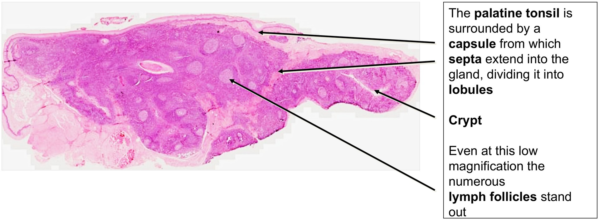
Palatine tonsil
Specimen Details:
Specimen Details:
Organ: Palatine Tonsil
Origin: Human
Staining: Hematoxylin-Eosin (H&E)
Method and Specimen Description:
Conventional histological section stained with a general overview stain (H&E).
Objective of the Examination:
To study the microscopic structure of the palatine tonsil, including its epithelial covering, crypts, lymphoid tissue organization, and stromal framework.
Special Features of the Specimen:
The palatine tonsil forms part of the pharyngeal lymphoid ring and, being covered by the epithelium of the oral cavity and pharynx, is considered part of the MALT (mucosa-associated lymphoid tissue) system.
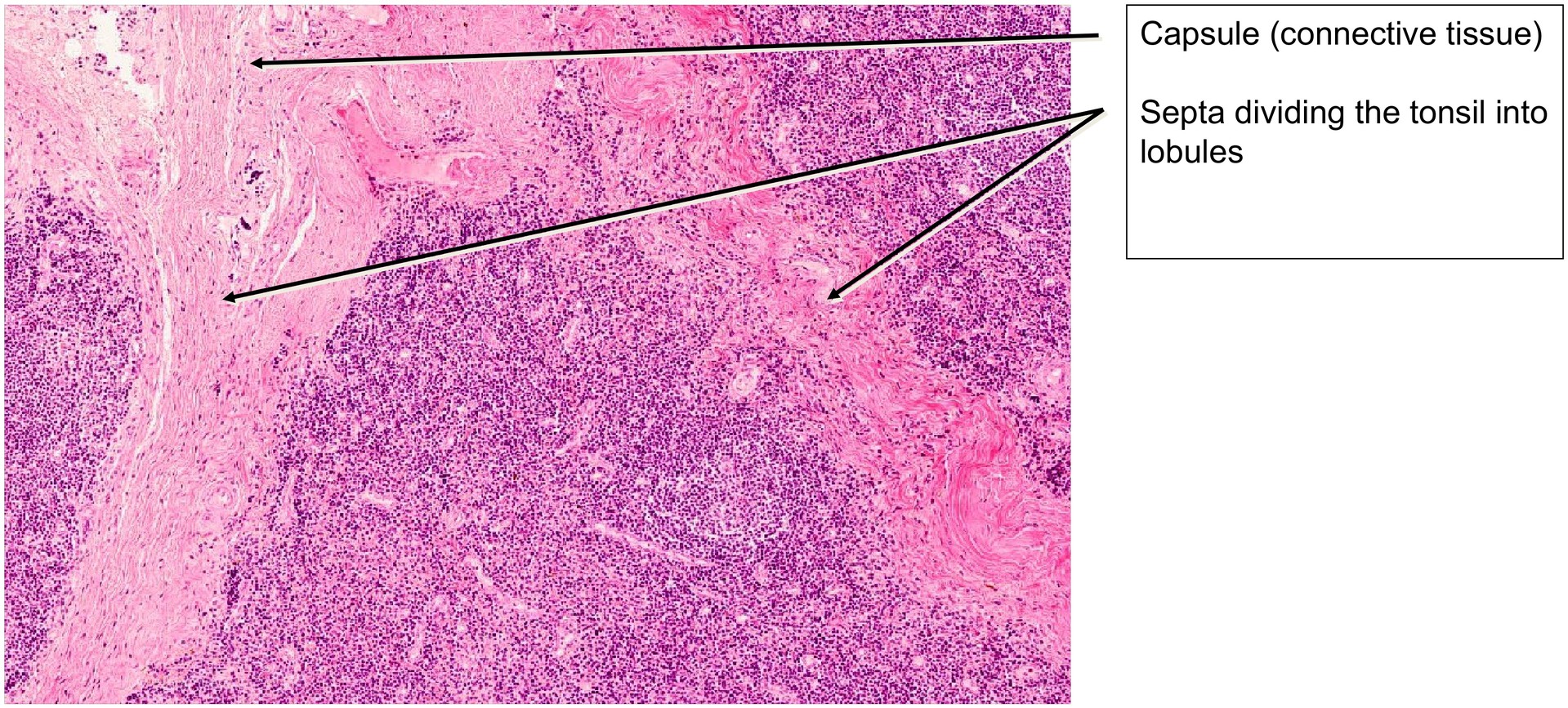
Like the lingual tonsil, it exhibits epithelial crypts, which in this case may extend even deeper into the organ. The tonsil is divided into lobules by fibrous connective tissue septa extending from a surrounding fibrous capsule.
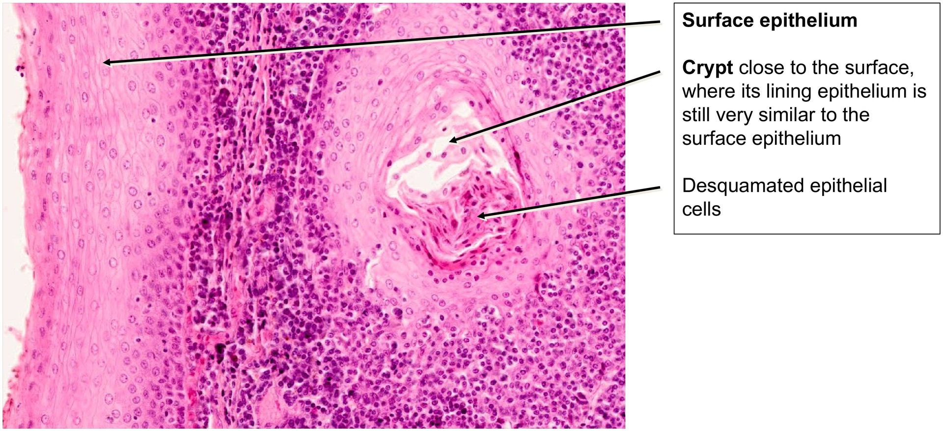
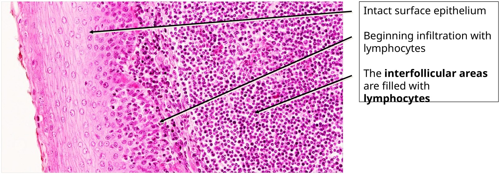
The epithelium of the crypts is heavily infiltrated by lymphocytes, often to the point where its epithelial structure is no longer clearly recognizable. The crypt lumina frequently contain cellular debris, shed epithelial cells, and degenerating lymphocytes.
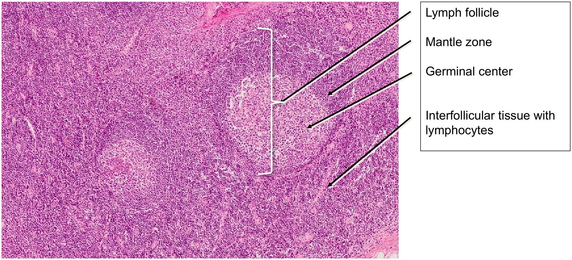
Within the lymphoid tissue:
- B-lymphocytes predominate in the lymphatic follicles, which display a light germinal center and a darker mantle zone (lymphocyte shell).
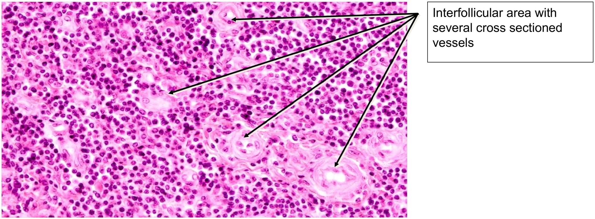
- T-lymphocytes are mainly located in the interfollicular regions, which also contain small blood vessels and capillaries.
Because of the high cellular density, the reticular (lymphoepithelial) framework of the tonsil is difficult to distinguish in the interfollicular areas.
Tasks:
- Identify the fibrous capsule and septa and recognize how they divide the tonsil into lobules.
- Compare the surface epithelium with the epithelium of the crypts.
- Determine the type of surface epithelium (hint: it corresponds to the oral mucosa type).
- Examine the lymphatic follicles and describe the difference between the germinal center and the mantle zone.
- Assess the contents of the crypts — note the presence of cell debris and infiltrating immune cells.
- Search for arterioles and other blood vessels in the interfollicular areas and compare the vascular pattern with that observed in the spleen.
License
University of Basel
Downloads