LYMPHATIC ORGANS (ANATOMICAL MICROSCOPY)
15.4
Marzipan, Cat 1
Preparation:
Preparation Details:
Organ: Spleen
Origin: Cat
Staining: Hematoxylin - Eosin (H&E)
Method and Specimen Description:
Conventional histological section stained with H&E for general morphological assessment.
Objective of the Examination:
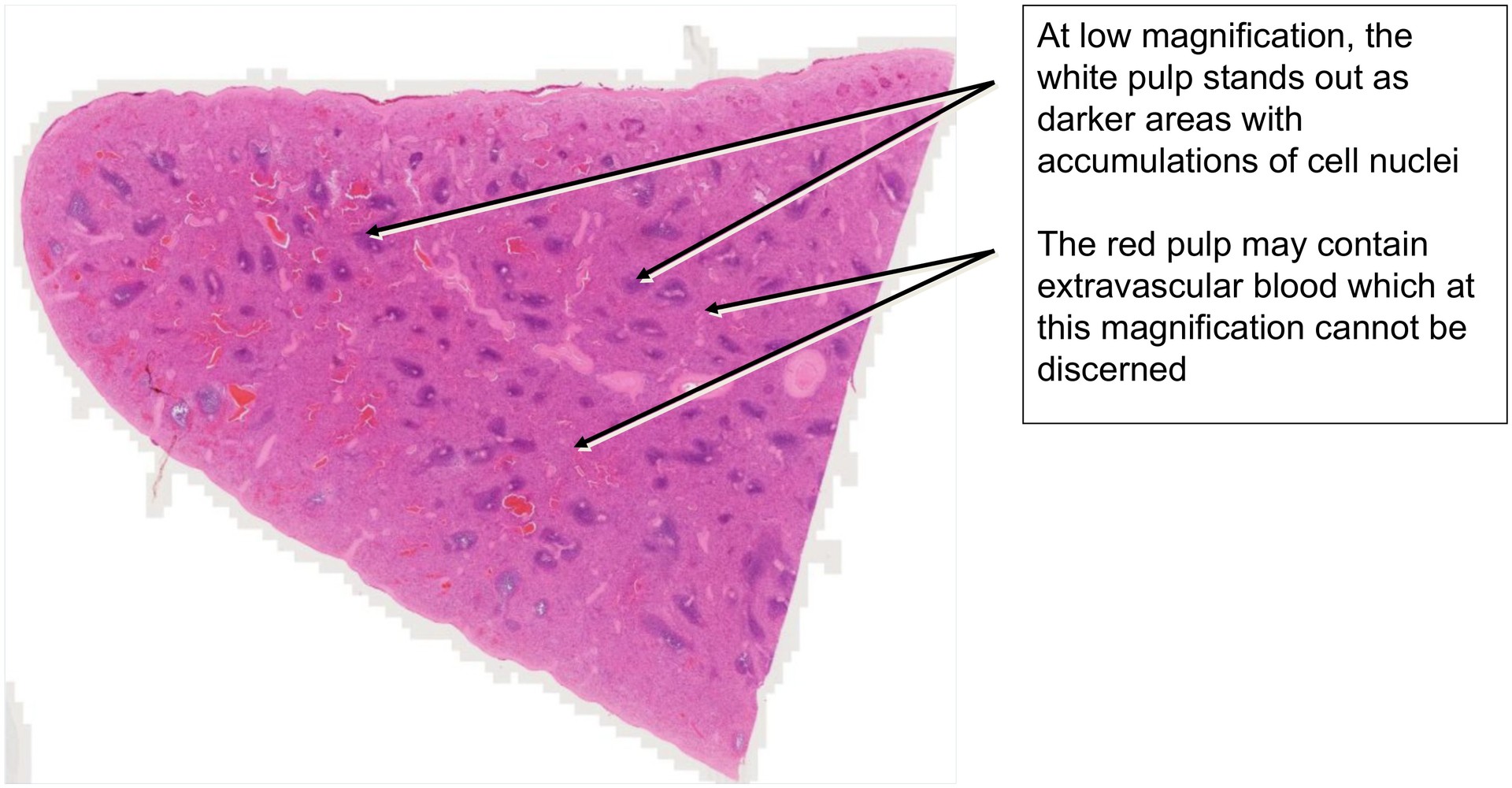
To study the spleen as a lymphatic organ incorporated as a filter station within the blood circulation. The spleen occupies a unique position due to its partly open blood circulation. The aim is to recognize and differentiate the white pulp and red pulp.
This preparation should be examined before the other spleen specimens (Spleen, Human and Spleen, Cat 2, stained with Gomöri).
Special Features of the Preparation:
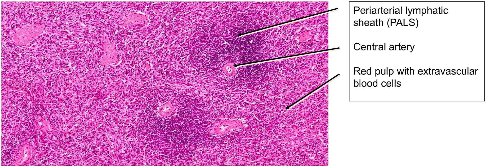
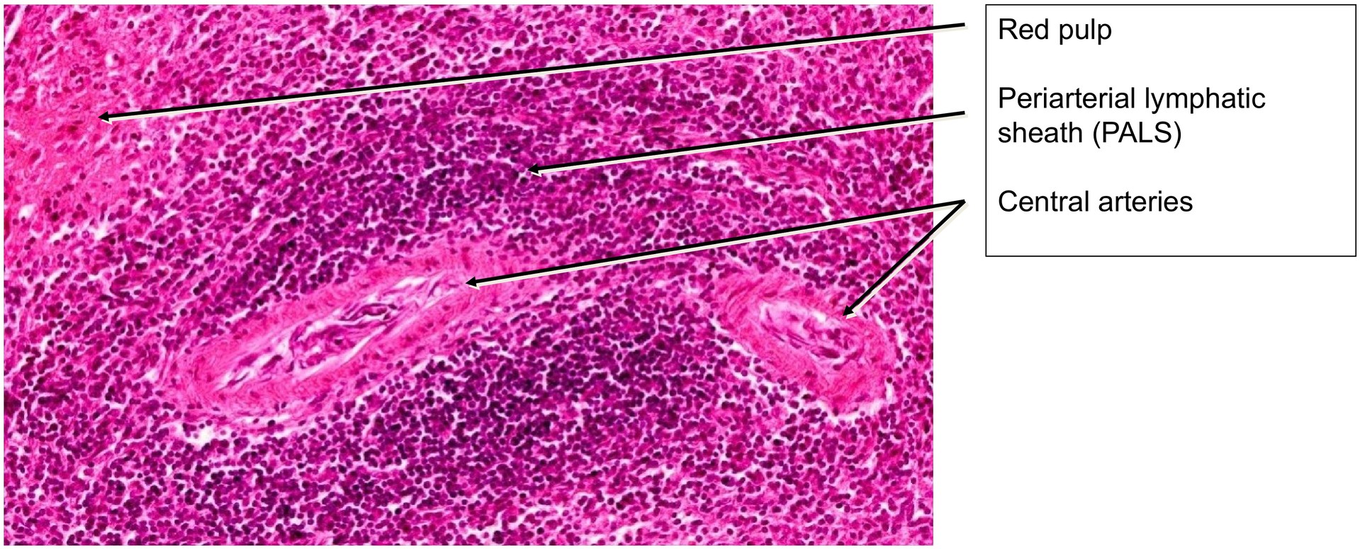
Even at low magnification, the white splenic pulp can be clearly identified as darker areas containing dense accumulations of cell nuclei. Larger, rounded accumulations represent lymph follicles, while smaller ones correspond either to tangential sections through the marginal regions of follicles or to periarterial lymphatic sheaths (PALS).
Together with the marginal zone adjacent to the follicles, these structures constitute the white pulp.
- The PALS contain T-lymphocytes.
- The lymph follicles and marginal zones contain predominantly B-lymphocytes.
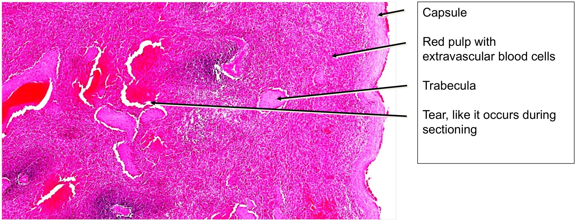
The red pulp is composed of splenic (pulp) cords and venous sinuses. Because part of the splenic blood flow is open, blood from the brush arterioles enters the reticular meshwork of the red pulp via sheathed capillaries before re-entering the sinusoids through endothelial gaps. As a result, extravascular blood can be observed in various locations.
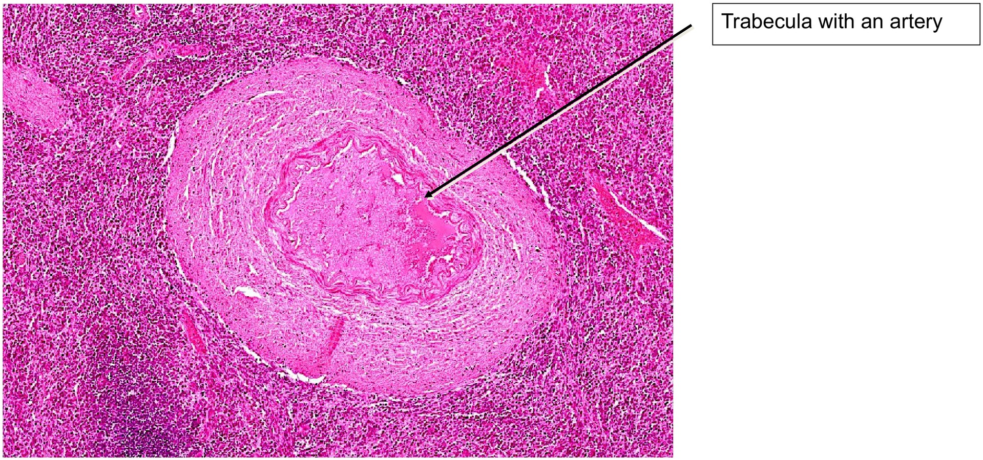
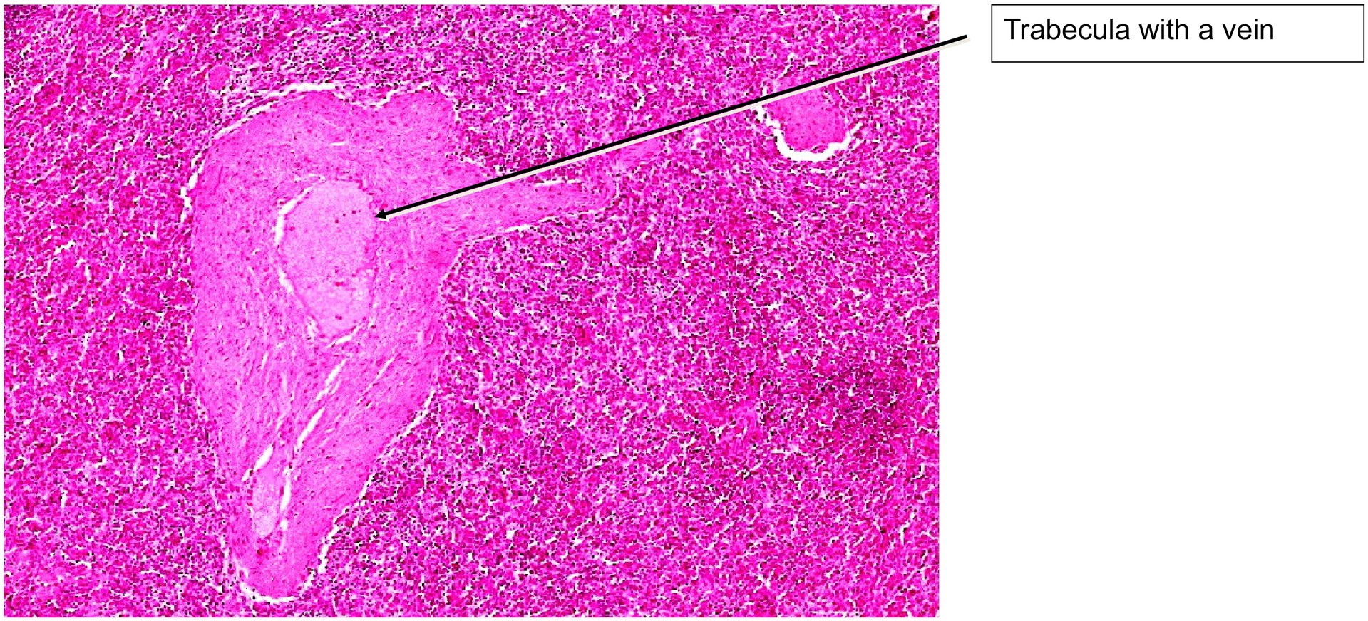
The stroma of the spleen consists of a dense connective tissue capsule and the trabeculae that extend inward from it. Trabeculae from the hilum convey vessels and nerves. The blood supply proceeds via trabecular arteries, from which central arteries branch. These central arteries are surrounded by periarterial lymphatic sheaths containing T-lymphocytes. The central arteries continue towards the lymph follicles, dividing into arterial capillaries (also termed sheathed capillaries).
Due to the differing consistencies within the red pulp (particularly where extravascular blood accumulates), the tissue may occasionally tear during sectioning.
Tasks:
- At low magnification, assess the parenchyma and stroma of the spleen, and distinguish between white pulp and red pulp.
- Identify the various vascular segments:
- Trabecular arteries and trabecular veins
- Central arteries
- Brush arteries
- Venous sinuses
- Locate areas containing extravascular blood, characteristic of the red pulp.
- Identify the periarterial lymphatic sheaths (PALS) surrounding the central arteries.
- Attempt to identify fibroblastic reticular cells, which form the cellular framework of the spleen.
- Trace the capsule and trabeculae, noting their composition of dense collagenous connective tissue.
License
University of Basel
Downloads