FEMALE REPRODUCTIVE ORGANS (ANATOMICAL MICROSCOPY)
10.12
Lactating breast tissue
PREPARATION DETAILS:
Organ: Lactating breast tissues
Origin: Human
Staining: Azan
METHOD AND SPECIMEN DESCRIPTION:
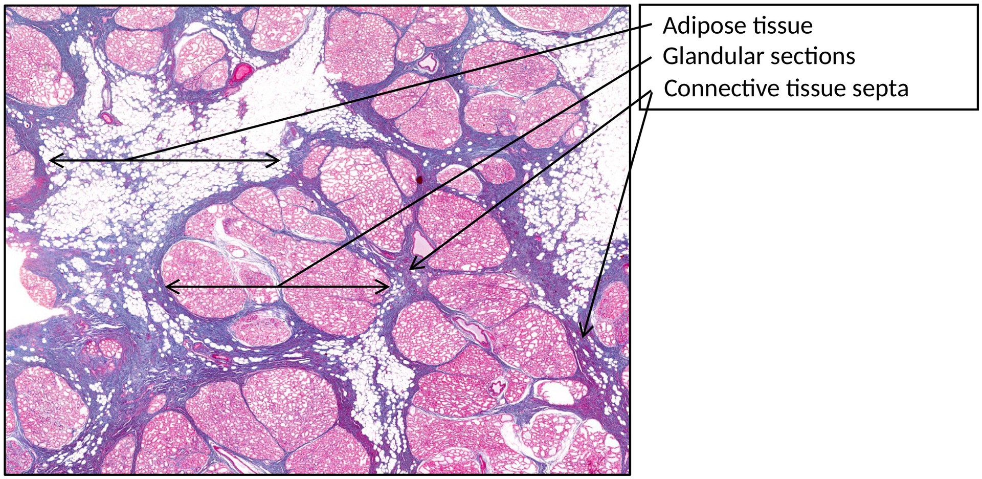
Histological section of a human lactating mammary gland. The Azan stain clearly differentiates connective tissue (blue) from epithelial and secretory components, allowing easy comparison with the non-lactating breast.
OBJECTIVE OF THE EXAMINATION:
To examine the structure of the lactating mammary gland and compare it with that of the non-lactating breast.
SPECIAL FEATURES OF THE PREPARATION:
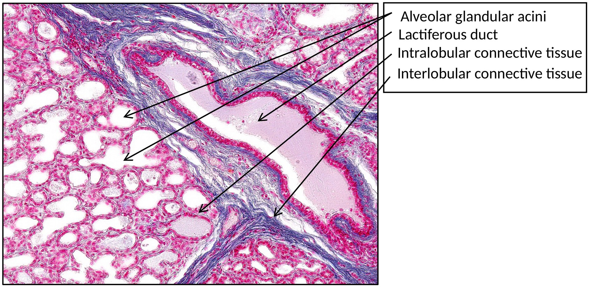
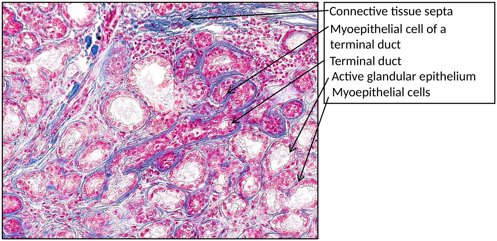
The lactating breast shows a marked increase in glandular tissue, consisting of numerous secretory alveoli (acini) lined by cuboidal to columnar glandular epithelial cells.
-
Milk production occurs within these epithelial cells and is released into the alveolar lumen by two mechanisms:
-
Apocrine secretion, in which part of the cell membrane and cytoplasm are released with the milk fat.
-
Merocrine secretion, in which milk proteins are secreted by exocytosis.
-
-
The terminal ducts collect the secretions and convey them to the lactiferous ducts.
-
Myoepithelial cells are present beneath the glandular epithelium and along the ducts; their contraction assists in the ejection of milk.
-
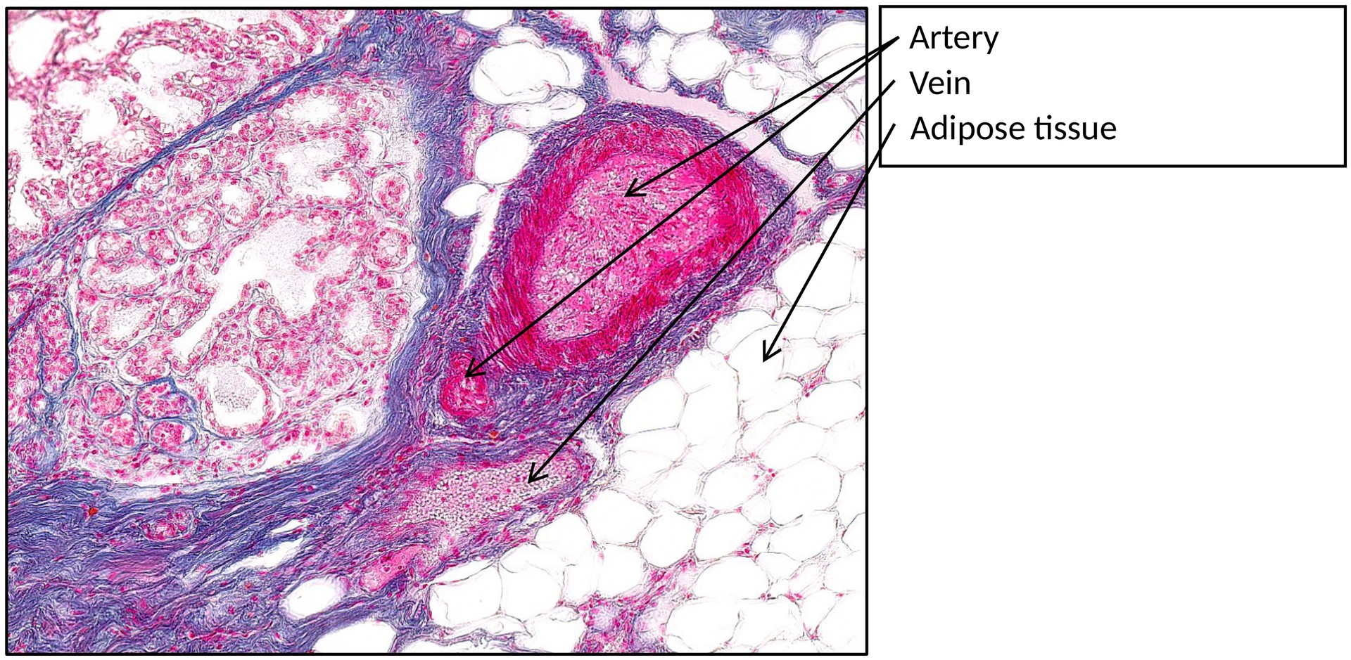
The glandular parenchyma is organized into lobules separated by connective tissue septa, with adipose tissue interspersed between lobules.
-
Compared to the non-lactating breast, there is a greatly reduced proportion of connective tissue and a corresponding expansion of glandular elements.
TASKS:
-
Identify the glandular tissue and the intervening connective tissue.
-
Observe the lobular organization of the mammary gland.
-
Identify the alveolar epithelial cells, myoepithelial cells, and terminal ducts.
-
Compare the amount of glandular and stromal tissue with that of the non-lactating breast.
License
Universität Basel