RESPIRATORY ORGANS (ANATOMICAL MICROSCOPY)
8.4
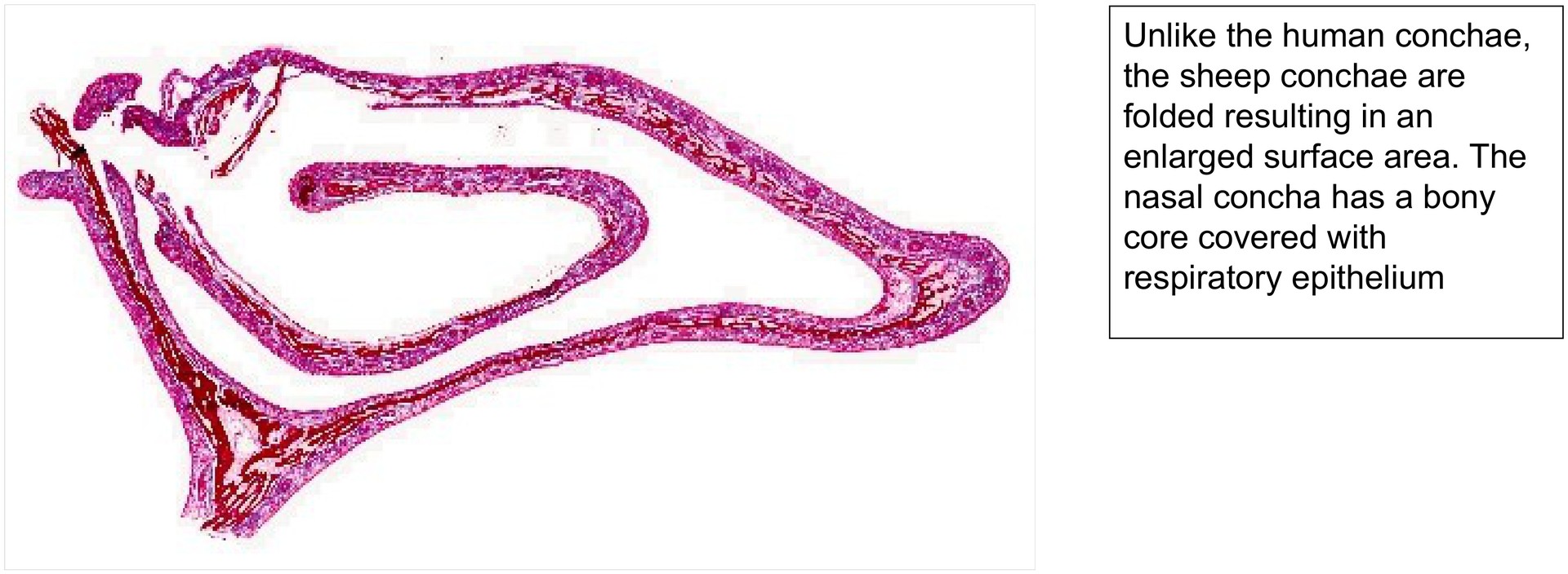
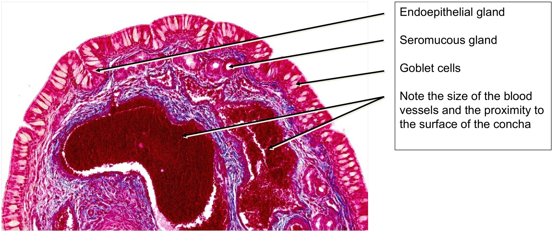
Nasal concha
Preparation:
Preparation Details:
Organ: Nasal Concha
Origin: Sheep
Staining: Azan
Method and Specimen Description:
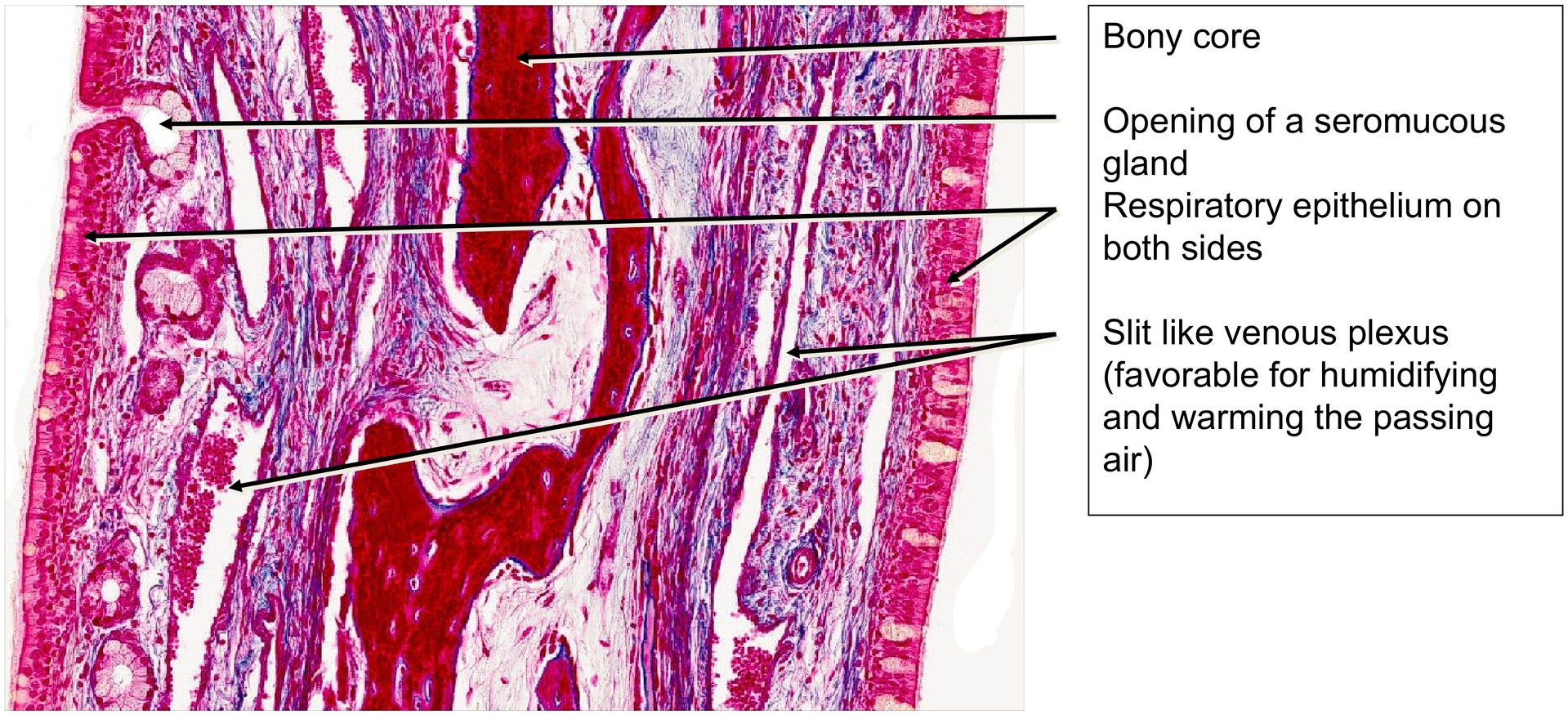
The nasal concha has a bony core, and therefore this specimen was demineralized before sectioning. Azan staining colors the epithelium, erythrocytes, and mature bone in shades of red, while connective tissue appears blue.
Objective of the Investigation:
To understand the structure of the nasal concha and its role within the respiratory tract, particularly in warming, humidifying, and filtering inhaled air, aided by its vascular network and respiratory epithelium.
Special Features of the Preparation:
The nasal conchae increase the surface area of the nasal cavity, thereby facilitating their functions in conditioning the inhaled air — warming, humidifying, and cleaning it — while also contributing to the creation of a largely laminar airflow. Although the conchae of sheep differ in shape from those of humans, their function and histological structure are comparable.
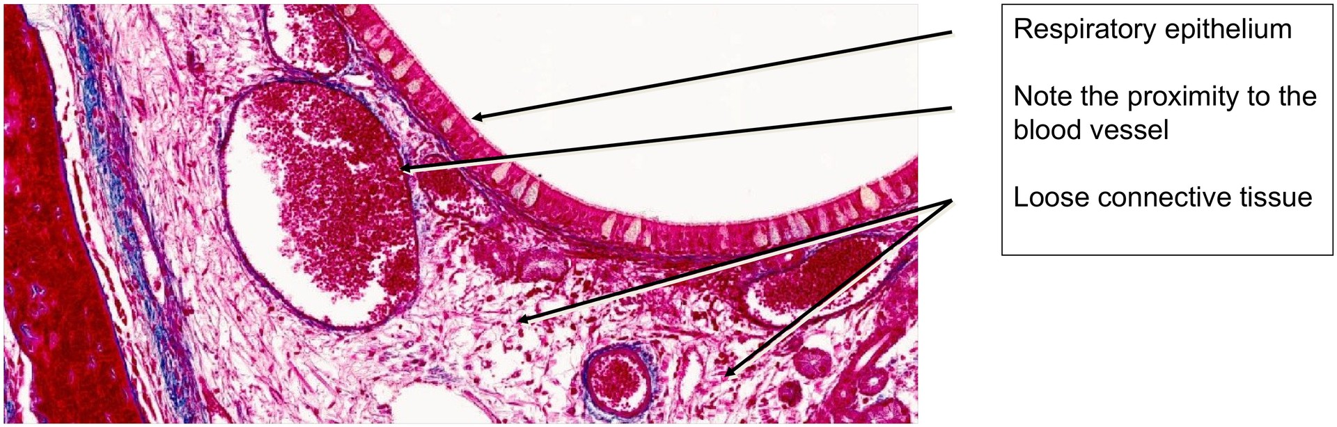
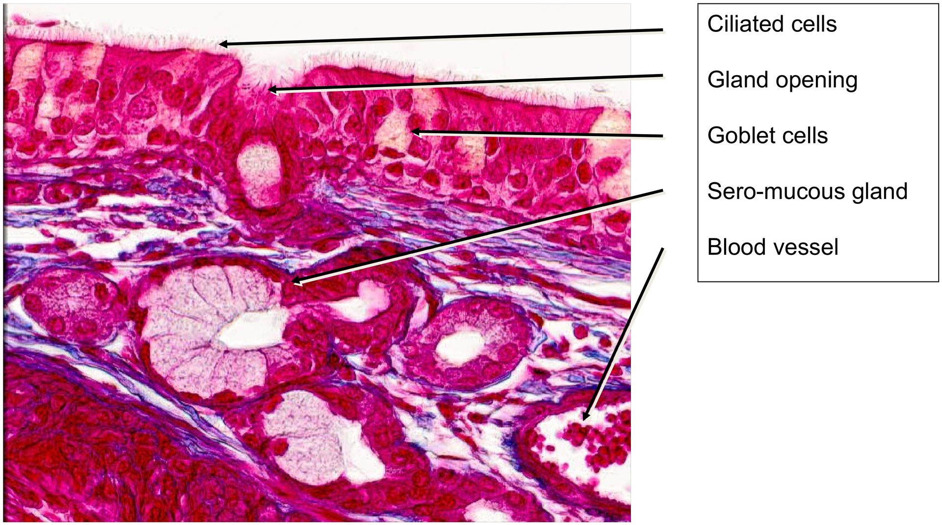
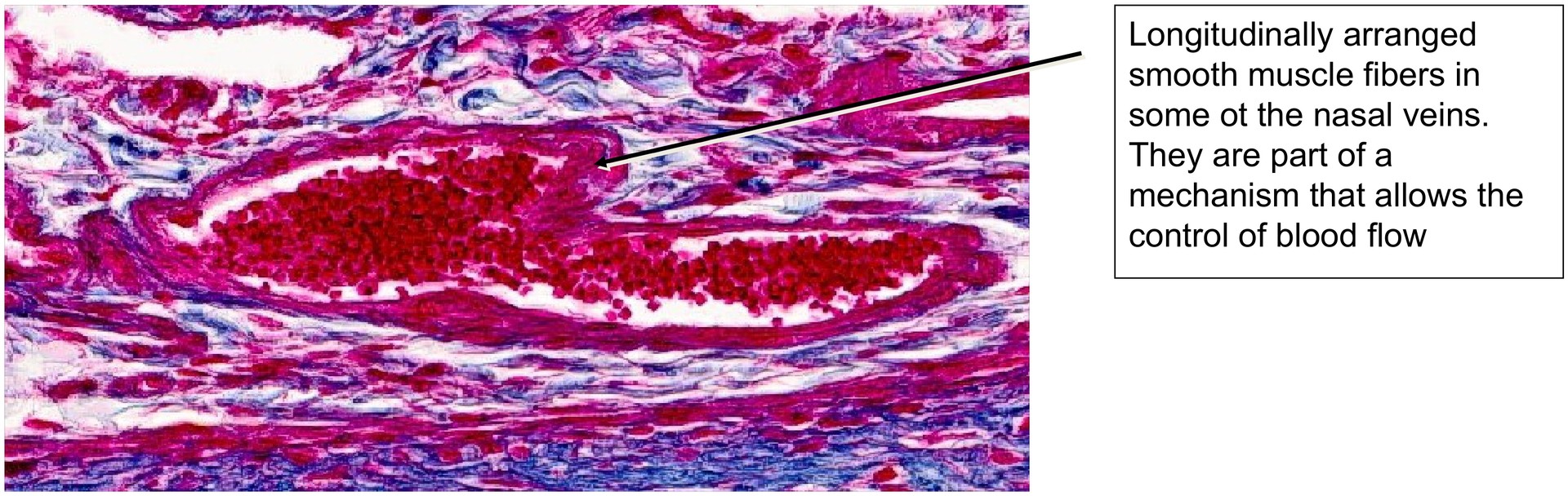
The surface is lined by respiratory epithelium, consisting mainly of ciliated columnar cells and goblet cells. Beneath this, numerous venous plexuses and arteriovenous anastomoses regulate blood flow through the capillary network, assisting in the warming of inhaled air. In addition, seromucous nasal glands open onto the epithelial surface, secreting both serous and mucous components that help moisten and cleanse the air.
The dense vascular plexuses located directly beneath the epithelium are responsible for warming the air but also explain the susceptibility to nosebleeds (epistaxis) in humans following trauma or inflammation. Endoepithelial glands, such as goblet cells, and clusters of goblet cells in small depressions, are also characteristic of the respiratory epithelium of the nasal concha.
The olfactory region is not present in this specimen. During normal breathing, airflow remains largely laminar and passes below the conchae. However, during sniffing, air is directed more forcefully upwards, allowing odorous molecules to reach the olfactory epithelium.
Tasks:
• Examine the specimen at low magnification to observe the overall shape of the nasal concha.
• Identify the respiratory epithelium and its main components.
• Locate areas with a prominent blood supply and note the proximity of vessels to the epithelial surface.
• Identify the nasal glands beneath the respiratory epithelium and explain why they are termed seromucous.
• Examine the respiratory epithelium under high magnification and distinguish goblet cells from ciliated cells.
• Identify the tissue stained strongly red in the centre between two epithelial layers — what is its nature?
License
University of Basel
Downloads