EPITHELIUM (GENERAL HISTOLOGY)
1.3
Stratified Keratinized Squamous Epithelium (Finger / Skin)
Preparation:
Preparation Details:
Organ: Skin (finger)
Origin: Human
Staining: Hematoxylin - Eosin (H&E)
Method and Specimen Description:
This is a routine histological section through the fingertip, stained with hematoxylin and eosin. The section demonstrates the structure of thick skin, which is subject to considerable mechanical stress.
Objective of the Examination:
To understand the structure of a multilayered, highly keratinized stratified squamous epithelium (the epidermis) and its adaptations to mechanical load and prevention of water loss. Additionally, to study the dermis (corium) with its collagenous connective tissue, and the subcutaneous tissue (subcutis) containing adipose tissue.
Special Features of the Preparation:
General Overview (low magnification)
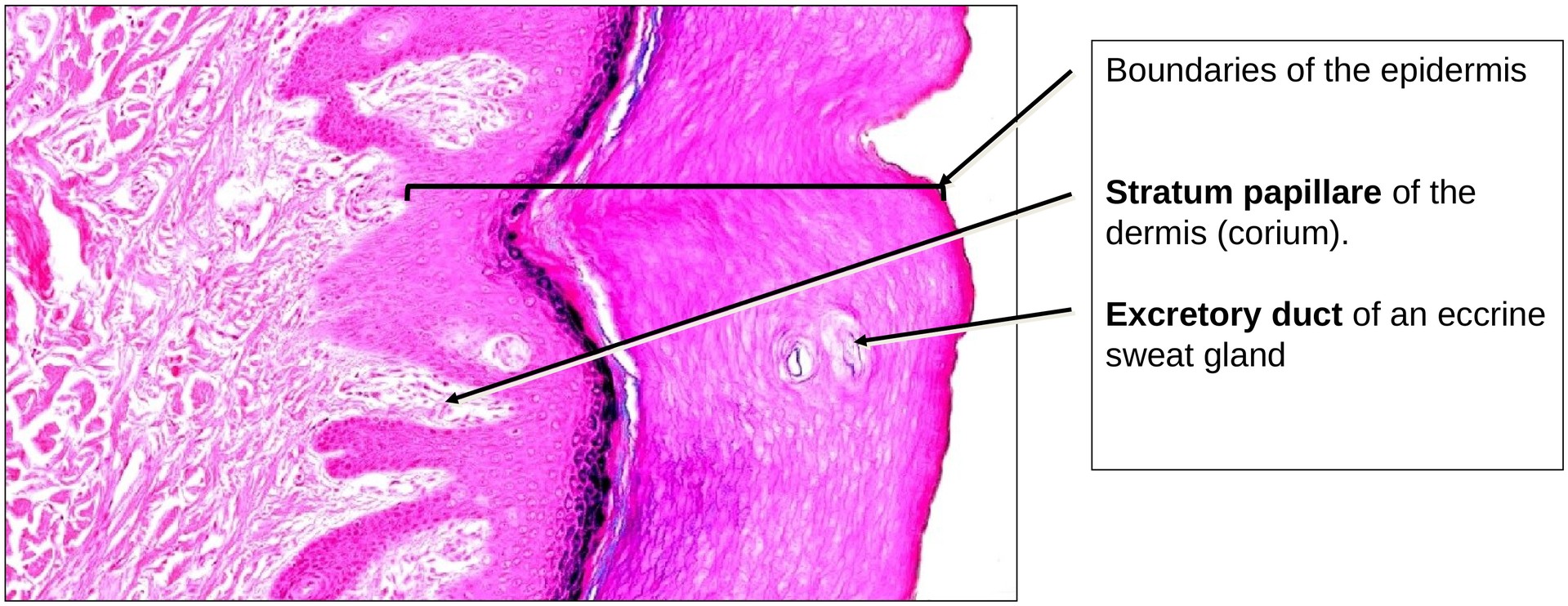
The epidermis and underlying connective tissue display a characteristic interlocking pattern formed by epidermal ridges and dermal papillae. This interdigitation reflects adaptation to mechanical stress, strengthening the attachment between the epithelium and the dermis.
Structure of the Epidermis (medium and high magnification)
From the basement membrane to the surface, the following layers can be identified:
-
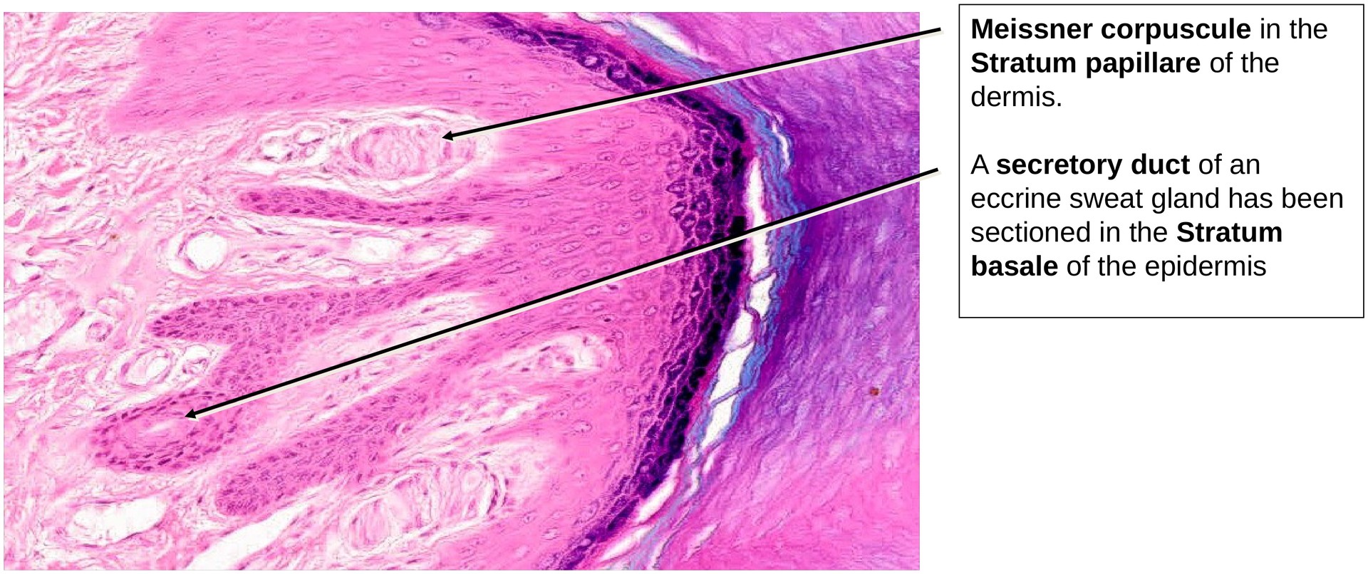
Stratum basale: Single layer of columnar or cuboidal cells forming the base of the epidermal ridges, firmly attached to the basement membrane.
-
Stratum spinosum: Several layers of large, polyhedral cells with prominent nuclei and intercellular bridges (desmosomes). The cells are connected by these desmosomes, which appear as spiny projections in light microscopy.
-
Stratum granulosum: Four to six layers of flattened cells containing dark basophilic keratohyalin granules. These granules contribute to keratin formation.
-
Stratum lucidum: A thin, lightly stained, translucent layer present only in thick skin. The cells are flattened, lack nuclei, and show no distinct boundaries.
-
Stratum corneum: The outermost layer consisting of dead, flattened, keratinized cells. Cellular structures and boundaries are no longer visible. Surface flaking corresponds to desquamation, in which superficial cells are shed and replaced by new cells from underlying layers.
Openings of the eccrine sweat gland ducts pass through the epidermis, sometimes visible as spiraling channels in longitudinal sections.
Structure of the Dermis (Corium) (medium and high magnification)
The dermis is divided into two layers:
-
Papillary dermis (stratum papillare): Contains dermal papillae that interlock with the epidermal ridges. The connective tissue here is loose, with capillaries influencing skin color. Meissner’s tactile corpuscles, responsible for light touch sensation, are located within these papillae.
-
Reticular dermis (stratum reticulare): Composed mainly of dense irregular connective tissue rich in collagen fibers, providing strength and elasticity.
Subcutis (Hypodermis):
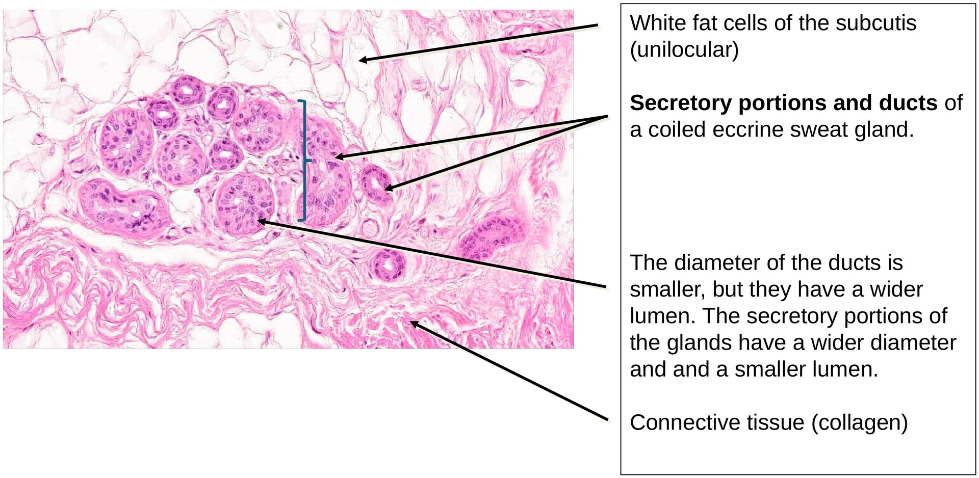
The subcutis consists predominantly of unilocular (white) adipocytes, arranged in lobules separated by fibrous connective tissue septa.\ Blood vessels and nerves run within these connective tissue tracts. Eccrine sweat glands are found at the junction of the dermis and subcutis; their coiled secretory portions lie deep in the subcutis, while their ducts ascend through the dermis and epidermis to open as sweat pores on the skin surface.
Tasks:
• Identify and divide the skin into its three principal layers: epidermis, dermis, and subcutis.
• Determine the individual layers of the epidermis from the stratum basale to the stratum corneum.
• Locate the excretory ducts of sweat glands within the epidermis and their glandular bodies in the subcutis.
• Distinguish between the papillary and reticular layers of the dermis.
• Identify Meissner’s tactile corpuscles in the dermal papillae.
• Observe the fat cells in the subcutis, and locate vessels and nerves within the connective tissue septa.
License
University of Basel
Downloads