EPITHELIUM (GENERAL HISTOLOGY)
1.4
Respiratory epithelium (trachea)
Specimen:
Specimen Details:
Organ: Trachea
Origin: Human
Staining: Azan
Method and Specimen Description:
This is a routine histological section of the trachea stained with Azan, a trichrome method that stains cell nuclei and muscle fibers red, and connective tissue blue.
The trachea is maintained open by C-shaped cartilaginous rings, some of which are included in this section. Because of the differing consistencies of cartilage and surrounding soft tissues, slight wrinkling artefacts may occur. In such areas, the image may appear blurred due to scanning limitations.
Objective of the Examination:
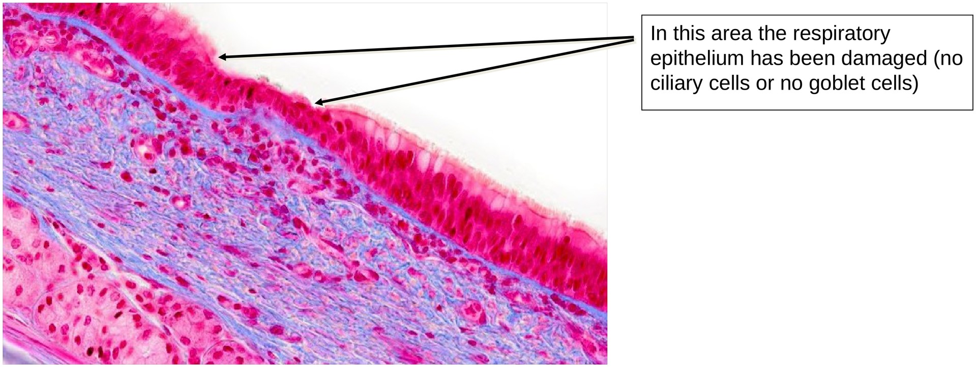
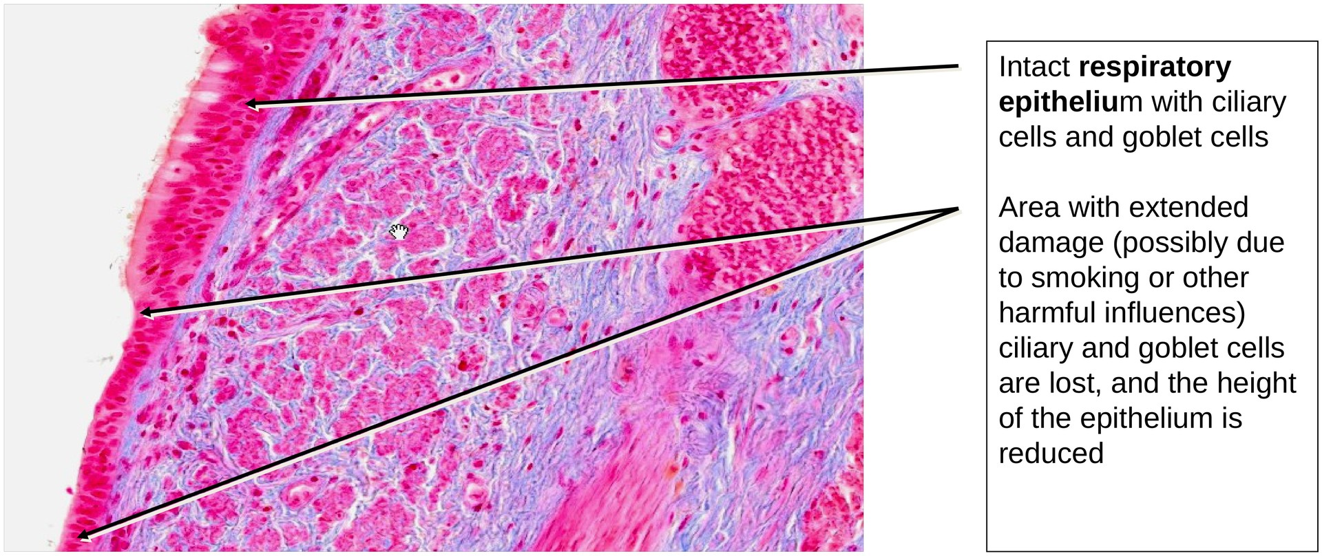
To study the structure of a pseudostratified ciliated columnar epithelium with goblet cells, collectively termed the respiratory epithelium. Additionally, to understand how various harmful influences (e.g. chronic irritation from smoke or pollutants) can cause loss of cilia and goblet cells, leading to epithelial degeneration or metaplasia.
Specimen Features:
General Overview (low magnification)
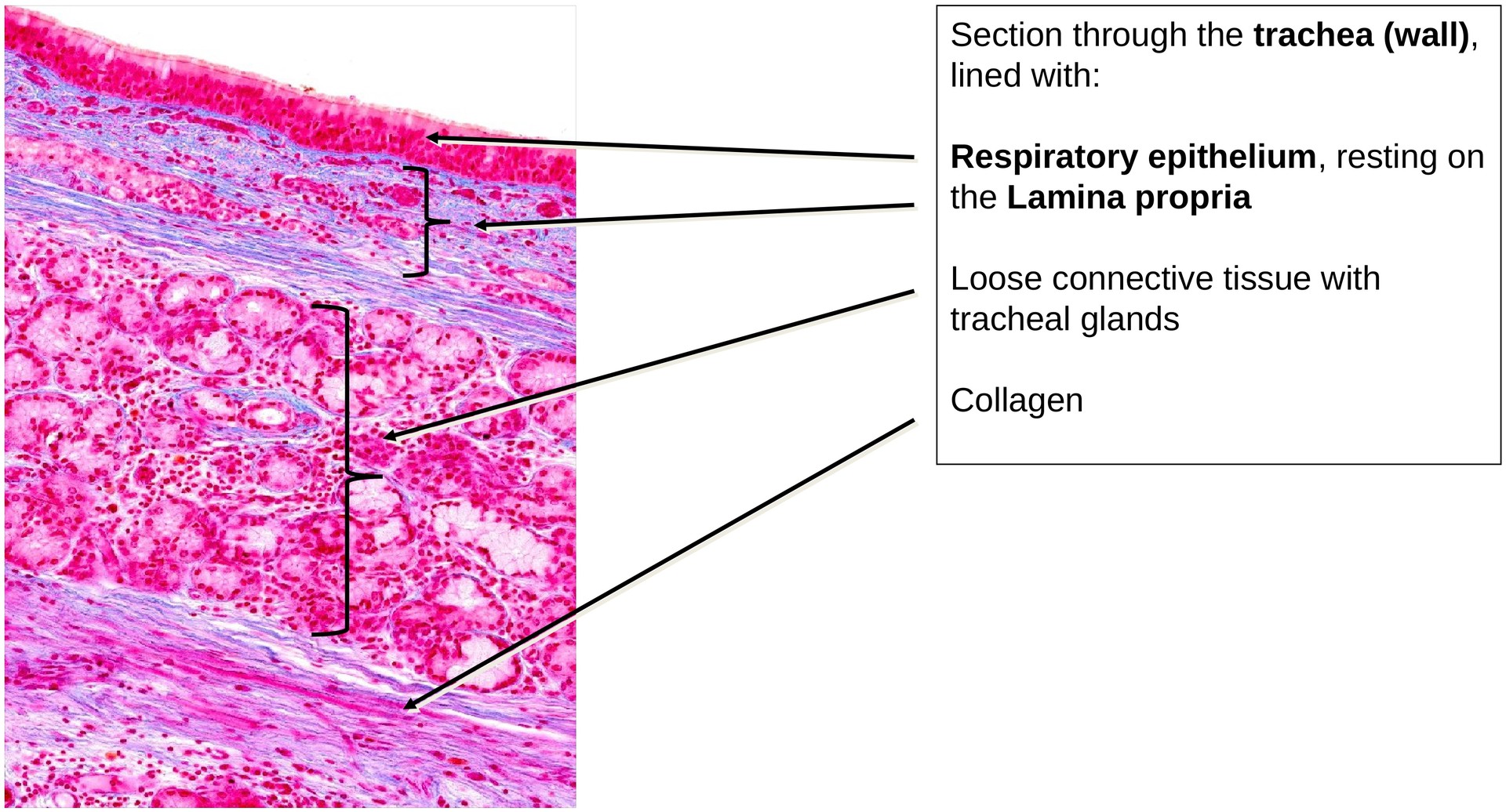
This transverse section through the tubular tracheal wall allows examination of its epithelial lining—the respiratory epithelium. The overall wall structure of the trachea (including cartilage and glands) will be studied in more detail in the context of the respiratory system.
Microscopic Structure of the Epithelium (medium and high magnification)
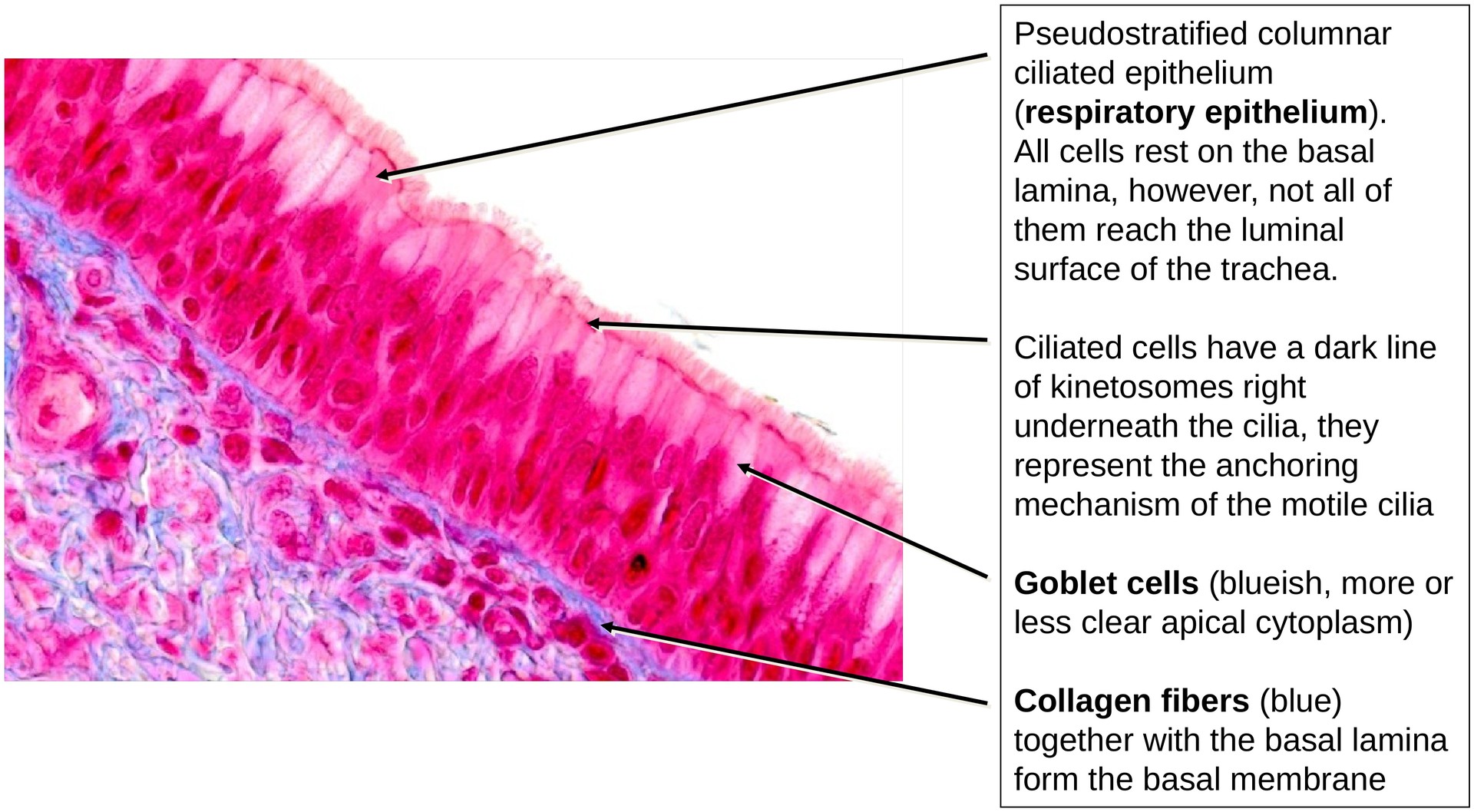
Select a region where the section passes perpendicularly through the epithelial surface. The epithelium appears uneven in height. In some areas, only two rows of nuclei are visible; in others, there are several rows. This arrangement is characteristic of pseudostratified epithelium—all cells rest on the basement membrane, but not all reach the luminal surface.
-
Basal cells: Located near the basement membrane, with spherical nuclei; they serve as stem cells for epithelial renewal.
-
Ciliated columnar cells: Tall, slender cells with elongated nuclei; they extend to the surface and bear motile cilia on their apical membrane.
- Just beneath the cilia lies a dark, continuous basal line, representing the kinetosome (basal granule) zone anchoring the cilia.
-
Goblet cells: Interspersed among the ciliated cells, identifiable by their apically expanded shape and pale to bluish cytoplasm due to mucin content.
- These cells secrete mucus that traps inhaled particles, contributing to the mucociliary clearance mechanism.
Beneath the epithelium lies loose connective tissue containing tracheal glands. These are tubulo-acinar glands with bluish-stained tubular portions in Azan sections. Occasionally, excretory ducts can be seen opening towards the epithelial surface.
Tasks:
• Follow the inner boundary of the trachea along the respiratory epithelium.
• Identify ciliated cells and locate goblet cells among them.
• Observe the kinetosome (basal granule) line in the apical cytoplasm of the ciliated cells.
• In goblet cells, look for mucus secretion and, where possible, discharge points at the epithelial surface.
• Identify any areas where the respiratory epithelium appears damaged or flattened, showing loss of cilia and goblet cells.
• Locate a tracheal gland within the connective tissue beneath the epithelium and describe its tubulo-acinar structure.
License
University of Basel
Downloads