FEMALE REPRODUCTIVE ORGANS (ANATOMICAL MICROSCOPY)
10.11
Ovary, cat
Specimen Details:
Specimen Details:
Organ: Ovary
Origin: Cat
Staining: Pasini
Method and Specimen Description:
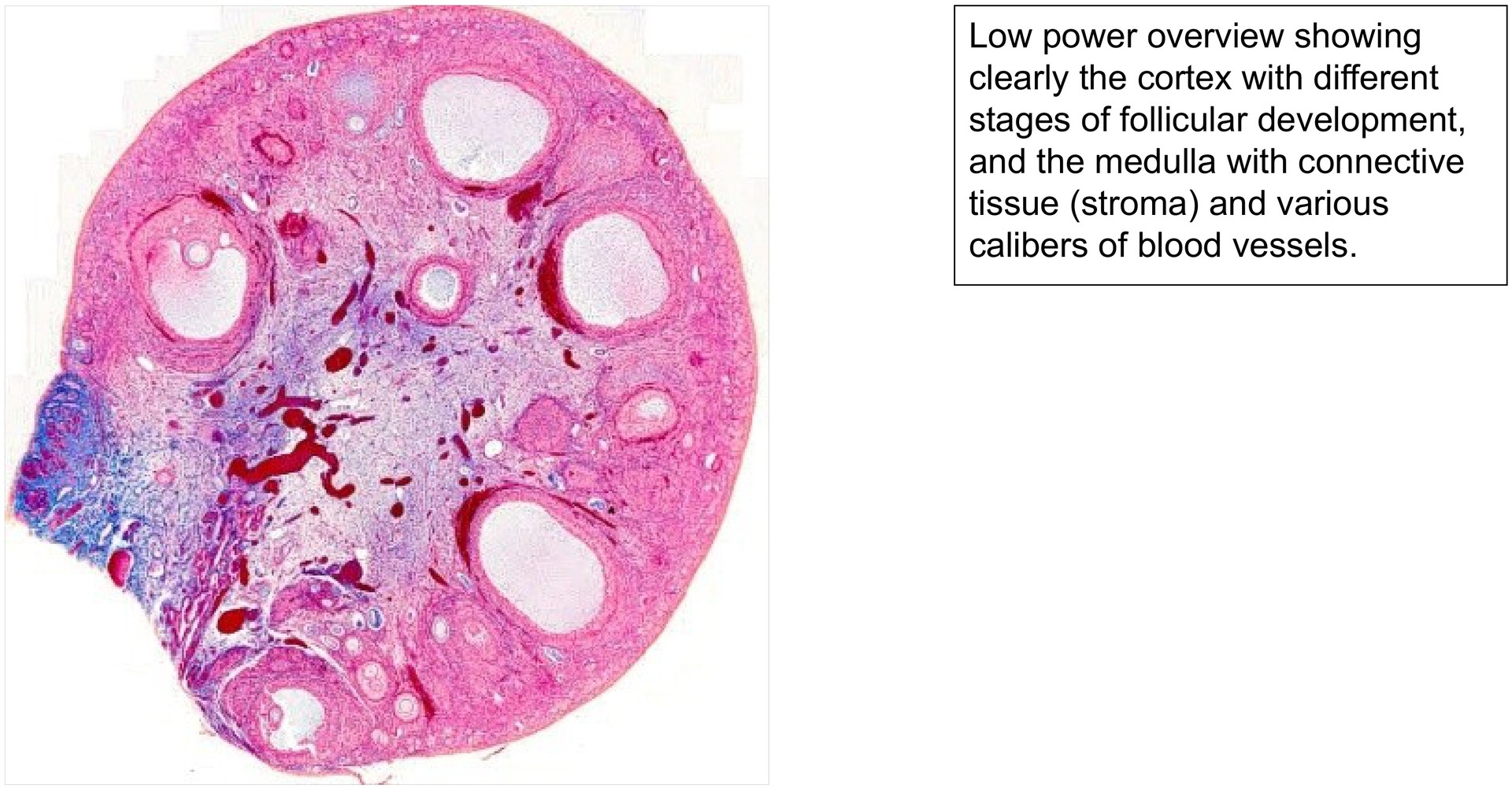
Histological section of a normal cat ovary, stained with Pasini (a trichrome stain comparable to Azan). The stain clearly differentiates connective tissue (blue) and blood (red), allowing the follicular and stromal structures to be identified with ease.
Objective of the Examination:
To study the structure of the feline ovary and to identify the different stages of follicular development. In the cat, multiple follicles often mature simultaneously, in contrast to the dominant single follicle typical of the human ovary.
Special Features of the Specimen:
At low magnification, the ovary shows a clear distinction between:
-
the cortex, containing follicles at various stages of development, and
-
the medulla, containing mainly blood vessels, connective tissue, and nerves, but typically no follicles.
Within the cortex, follicles can be observed in successive stages of maturation:
-
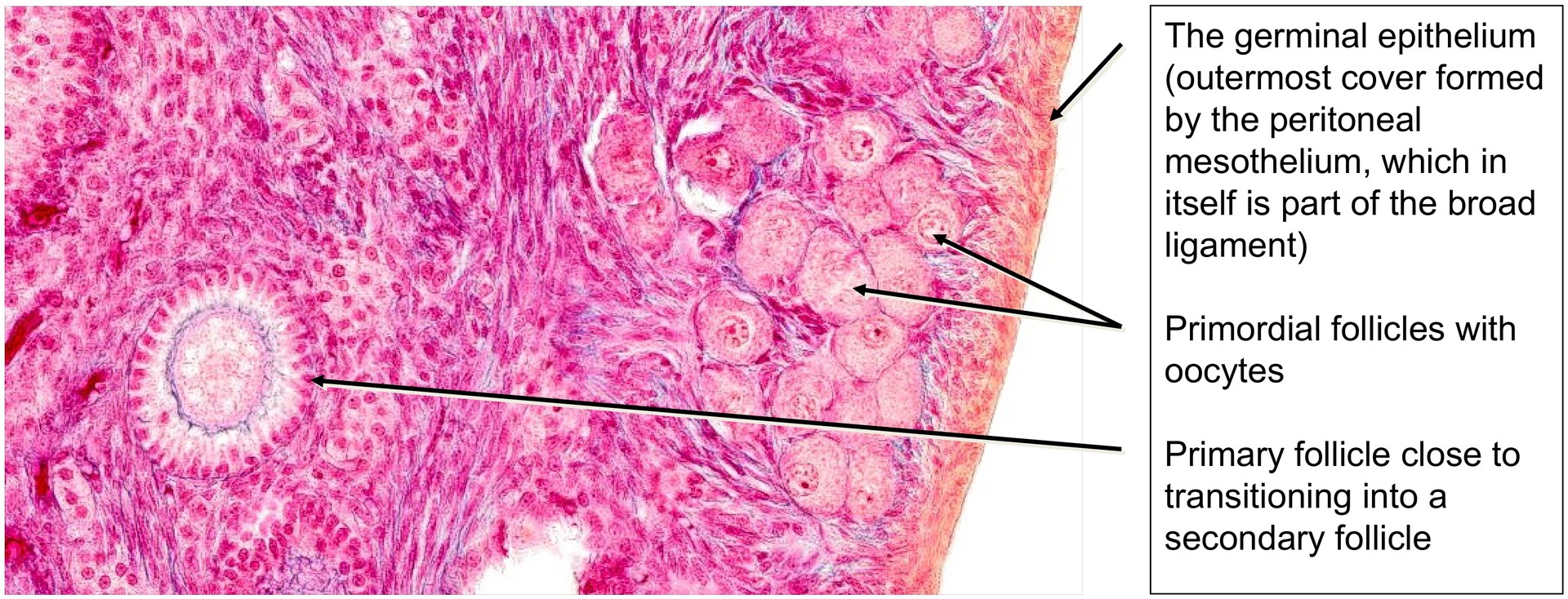
Primordial follicles: located near the ovarian surface, each consisting of a primary oocyte surrounded by a single layer of flattened follicular (squamous) cells. The follicular epithelium may appear incomplete in tangential sections.
-
Primary follicles: the oocyte is enclosed by a single layer of cuboidal follicular cells, which form the follicular epithelium.
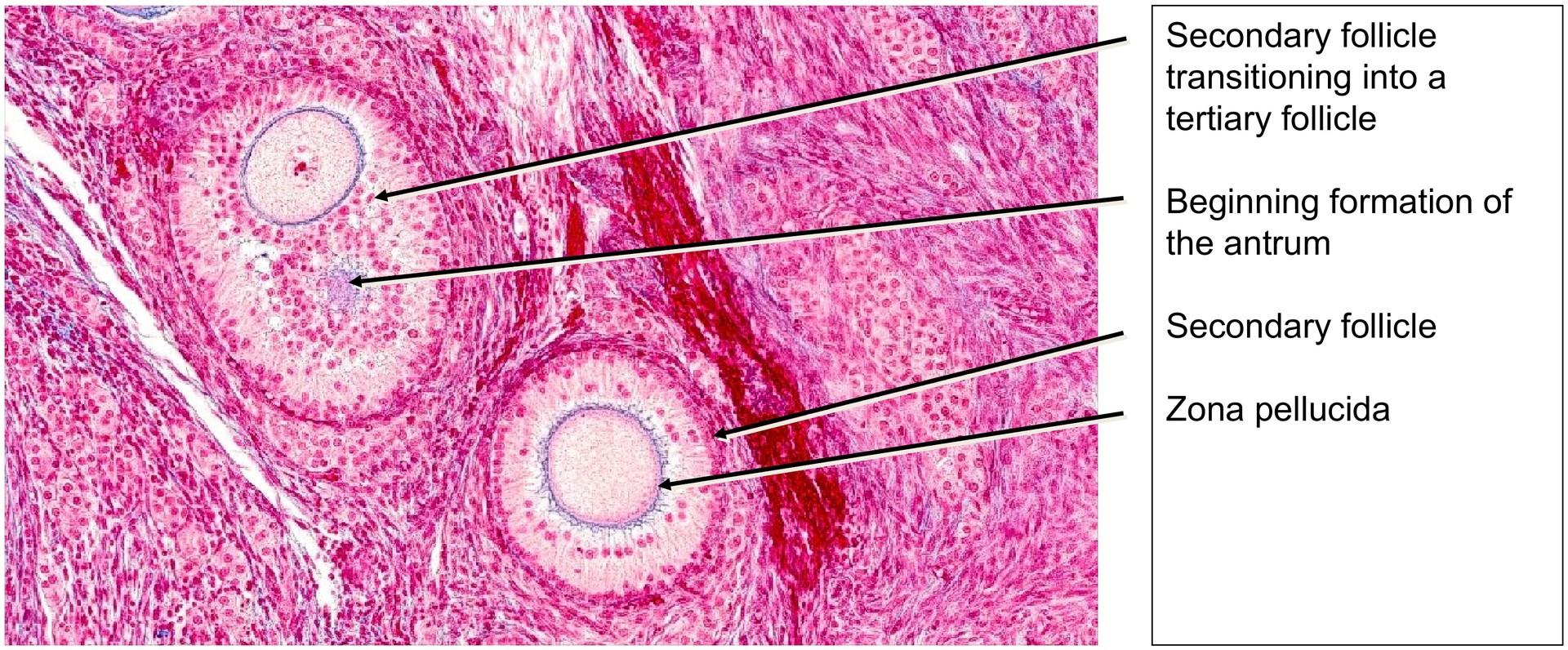
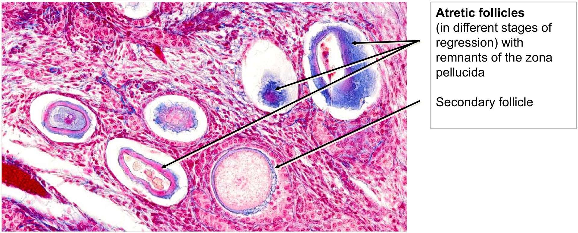
-
Secondary follicles: show a multi-layered follicular epithelium (the granulosa cell layer) and a distinct zona pellucida, the glycoprotein layer separating the oocyte from the follicular cells.
-
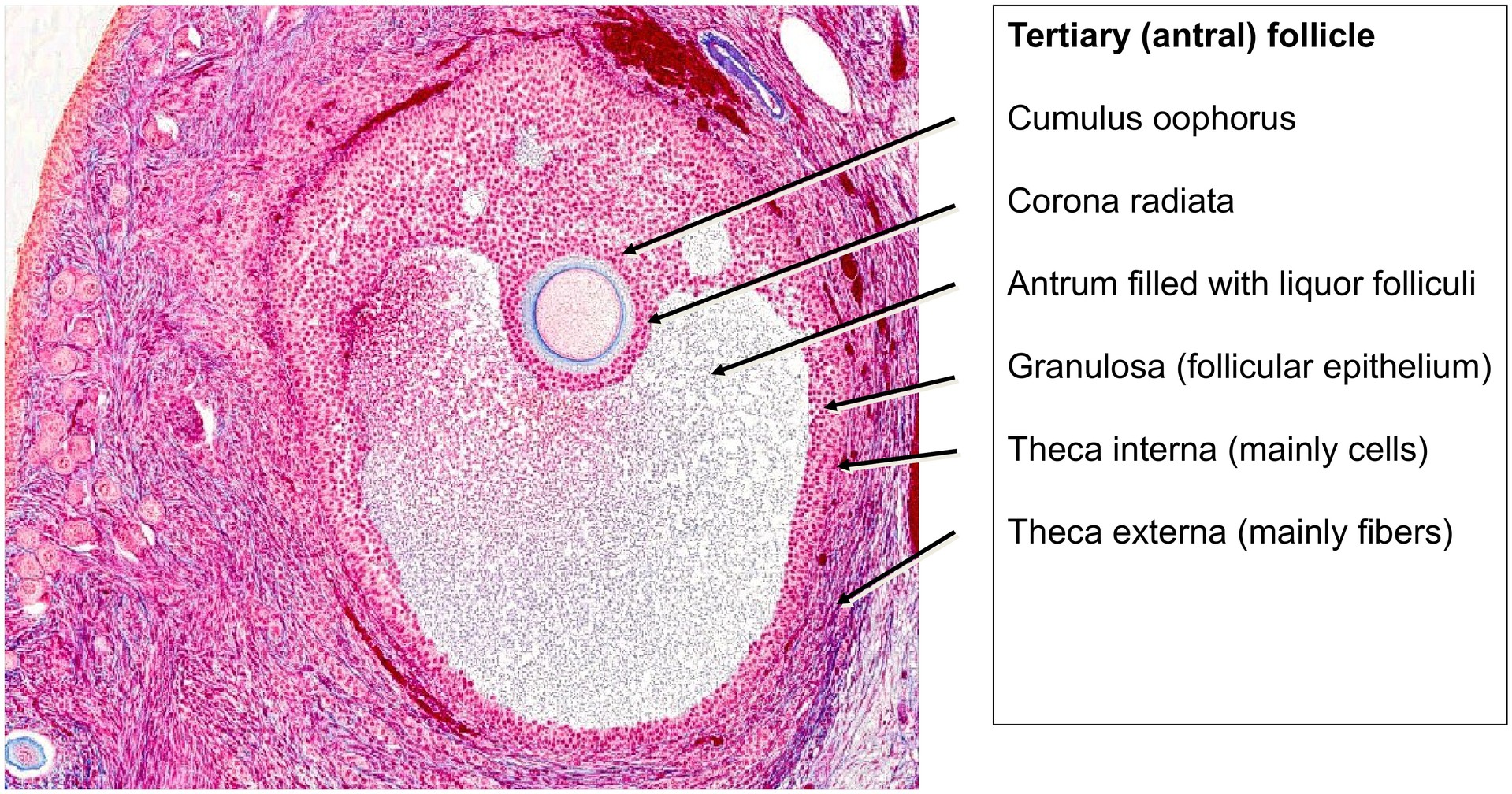
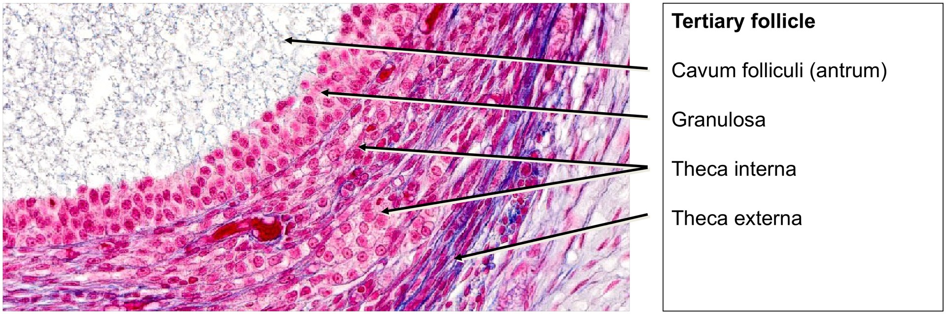
Tertiary (antral) follicles: contain a fluid-filled cavity (antrum) filled with liquor folliculi. The oocyte is located on a mound of granulosa cells, the cumulus oophorus, and is surrounded by the corona radiata, consisting of the innermost granulosa cells closely adherent to the zona pellucida.
Externally, the tertiary follicles are surrounded by two distinct thecal layers:
-
the theca interna, composed of endocrine, steroidogenic cells with pale cytoplasm, and
-
the theca externa, consisting mainly of fibrous connective tissue and smooth muscle cells.
Atretic follicles in various stages of regression are also visible. These represent follicles that have ceased development and are undergoing degeneration and resorption.
In some follicles, theca cells have differentiated into theca lutein cells, characterized by a larger size and light cytoplasm, indicating luteinization following ovulation.
The ovary is externally covered by the germinal epithelium, a continuation of the peritoneal mesothelium forming part of the broad ligament.
Tasks:
• Identify the cortex and medulla, and describe the main structures observed in each.
• Locate and compare the different stages of follicular development: primordial, primary, secondary, and tertiary follicles. Describe the morphological differences.
• Identify the corona radiata—which specific cells does it include?
• Distinguish between the theca interna and theca externa. How do they differ in structure and function?
• Identify atretic follicles. What is their role in ovarian physiology?
• Locate the zona pellucida in developing follicles.
• Identify granulosa cells. What important substances or hormones originate from these cells?
License
University of Basel
Downloads