SENSORY ORGANS (ANATOMICAL MICROSCOPY)
17.3
Inner ear (Organ of Corti)
Preparation:
Preparation Details:
Organ: Cochlea
Origin: Guinea pig
Staining: Haematoxylin - Eosin (H&E)
Method and Specimen Description:
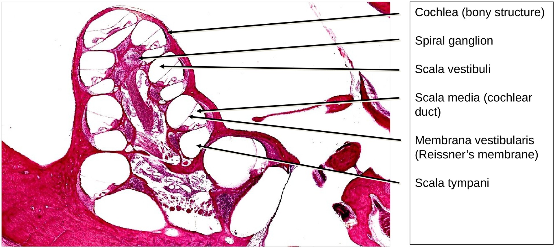
Because of the extensive bony structures within the cochlea, this preparation was decalcified prior to sectioning — typically achieved using acid or chelating agents. A guinea pig tympanic cavity was used, as its cochlea has more turns than that of a human, clearly illustrating the characteristic spiral (the cochlea, or “snail shell”).
Objective of the Examination:
To study the organ of Corti, including its relationship with the bony and membranous labyrinths, the cochlear turns, and the spiral ganglion.
Special Features of the Specimen:
In the overview, the cochlea is easily recognizable by its spiral shape within the tympanic cavity. At its center lies the bony core, or modiolus—a structure reminiscent of a snail shell with the outer whorls removed. The cochlea and vestibule together constitute the bony labyrinth.
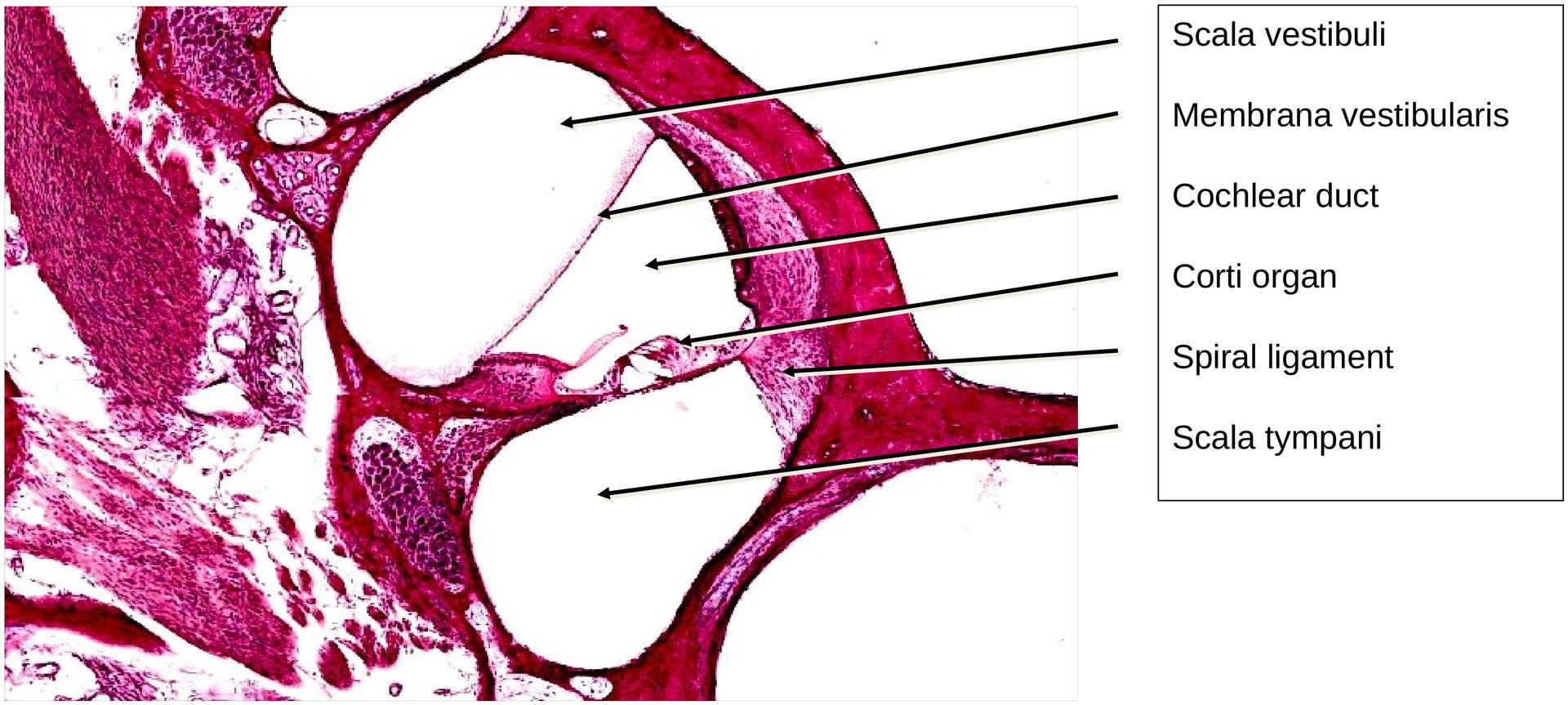
Inside this bony labyrinth lies the membranous labyrinth, a delicate connective tissue-lined system that fits within it like the lining of a garment. It consists of the scala vestibuli and scala tympani, both filled with perilymph, and the cochlear duct (scala media) located between them, which contains endolymph and houses the organ of Corti.
The cochlear duct (scala media) with the organ of Corti is bounded by:
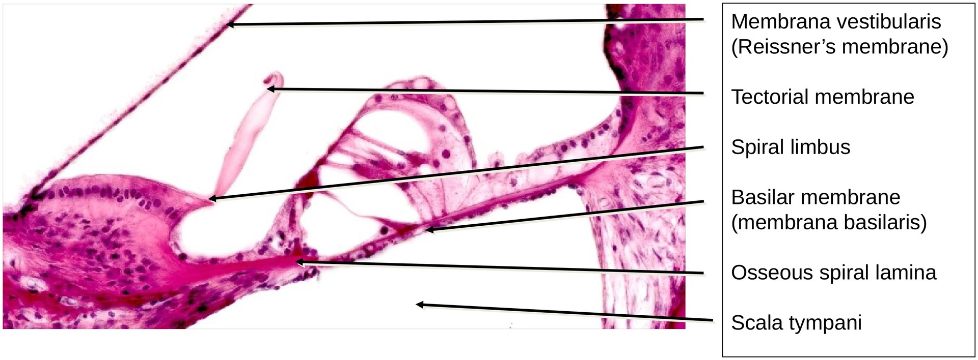
- Reissner’s membrane (membrana vestibularis) — separating it from the scala vestibuli,
- Basilar membrane (membrana basilaris) — separating it from the scala tympani.
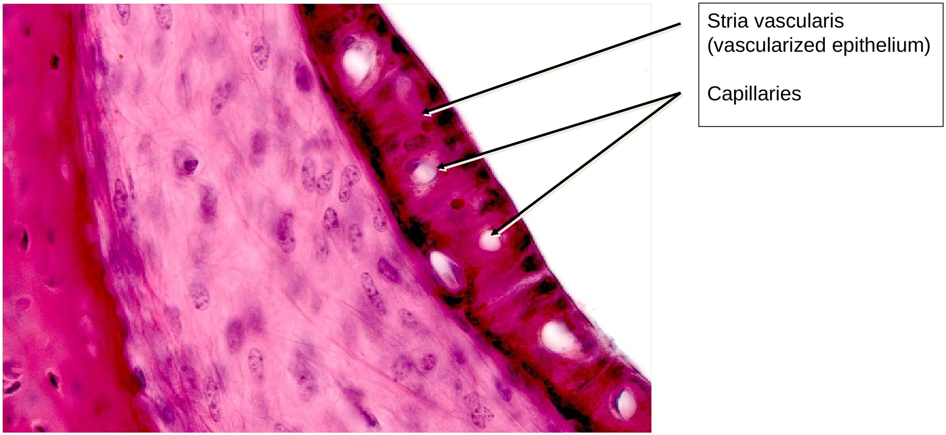
Laterally, the duct is bordered by the spiral ligament and its specialized epithelial covering, the stria vascularis. The stria vascularis is responsible for producing endolymph, a potassium-rich fluid essential for hair cell function. It is the only vascularized epithelium in the body. The spiral ligament itself is composed of connective tissue containing specialized fibroblasts.
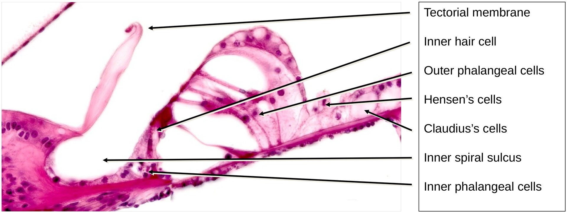
Organ of Corti:
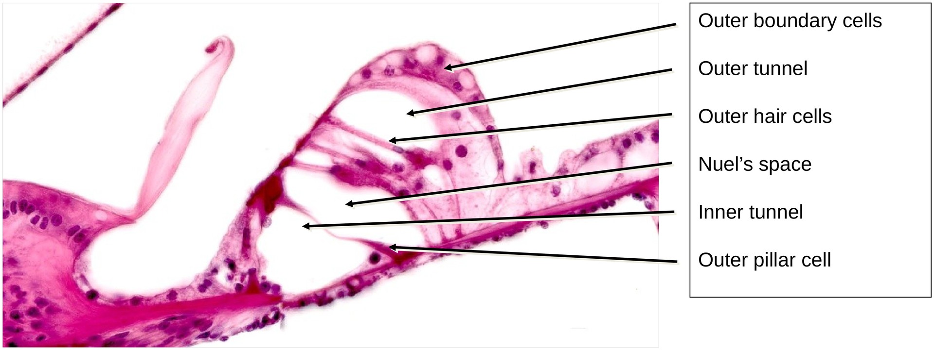
The organ of Corti is the sensory epithelium responsible for sound transduction. It contains:
- Inner hair cells (a single row), and
- Outer hair cells (three to five rows), each covered with stereocilia that normally make contact with the tectorial membrane (membrana tectoria). In this preparation, due to decalcification and fixation, the tectorial membrane has detached from the hair cells.
The tectorial membrane is an acellular gelatinous layer, anchored to the spiral limbus, and normally extends over the entire organ of Corti.
Supporting cells include:
- Inner and outer phalangeal cells supporting the hair cells,
- Inner and outer pillar cells, which together form the inner tunnel,
- Hensen’s cells and Claudius’ cells, located laterally beyond the outer hair cells, and
- The outer boundary cells forming the external spiral sulcus.
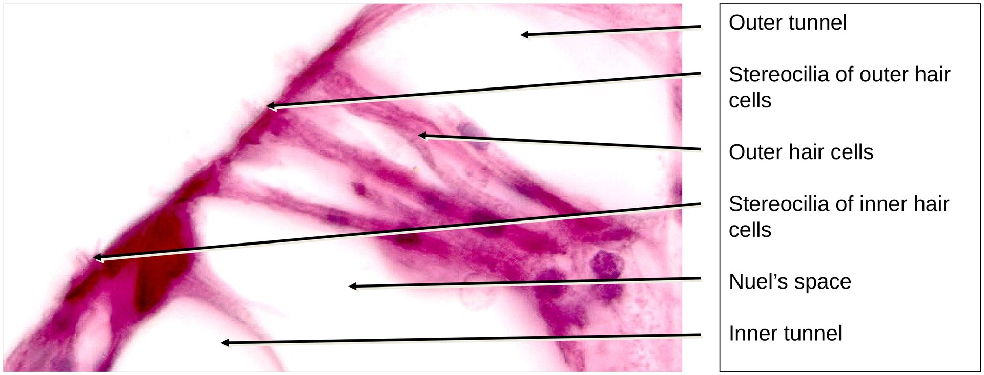
The organ of Corti is traversed by several fluid-filled spaces:
- Inner tunnel, bounded by the inner and outer pillar cells,
- Nuel’s space, between the outer pillar cells and outer hair cells, and
- Outer tunnel, between the outer boundary cells and outer hair cells.
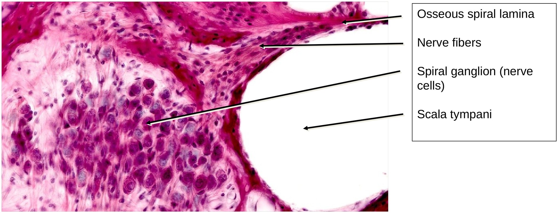
The basilar membrane, on which the organ of Corti rests, extends between the osseous spiral lamina and the spiral ligament. It is composed of collagen fibers and supports the epithelium of the scala tympani on its lower surface.
Spiral Ganglion:
Nerve fibers arising from the hair cells pass through the region below the osseous spiral lamina and enter the spiral ganglion, located within the modiolus. From here, the fibers converge to form the cochlear division of the vestibulocochlear nerve (cranial nerve VIII).
Tasks:
Identify the following structures and answer the questions based on the specimen:
- Tympanic cavity and bony labyrinth – which structures are located within the bony labyrinth?
- Scala vestibuli, scala tympani, and cochlear duct – in which of these is the organ of Corti located?
- Which structure produces the endolymph? What structures bound the cochlear duct?
- Adjacent to which space does the basilar membrane lie?
- Where are the inner tunnel, outer tunnel, and Nuel’s space located?
- Which cells are normally in contact with the tectorial membrane?
- Identify the spiral ganglion, osseous spiral lamina, spiral limbus, and spiral ligament.
License
University of Basel
Downloads