ENDOCRINE ORGANS (ANATOMICAL MICROSCOPY)
9.2
Pituitary gland
Specimen Details:
Specimen Details:
Organ: Hypophysis
Origin: Human
Staining: Azan
Method and Specimen Description:
Standard histological specimen stained with Azan, which allows differentiation of acidophilic, basophilic, and chromophobic cells.
Objective of the Examination:
To study the hypophysis, its subdivision into the adenohypophysis and neurohypophysis, and to identify the principal pituitary cell types.
Special Features of the Specimen:
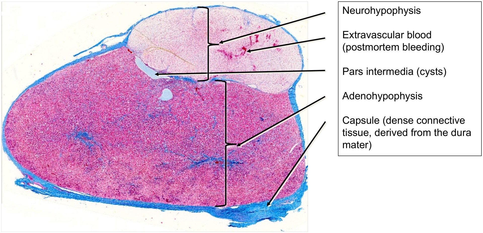
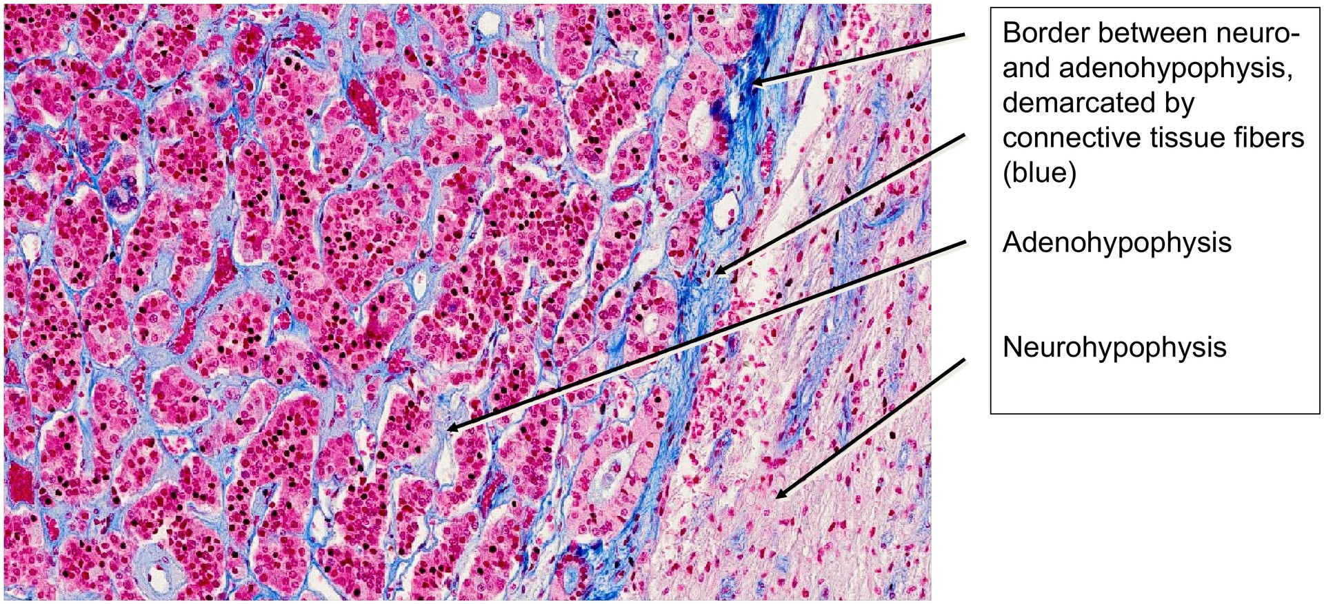
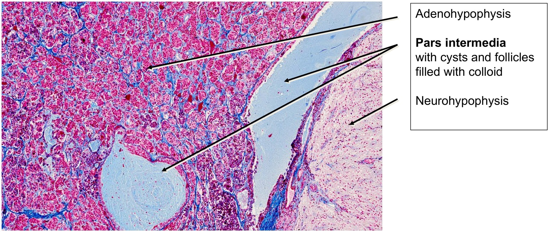
At low magnification, it is immediately apparent that the pituitary gland consists of two major components: the adenohypophysis (anterior lobe) and the neurohypophysis (posterior lobe). The gland is enclosed by a dense fibrous capsule, derived from the dura mater of the meninges.
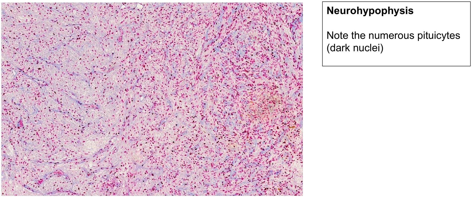
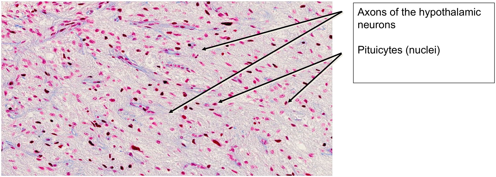
The neurohypophysis is smaller than the adenohypophysis and, consistent with its origin from neuroectoderm of the diencephalon (the floor of the third ventricle), it is composed of nervous tissue. The cell nuclei visible here belong to specialized glial cells termed pituicytes. Only the axons of hypothalamic neurons extend into the neurohypophysis; the corresponding neuron cell bodies reside in the supraoptic and paraventricular nuclei of the hypothalamus.
These axons transport the hypothalamic hormones oxytocin and antidiuretic hormone (ADH, vasopressin) to the posterior pituitary, where they are stored and released when required. The characteristic axonal swellings known as Herring bodies, which contain hormone-filled vesicles, are not demonstrable in this specimen due to the Azan stain.
In human autopsy specimens, postmortem changes can often be seen as extravascular blood within the neurohypophysis, an artefact not present in vivo.
Immediately adjacent to the neurohypophysis lies the pars intermedia, a narrow zone of the adenohypophysis containing cysts and follicles with colloid. These represent remnants of Rathke’s pouch, an invagination of oral ectoderm from the roof of the developing pharynx during embryogenesis. The colloid material within these structures likely contains POMC (pro-opiomelanocortin), a precursor of MSH (melanocyte-stimulating hormone). Since MSH is of limited physiological importance in humans, the pars intermedia may be only rudimentarily developed; however, it is well-defined in this specimen.
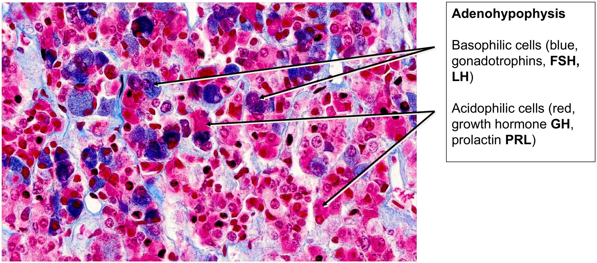
Within the adenohypophysis, three principal cell types can be distinguished by Azan staining:
-
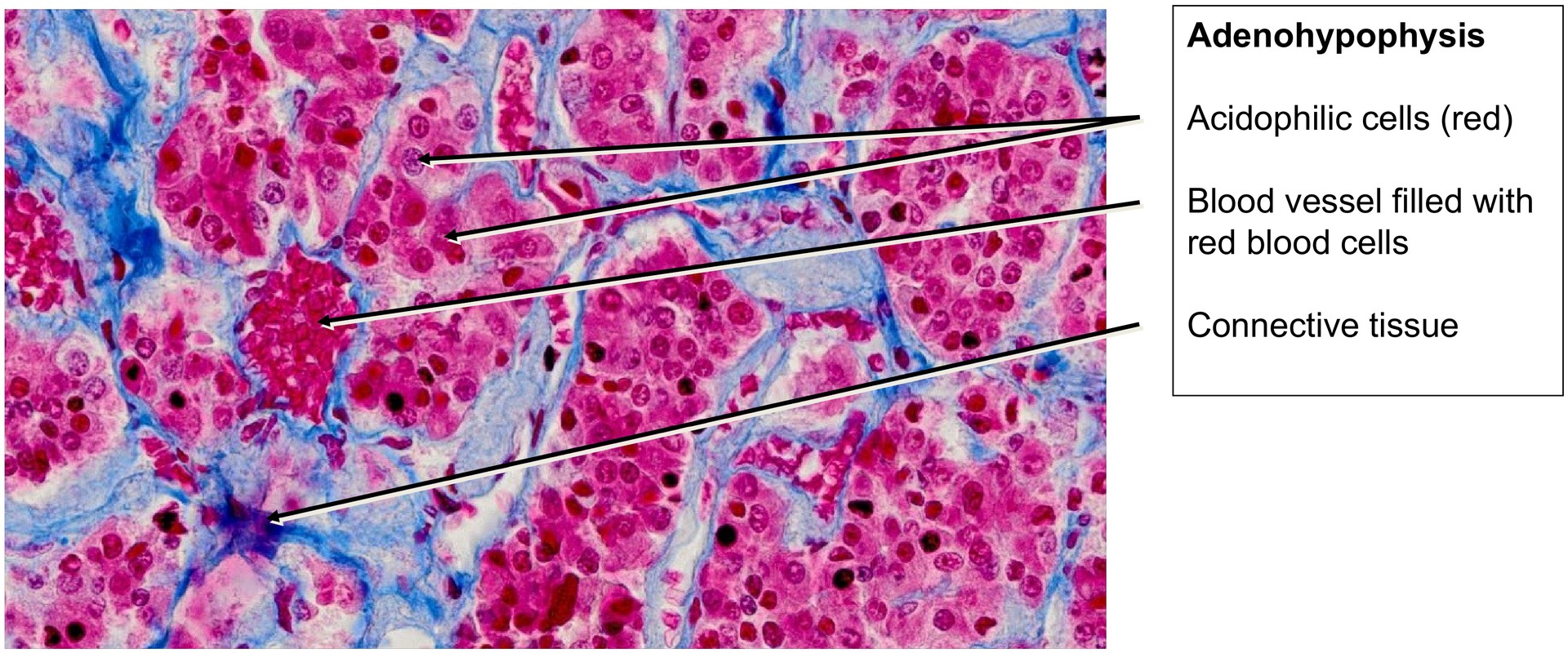
Acidophilic cells – stain red and produce growth hormone (GH, somatotropin) and prolactin (PRL).
-
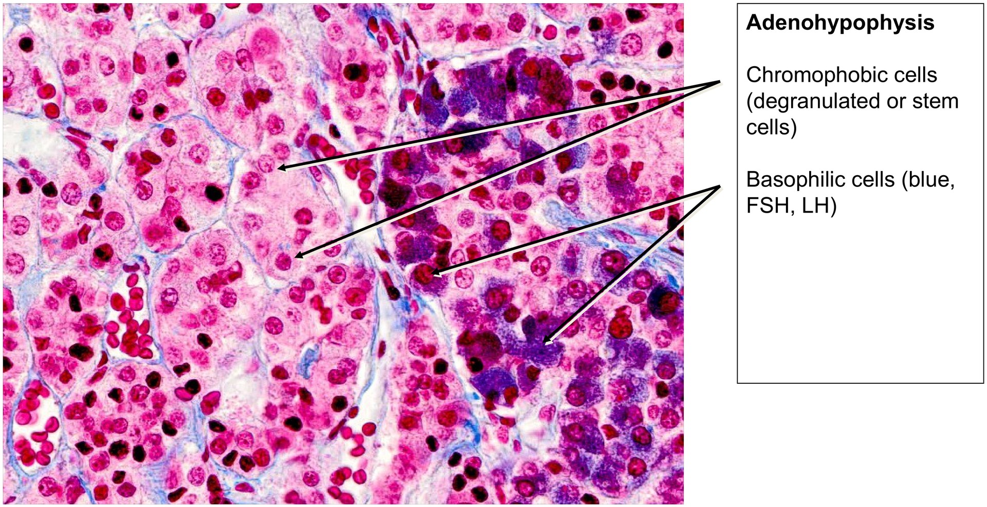
Basophilic cells – stain blue and secrete gonadotrophins (FSH, LH) and thyrotrophin (TSH), as well as ACTH.
-
Chromophobic cells – stain weakly or not at all; they likely represent degranulated cells or stem cells capable of differentiating into the other two types.
Although these cell types are intermingled, regional clustering is commonly observed.
Tasks:
-
Identify the two main parts of the gland at low magnification: the adenohypophysis and neurohypophysis.
-
Examine the capsule and note its dense fibrous connective tissue.
-
At higher magnification, locate pituicytes in the neurohypophysis.
-
In the neurohypophysis, identify extravascular blood and axons of hypothalamic neurons.
-
Observe the boundary region between the adenohypophysis and neurohypophysis. What structures are present in this intermediate zone?
-
Locate the three cell types in the adenohypophysis: chromophobic, acidophilic, and basophilic.
-
Note the non-uniform distribution of these cells throughout the adenohypophysis.
License
University of Basel
Downloads