ADDITIONAL INFORMATION
21.1
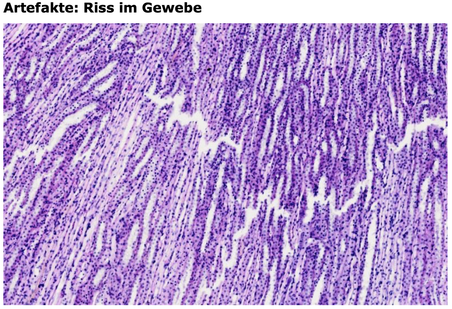
Artifacts
The production of histological sections involves a variety of individual steps — from tissue removal, fixation, and dehydration, to embedding in sectionable wax, cutting on a microtome, and finally, the specific staining of the sections.
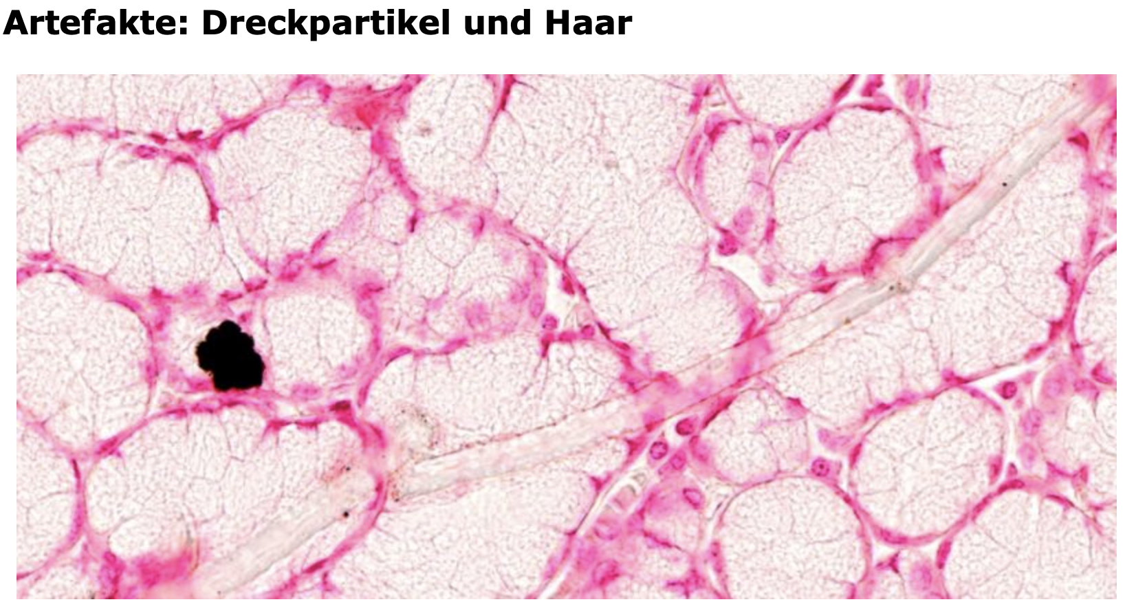
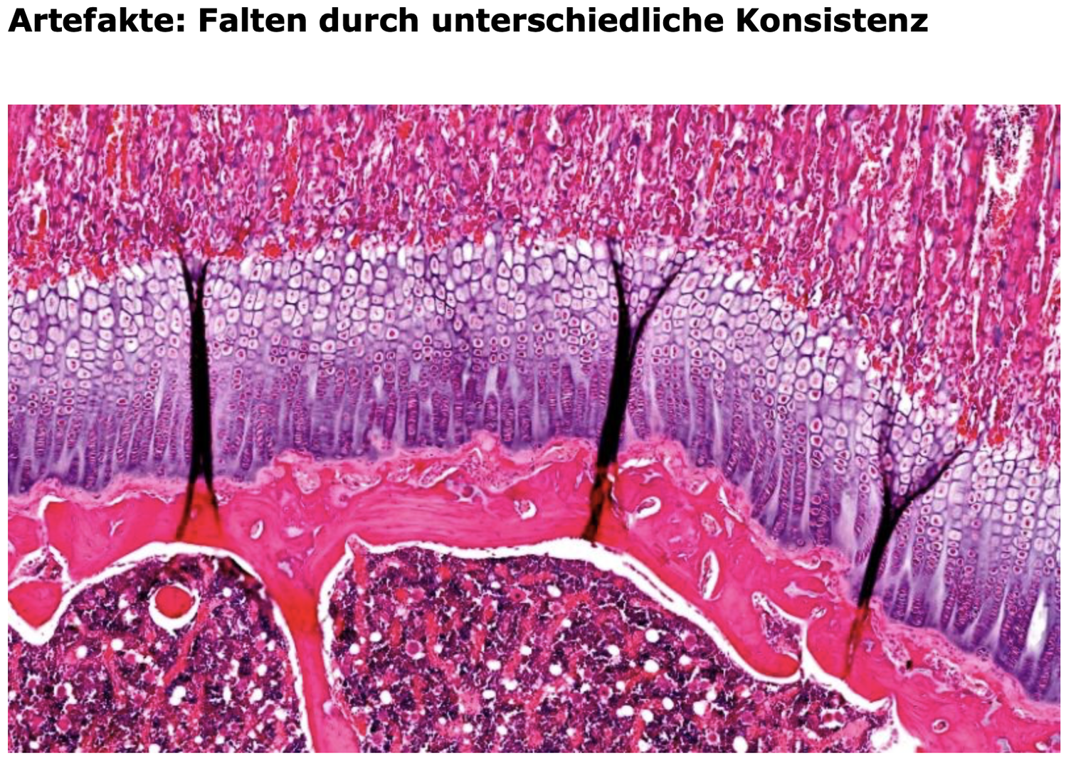
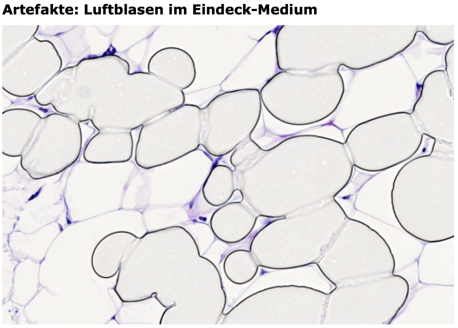
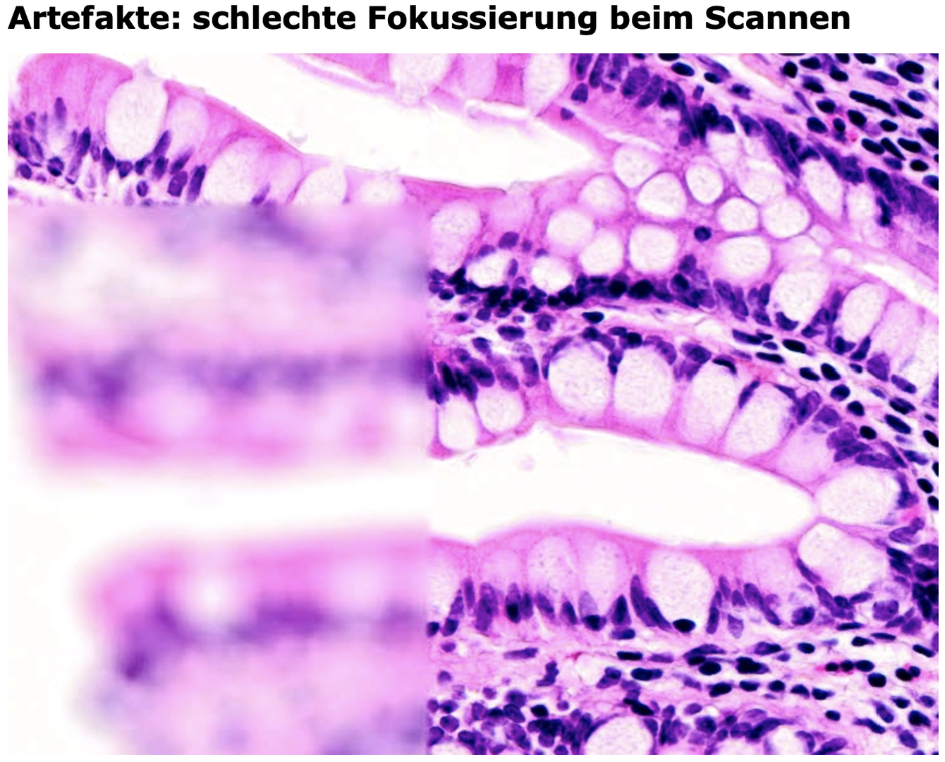
During all these steps, artifacts may occur. For example, differences in tissue consistency during cutting can result in folds, cracks, or gaps. In addition, air bubbles or precipitated dye crystals may occasionally be visible on the slides. The automated scanning process can also, in rare cases, produce blurred regions. In almost all instances, such artifacts can be avoided or circumvented by examining other areas of the specimen.
Many of the preparations in this collection consist of several thousand individual images, and in such large datasets, artifacts are not always completely avoidable. At some locations, explicit notes draw attention to these artifacts.
Below are some overview images illustrating possible artifacts:
License
University of Basel