DIGESTIVE ORGANS: GASTROINTESTINAL TRACT (ANATOMICAL MICROSCOPY)
19.4
Ileum, Rabbit
Specimen:
Specimen Details:
Organ: Ileum
Origin: Rabbit
Staining: Hematoxylin - Eosin (H&E)
Method and Specimen Description:
Normal histological section stained with the routine overview stain (H&E).
Objective of the Examination:
To study the Peyer’s patches and their role in the immune function of the ileum, as well as to identify the dome epithelium and high endothelial venules (HEVs).
Specific Features of the Specimen:
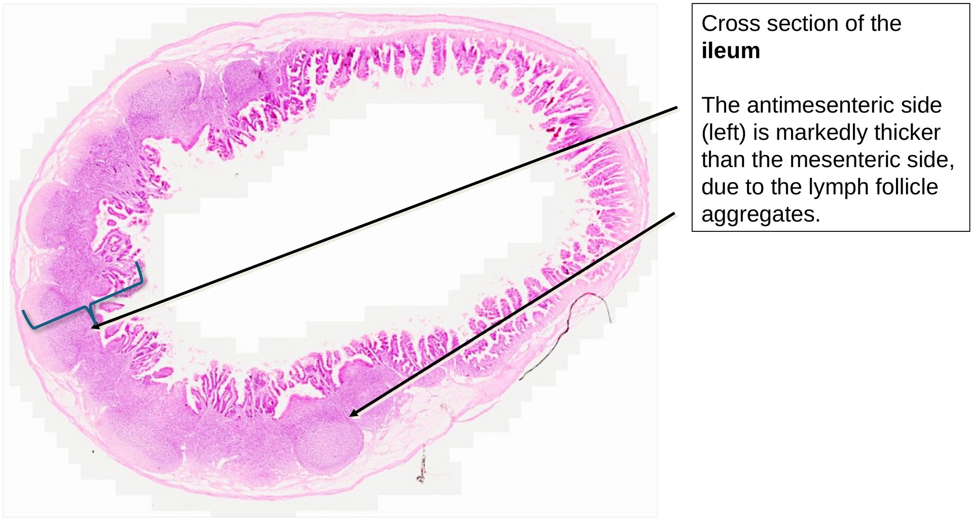
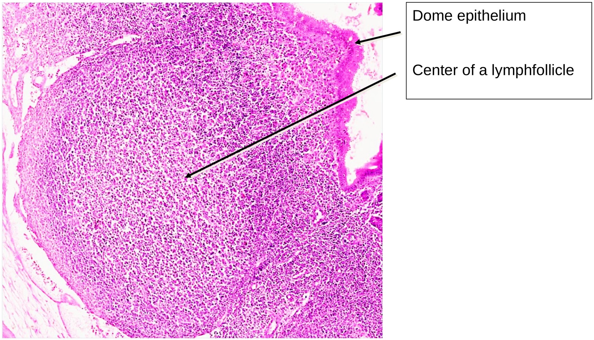
At low magnification, the uneven wall structure of the terminal ileum is evident. The wall is thicker on the antimesenteric side than on the mesenteric side. Higher magnification reveals that this thickening results from aggregated lymphoid follicles (Peyer’s patches).
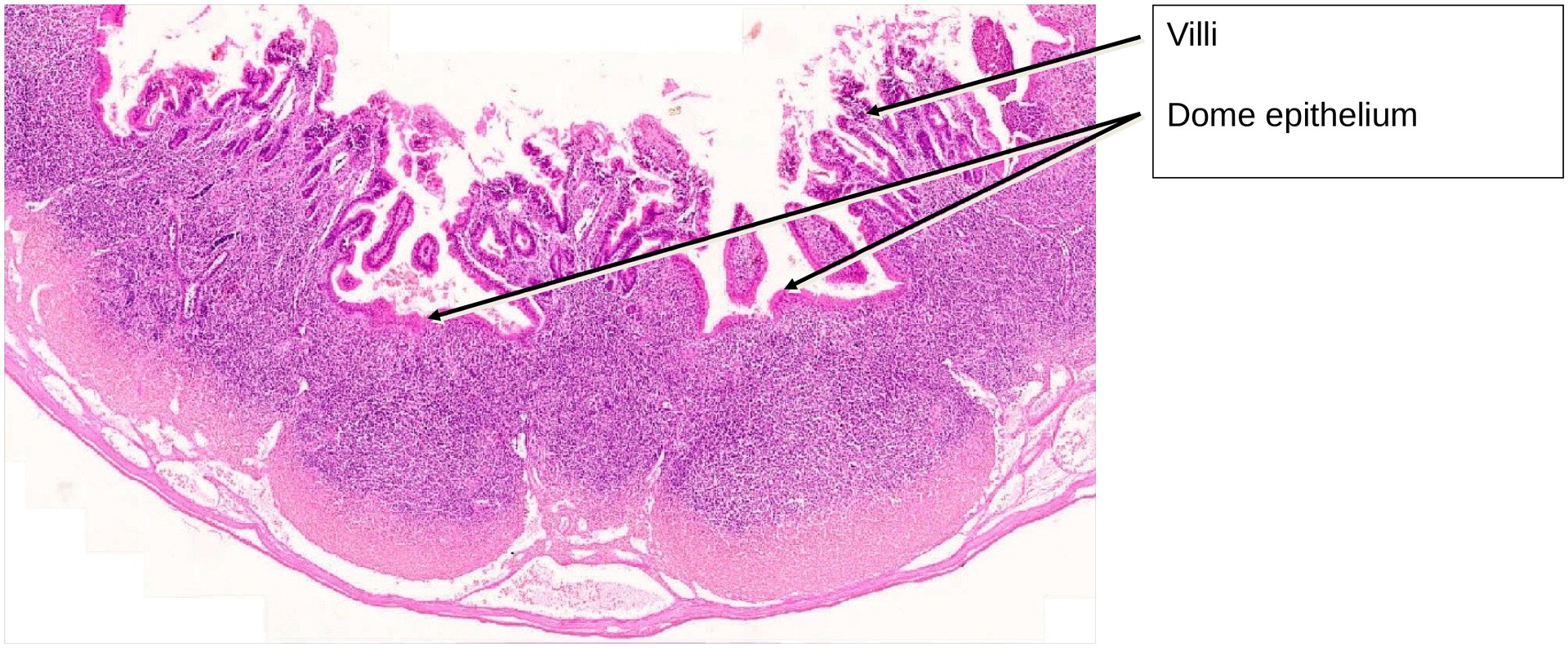
These follicles primarily arise within the lamina propria of the tunica mucosa, but may extend through the muscularis mucosae and into the submucosa. The lymphoid tissue is covered towards the intestinal lumen by a specialized dome epithelium (follicle-associated epithelium), which contains numerous free immune cells and overlies the dome region of each follicle.
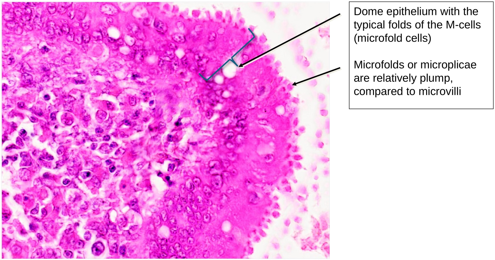
The dome epithelium arches over the follicles and is composed of enterocytes interspersed with M cells. The latter are characterized by their microfolds (microplicae) on the apical surface — hence the name M cells. These cells are crucial for antigen transport from the intestinal lumen to underlying immunocompetent cells, thereby initiating local immune responses. The microfolds impart a distinctive morphology to the dome epithelium, which can be clearly seen in the provided images.
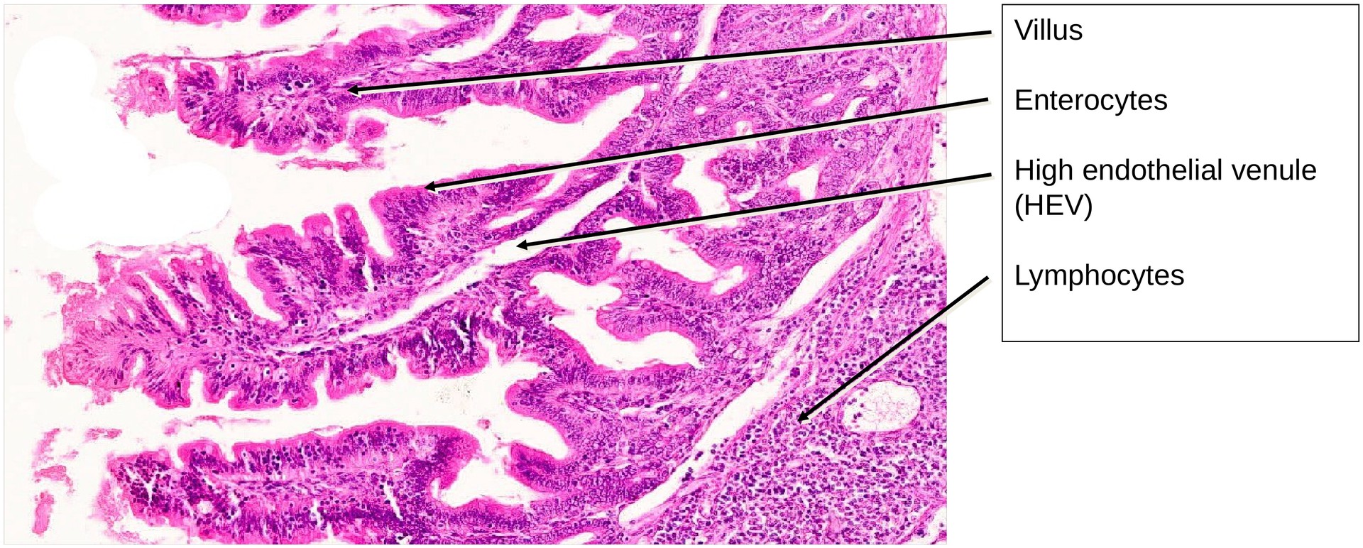
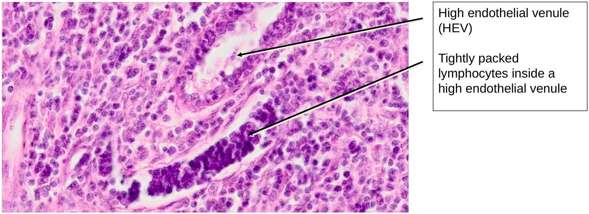
A notable vascular feature of the ileum is the presence of high endothelial venules (HEVs) — small veins lined with cuboidal (columnar) endothelium. These vessels often appear densely filled with lymphocytes, as they serve as sites of lymphocyte migration (diapedesis) into the surrounding lymphoid tissue. HEVs must be distinguished from crypts, which can appear similar in cross-section.
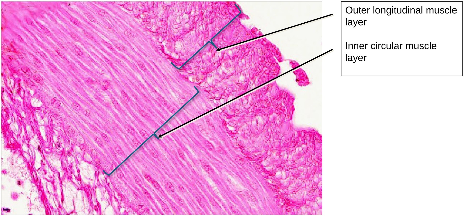
Apart from these lymphoid structures, the ileum shows the typical histological organisation of the small intestine, though the tunica muscularis (both inner circular and outer longitudinal layers) is relatively thin, especially compared with the duodenum or jejunum. The villi are shorter and less numerous in this terminal section of the small intestine.
Tasks:
- Identify under low magnification which side of the specimen corresponds to the mesenteric and antimesenteric borders.
- Compare both sides: on which side are the aggregated lymphoid follicles (Peyer’s patches) located?
- Observe the height of the folds and villi — note that they are relatively low in the ileum.
- Locate the dome epithelium and identify M cells with their characteristic microfolds under higher magnification.
- Search for high endothelial venules (HEVs) and examine their endothelial lining and intraluminal lymphocytes.
License
University of Basel
Downloads