LYMPHATIC ORGANS (ANATOMICAL MICROSCOPY)
15.9
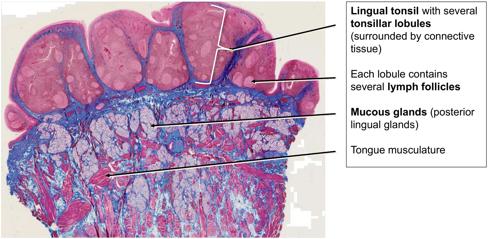
Lingual tonsil (base of tongue)
Specimen Details:
Specimen Details:
Organ: Base of the tongue
Origin: Human
Staining: Azan
Method and Specimen Description:
Conventional histological section stained with Azan, in which connective tissue appears blue, while epithelium, erythrocytes, and muscle cells stain red.
Objective of the Examination:
To study the structure of the lingual tonsil and its surrounding tissues at the base of the tongue, including the mucous glands and tongue musculature located in this region.
Special Features of the Specimen:
The lingual tonsil, as a component of the pharyngeal lymphoid ring, is directly connected to the surface epithelium and is therefore part of the MALT system (mucosa-associated lymphoid tissue).
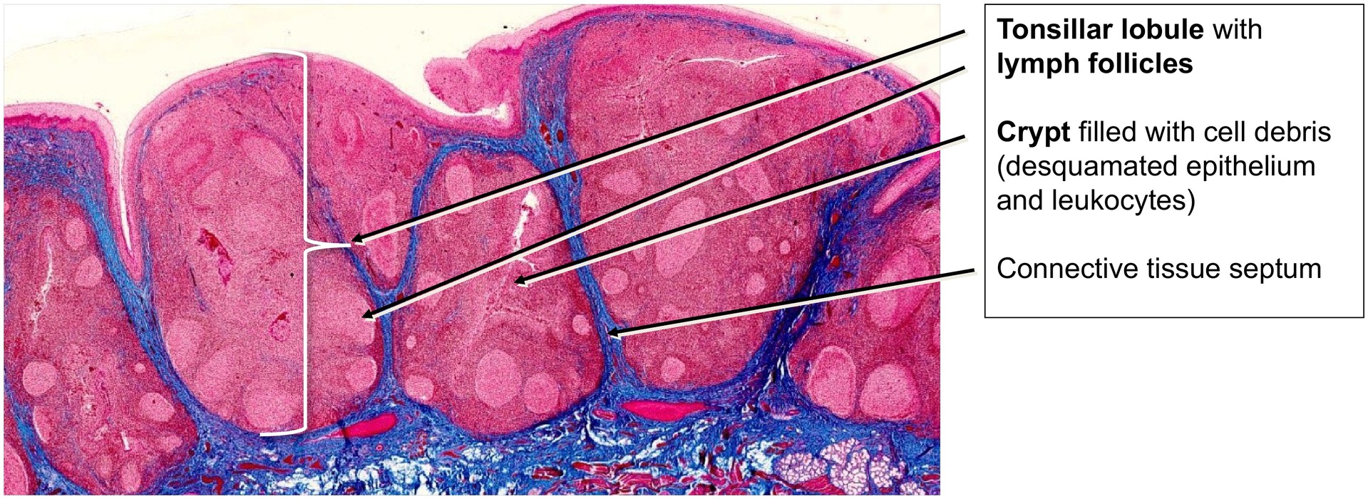
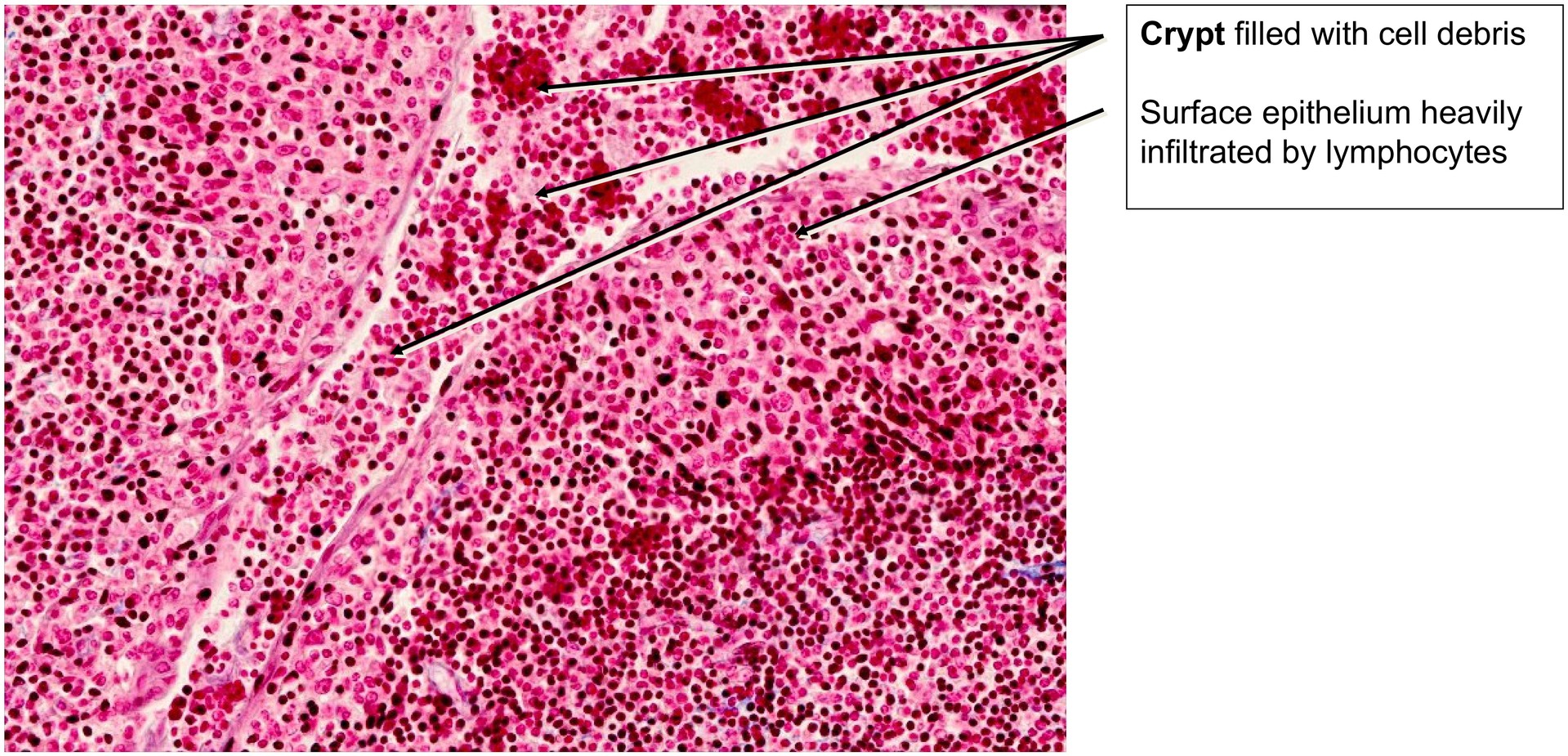
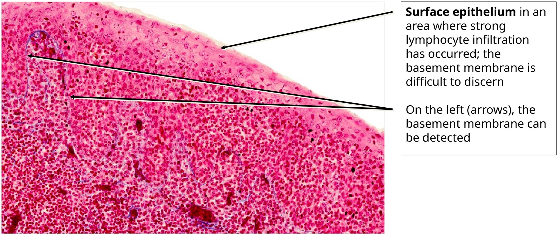
It is covered by a stratified, non-keratinized squamous epithelium, which may appear irregular or disrupted in some regions due to heavy infiltration by lymphocytes and other leukocytes migrating through it.
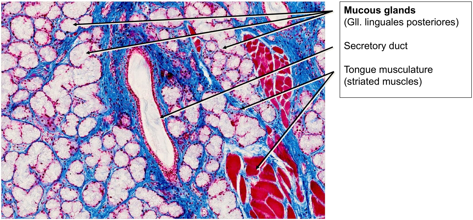
The surface epithelium invaginates deeply to form crypts. The posterior lingual glands (mucous glands) open into the bases of these crypts. Within the crypts, the epithelium is much thinner and no longer clearly recognizable as stratified non-keratinized squamous epithelium, typically consisting of only one to three cell layers.
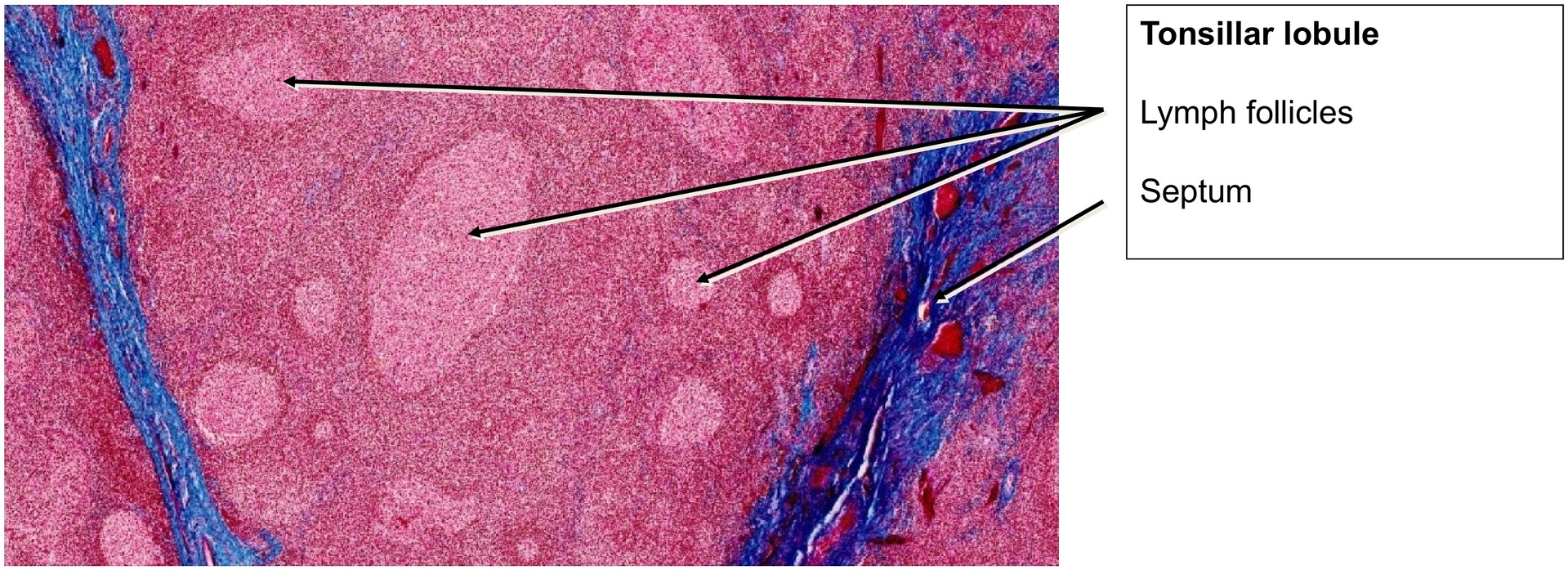
The lymphoid tissue is subdivided by connective tissue septa, which merge seamlessly with the connective tissue of the surrounding glands and tongue musculature. A zone of lymphatic tissue enclosed by connective tissue, with crypts opening to the surface, is termed a lingual bulge (Tonsillar lobule). Within this tissue:
- Lymphoid follicles contain predominantly B-lymphocytes.
- The interfollicular regions are rich in T-lymphocytes.
Tasks:
- Follow the surface epithelium over a long stretch and locate an area where it is heavily infiltrated by lymphocytes.
- Compare the surface epithelium with the epithelium of the crypts.
- The crypt epithelium is typically found at the center of a lingual bulge and differs markedly from the surface epithelium between bulges.
- Identify the type of surface epithelium and its individual layers.
- Locate several posterior lingual glands (mucous glands) and observe their secretory character.
- Examine the tongue musculature, noting the orientation of the muscle fibers.
- Identify the type of muscle tissue present (hint: skeletal muscle).
License
University of Basel
Downloads