SKIN AND APPENDAGES (ANATOMICAL MICROSCOPY)
13.1
Armpit
Specimen:
Specimen Details:
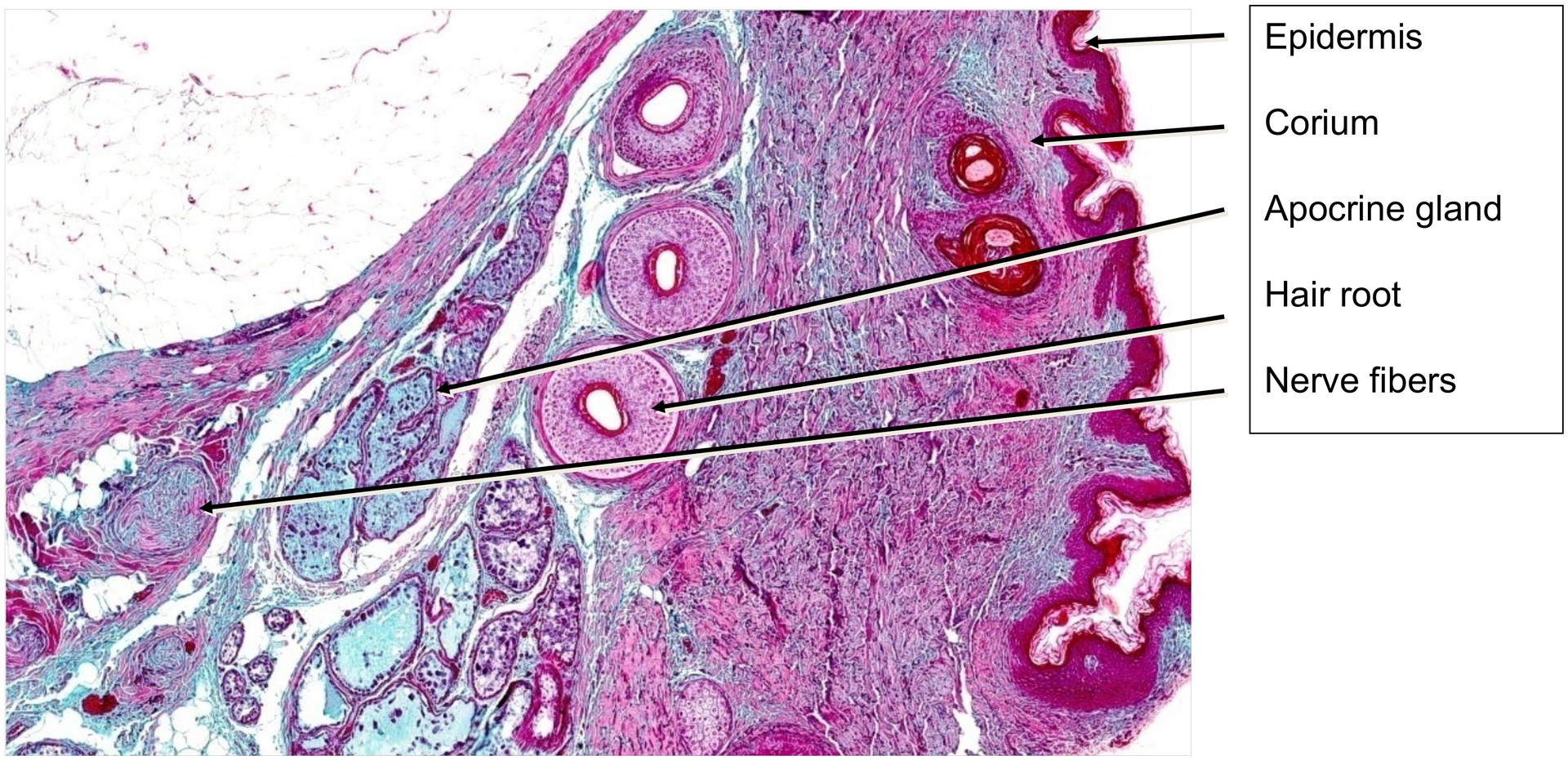
Organ: Skin, Axilla
Origin: Human
Staining: RFAL
Method and Specimen Description:
Normal histological section of axillary skin, stained with RFAL, which distinctly demonstrates elastic fibers.
Objective of the Examination:
To study the skin of the axilla with its specific gland types:
- Eccrine sweat glands, opening directly onto the skin surface,
- Apocrine sweat glands, and
- Holocrine sebaceous glands, both of which open into hair follicles.
Special Features of the Specimen:
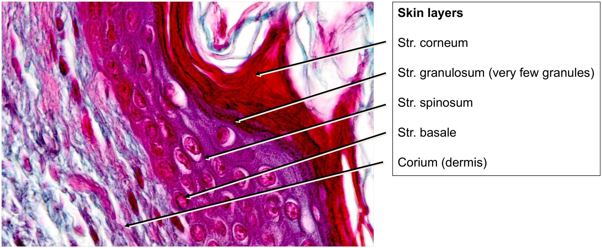
Although the typical epidermal layers are recognizable, they are comparatively thin in this region. This is because the axillary skin is exposed to minimal mechanical stress, and thus the stratum corneum is weakly developed. Consequently, the stratum granulosum is also poorly defined and can only be seen in a few areas.
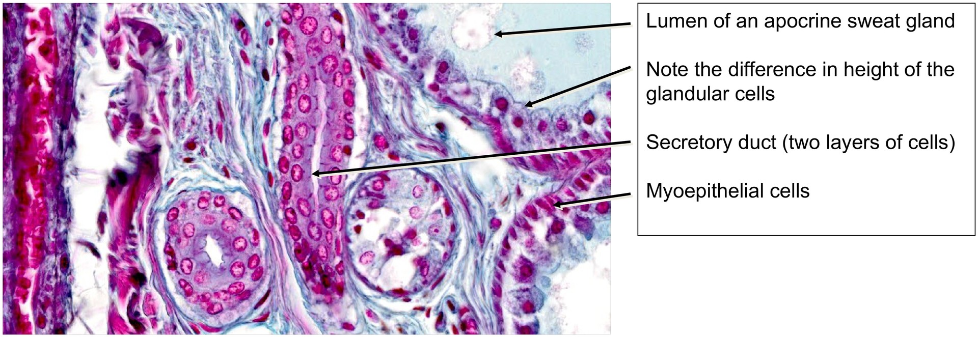
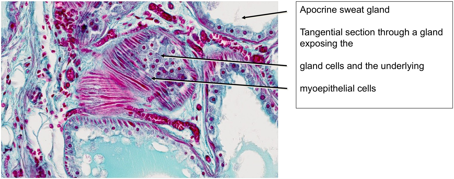
Numerous glands are present, most prominently the apocrine sweat (scent) glands. These become active under the influence of sex hormones during puberty.
- They exhibit a large lumen and a variable epithelial height, reflecting their merocrine secretion mechanism, during which parts of the apical cytoplasm are shed.
- Myoepithelial cells between the glandular epithelium and basement membrane facilitate accelerated secretion through contraction.
- Apocrine glands open into hair follicles, and their excretory ducts are bilayered and lack myoepithelial cells.
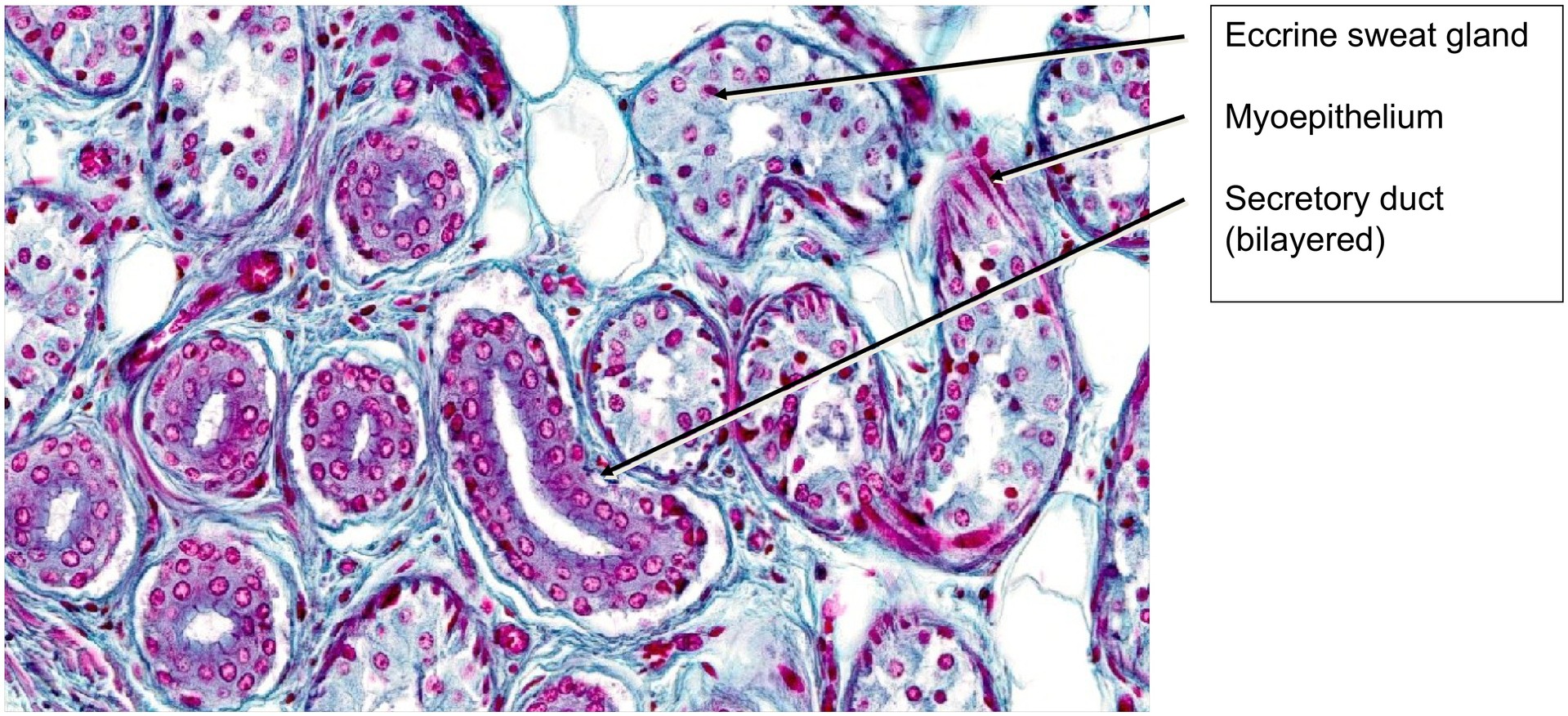
In contrast, the eccrine sweat glands open directly onto the skin surface via the epidermis.
- Only a few are present in this section.
- Their secretory cells are surrounded by myoepithelial cells, possess a narrow lumen, and have a uniform epithelial height.
- The ducts are bilayered.
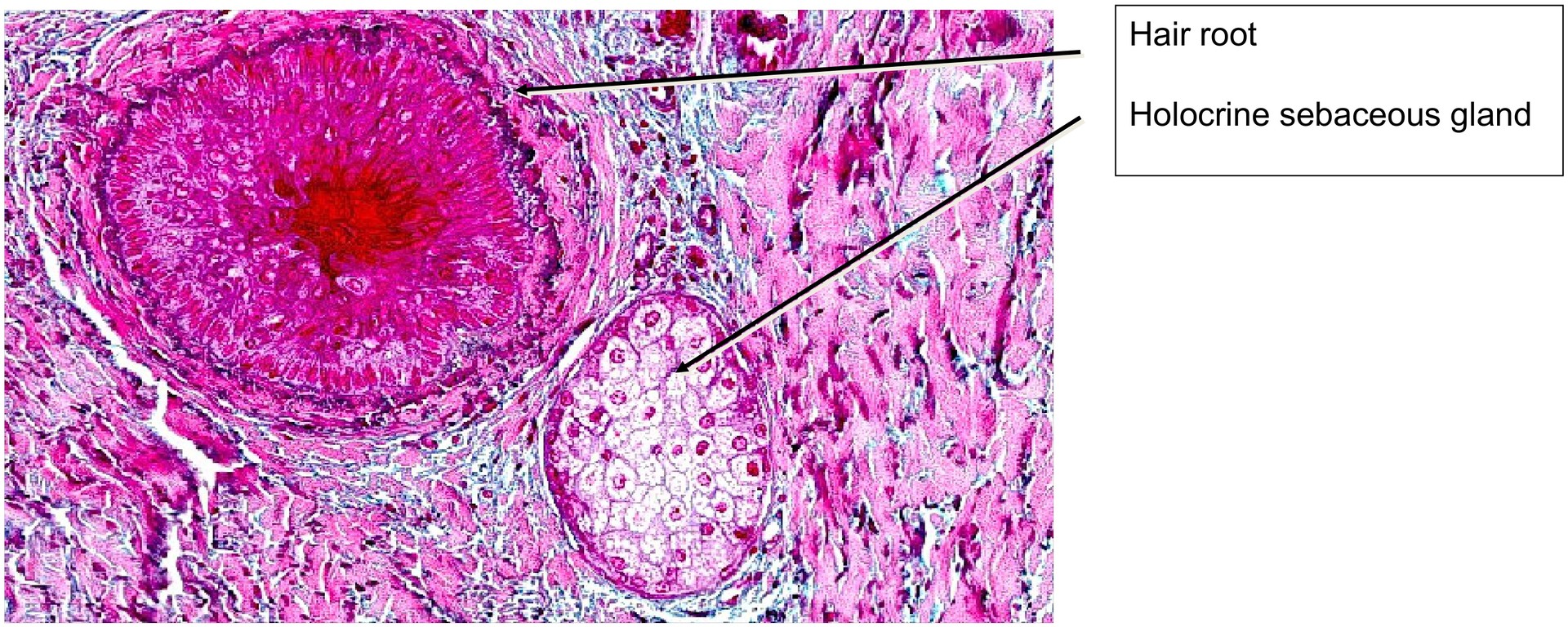
The holocrine sebaceous glands, located adjacent to the coarse axillary hairs, also open into hair follicles.
- They show a characteristic foamy cytoplasm due to lipid content.
- During holocrine secretion, entire cells disintegrate, releasing sebum.
- The arrector pili muscle beneath the sebaceous glands assists in sebum release during contraction.
As a reminder, the epidermal and dermal layers visible here (from outside to inside) are:
- Stratum corneum (thin)
- Stratum granulosum (weakly developed)
- Stratum spinosum
- Stratum basale
- Corium (dermis) with stratum papillare and stratum reticulare
- The interdigitation of epidermis and dermis is shallow due to limited stress.
- Subcutis, rich in adipose tissue.
Tasks:
- Identify the various layers of the epidermis and entire skin.
- Locate the stratum granulosum in the epidermis.
- Find hair follicles and associated hairs.
- Identify the three gland types: eccrine, apocrine, and holocrine.
- Search for myoepithelial cells – which gland type lacks them?
- Compare the lumina of apocrine and eccrine glands.
- Explain the mechanism for accelerated secretion in sebaceous glands.
- Note the abundance of elastic fibers in this specimen.
License
University of Basel
Downloads