DIGESTIVE ORGANS: GASTROINTESTINAL TRACT (ANATOMICAL MICROSCOPY)
19.9
Stomach (Fundus)
Specimen:
Specimen Details:
Specimen Details:
Organ: Stomach (Fundus) Origin: Cat Staining: Acridine Yellow
Method and Specimen Description:
Normal histological section stained with acridine yellow, which effectively stains mucus within the mucous cells of the gastric glands (accessory cells).
Objective of the Examination:
To study the heterocrine glands of the corpus and fundus regions of the stomach, identify the different gland cell types (chief, accessory, and parietal cells), and recognize the general layering of the gastrointestinal tract, including the gastric foveolae (pits).
Special Features of the Specimen:
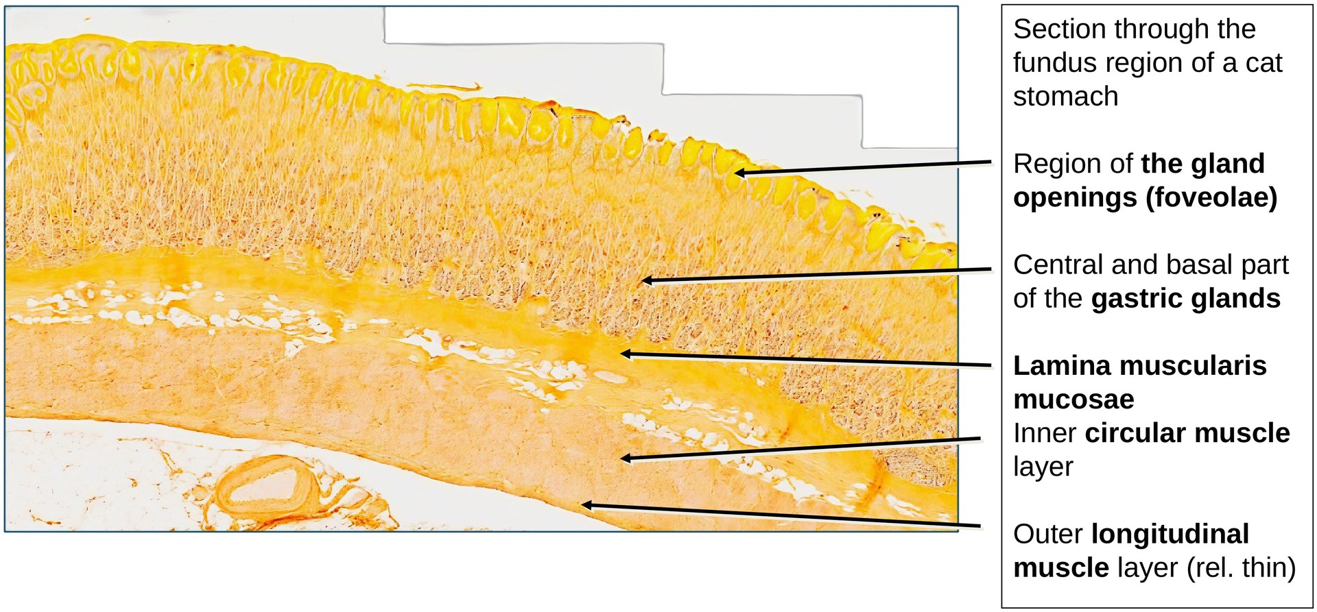
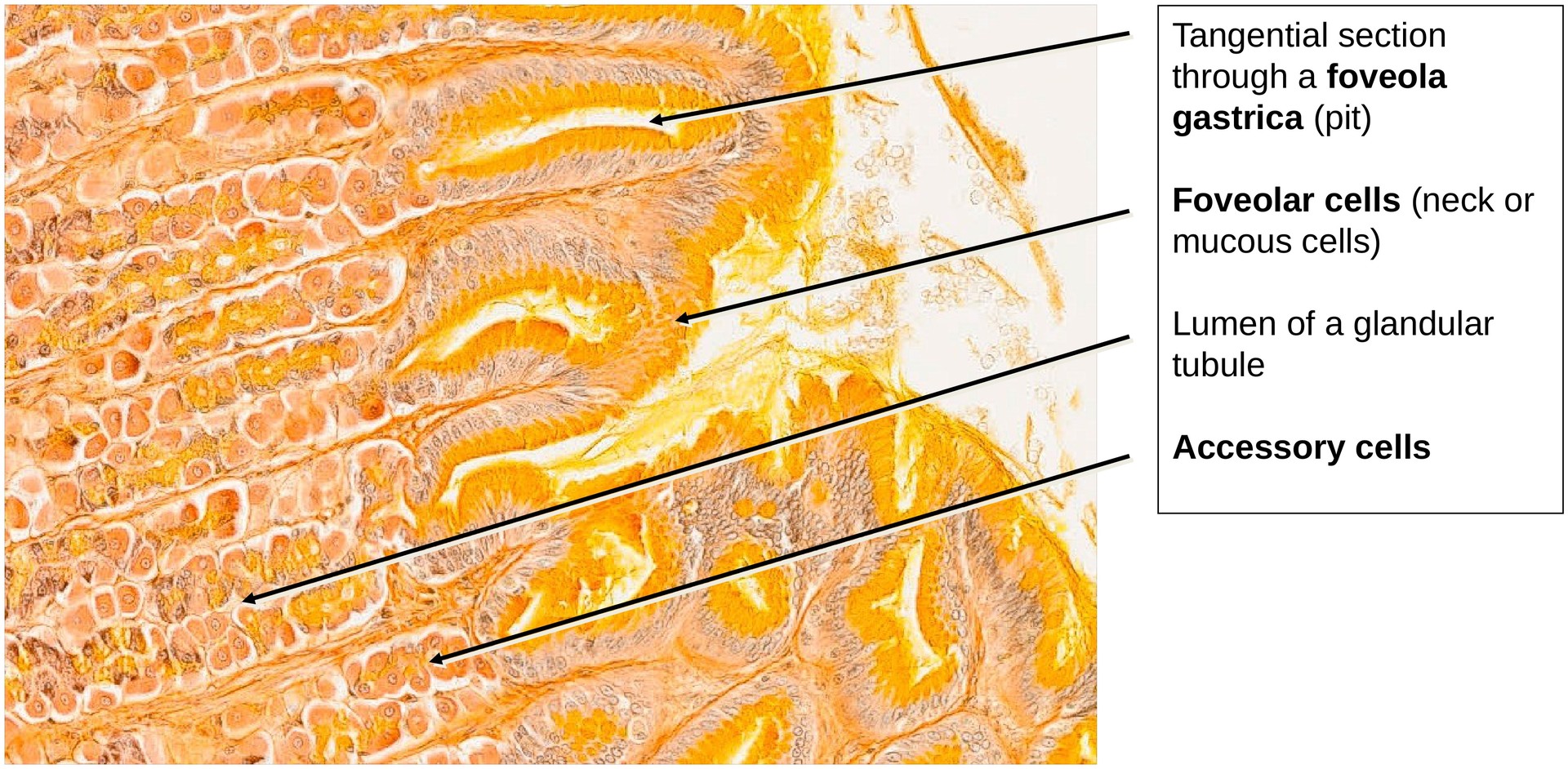
The gastric mucosa is formed by a single layer of columnar epithelium, which invaginates to form gastric foveolae (pits). These pits open into the gastric glands.
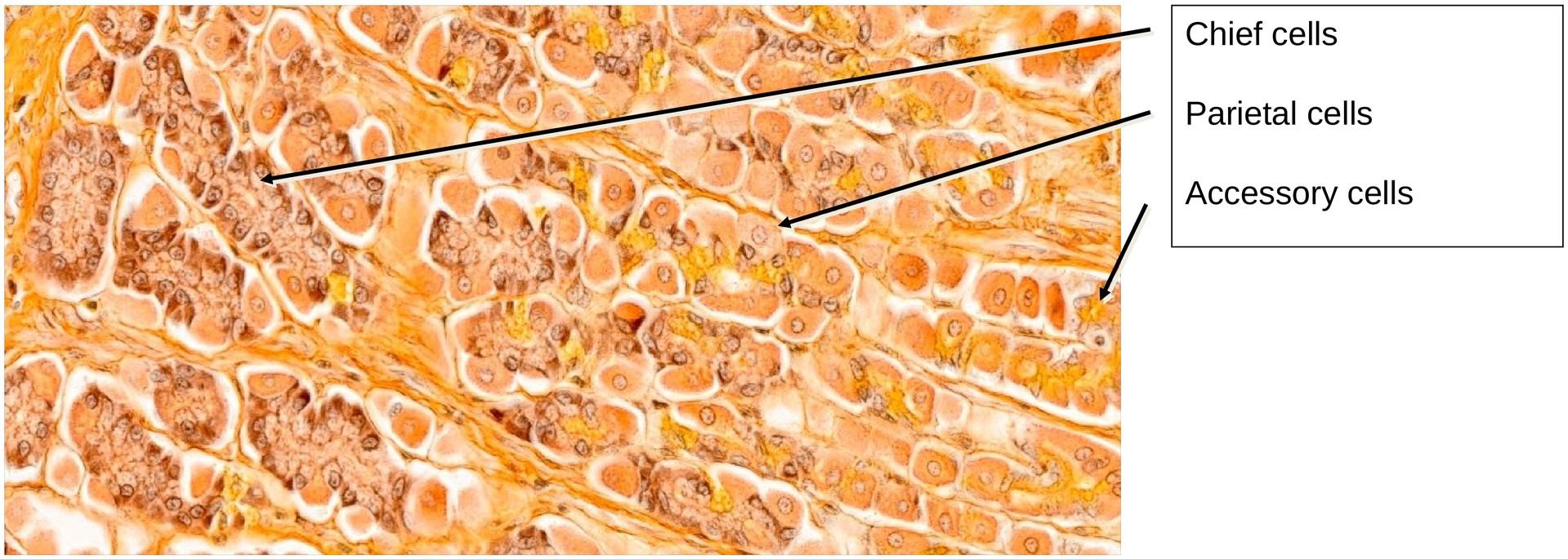
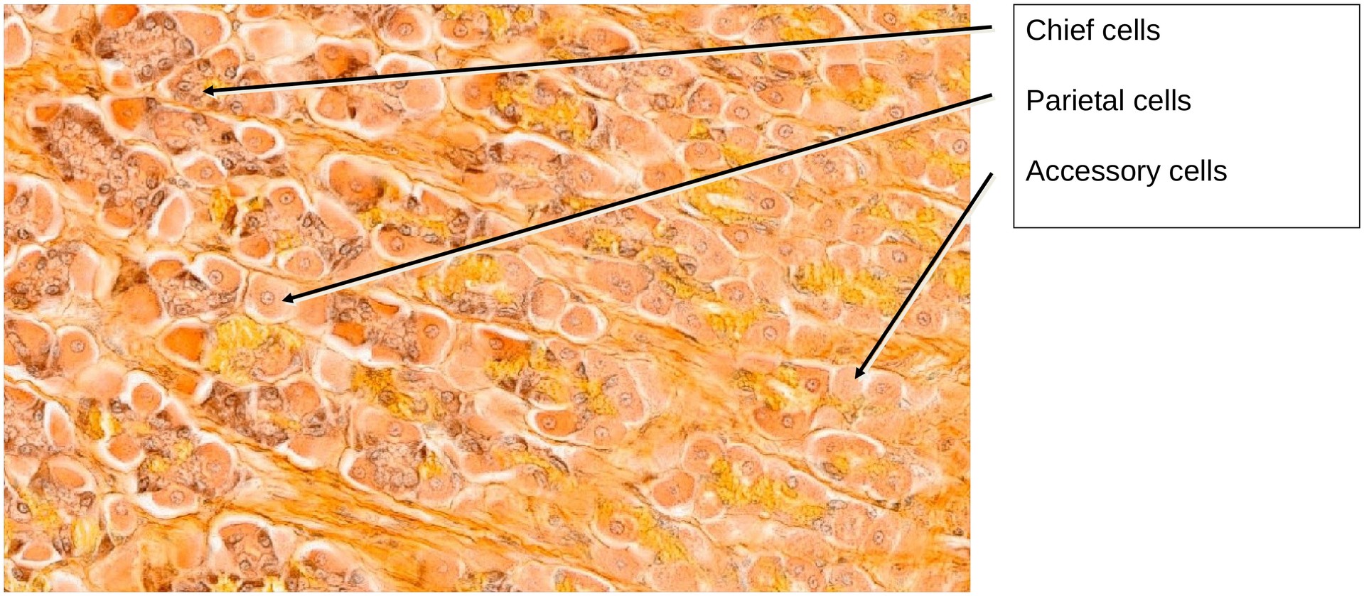
In the fundus and corpus regions, the glands are heterocrine, meaning they contain several distinct cell types that produce different secretions. By contrast, the glands in the cardiac and pyloric regions are homocrine, containing mainly mucous cells.
The gastric glands show an isthmus (a narrowing directly beneath the gastric foveola) and a main body. Occasionally, two glands may open into a single pit, and the glandular tubules often exhibit branching.
- In the isthmus and pit regions, accessory cells predominate — these secrete mucus to protect the mucosa from acid.
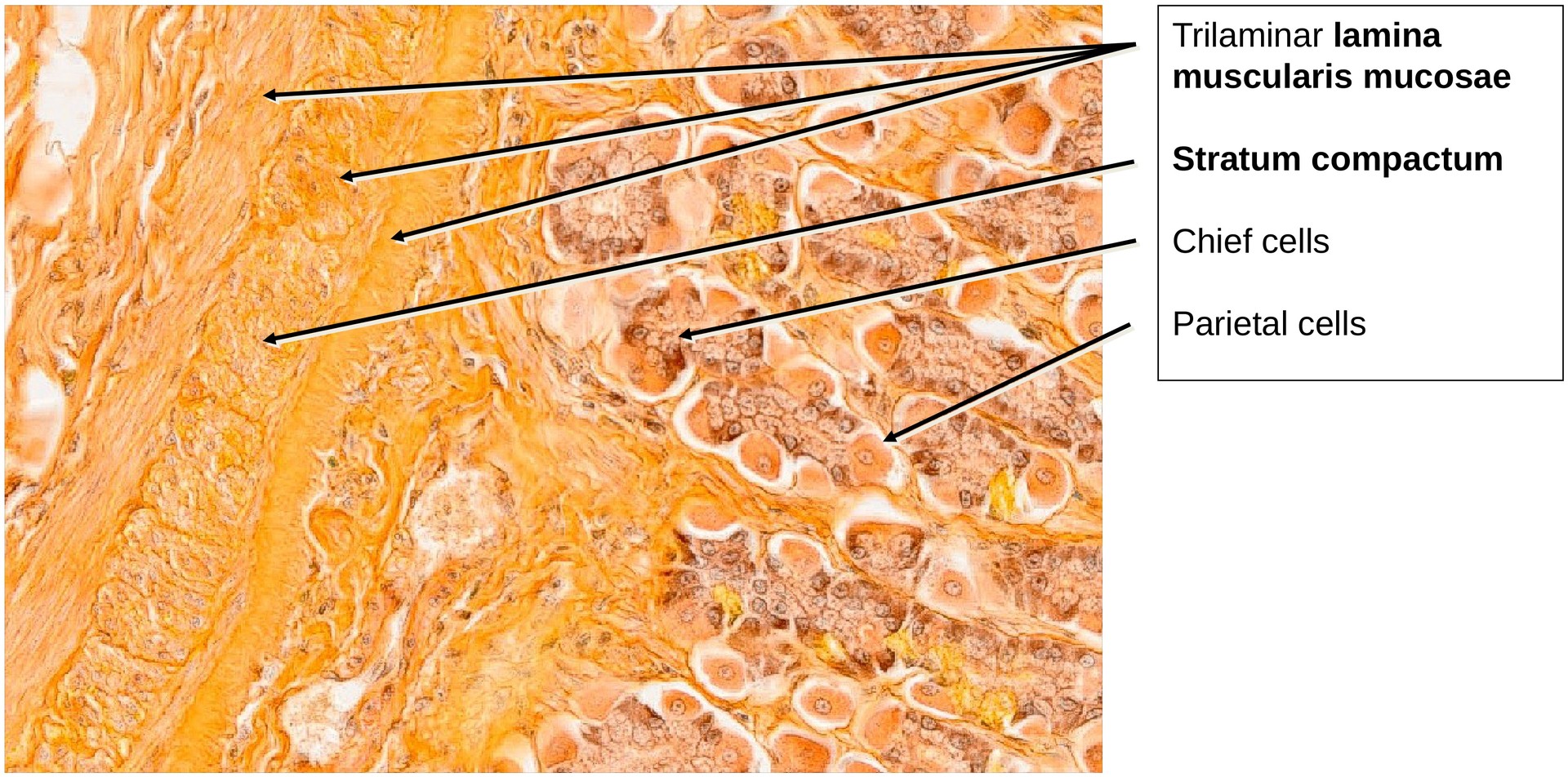
- In the main (central) part of the gland, numerous parietal cells are present — they produce hydrochloric acid (HCl) and intrinsic factor.
- In the basal part, mainly chief cells are found — they secrete pepsinogen, the inactive precursor of pepsin.
Other cell types, such as ECL cells (enterochromaffin-like cells), are not clearly distinguishable with acridine yellow staining.
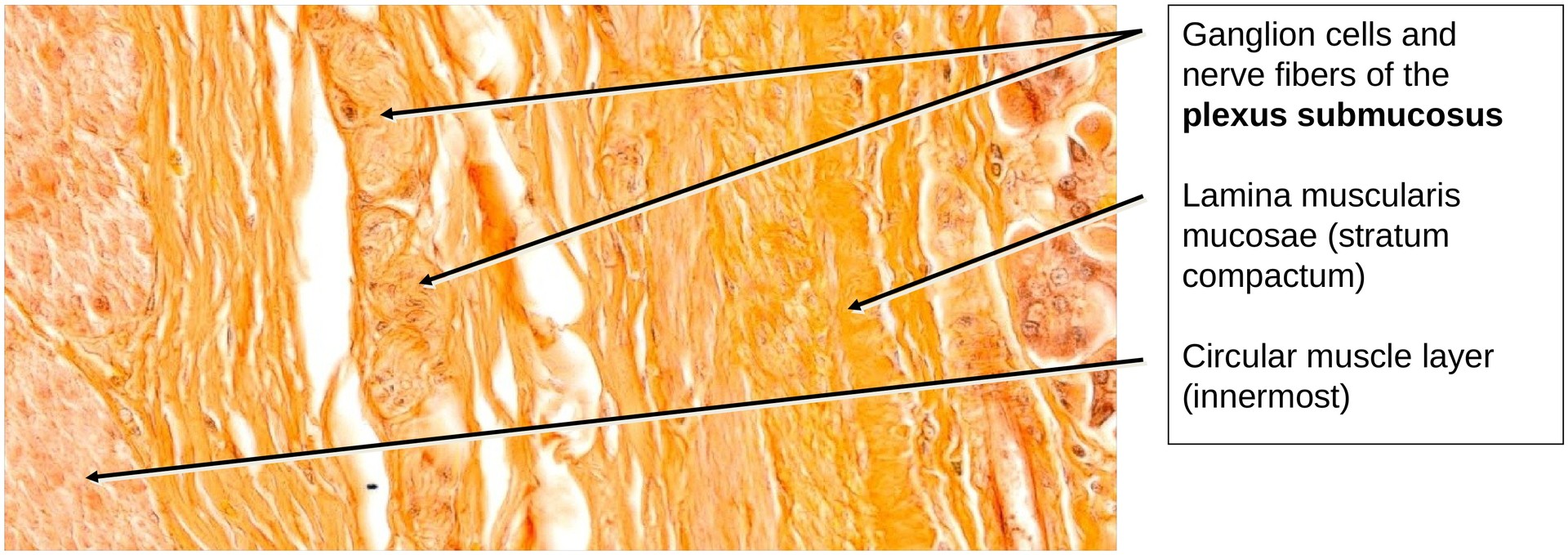
The lamina muscularis mucosae in carnivores (e.g., cat) is characterized by a stratum compactum, giving it a trilaminar appearance.
Within the tela submucosa, ganglion cells and nerve fibers of the submucosal (Meissner’s) plexus are visible. The submucosa itself is relatively thin, and the outer longitudinal muscle layer of the tunica muscularis is only weakly developed.
Tasks:
- Orient yourself within the major layers of the stomach wall: mucosa, submucosa, and muscularis.
- Examine the mucosa and explain why the glands of the fundus and corpus are described as heterocrine.
- Identify the three main gland cell types (chief, parietal, accessory) and note their distribution within the gland tubules.
- Locate examples of branching in the glandular tubules.
- Determine where the gastric foveolae (pits) are located and describe their appearance.
- Describe the structure and organization of the lamina muscularis mucosae, including its stratum compactum.
- Search for areas containing ganglion cells and nerve fibers of the submucosal plexus within the tela submucosa.
License
University of Basel
Downloads