BONES (GENERAL HISTOLOGY)
5.1
Lamellar bone (Tibia, Dog)
Specimen Details:
Specimen Details:
Organ: Long bone (Tibia)
Origin: Dog
Staining: Thionin after Schmorl
Method and Specimen Description:
To allow cryostat sectioning, the bone was first decalcified. The resulting cryosections are inevitably thicker than conventional histological sections.
With thionin staining according to Schmorl, the bone canals and lacunae—the sites occupied by osteocytes with their nuclei and processes—are clearly visualized.
Objective of the Investigation:
To study the structure of lamellar and compact bone, including:
-
the arrangement of bone lamellae,
-
the structure of osteocytes,
-
the organization of circumferential lamellae and interstitial systems,
-
and their relationship to the vascular supply.
Special Features of the Specimen:
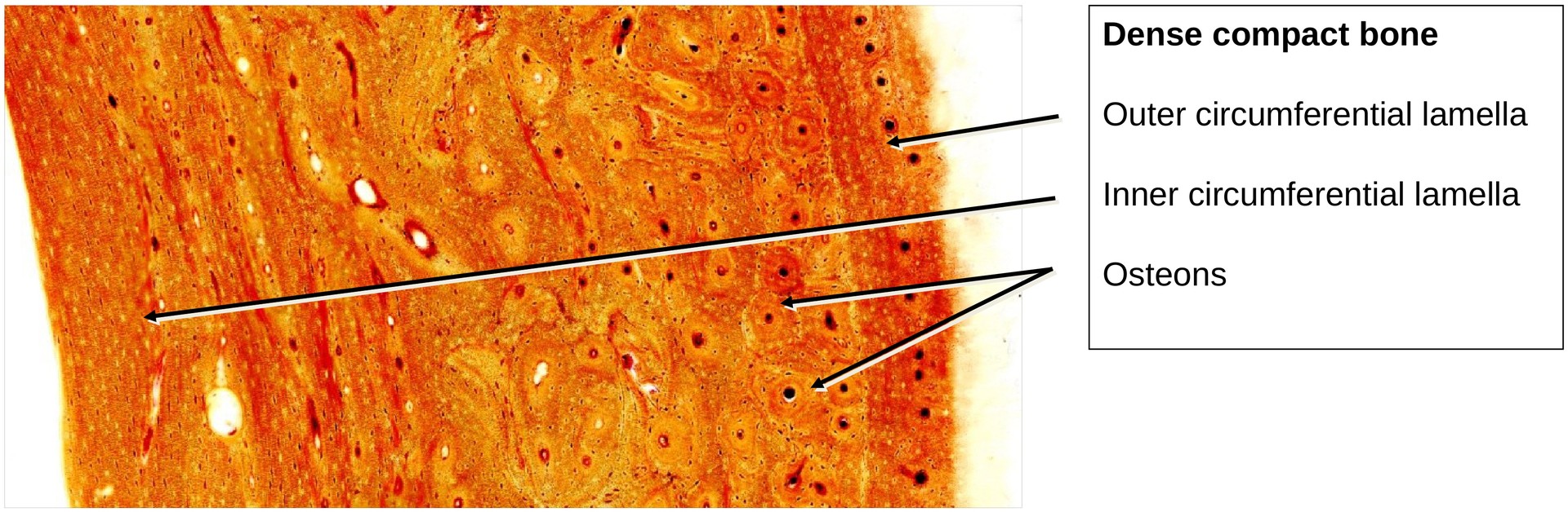
The shaft (diaphysis) of a long bone consists almost entirely of dense compact bone.
It is externally bounded by the outer circumferential lamella and internally, towards the marrow cavity, by the inner circumferential lamella. The thickness of these layers varies around the circumference. In some areas, the inner circumferential lamella may continue as spongy (cancellous) trabeculae projecting into the marrow cavity (though these are not visible in this specimen).
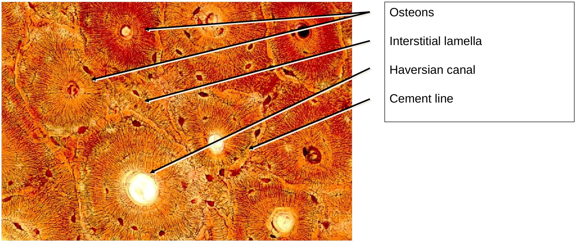
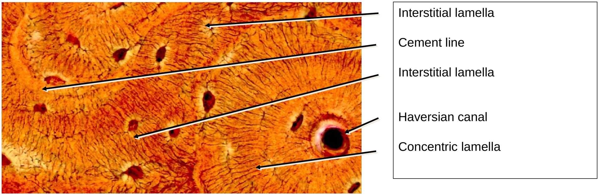
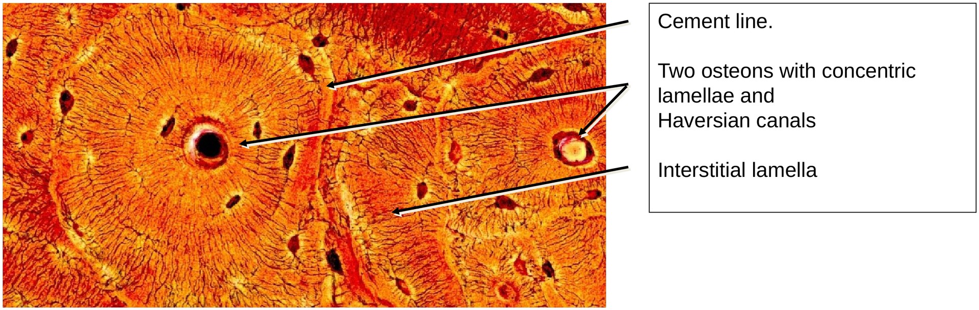
Between the inner and outer circumferential lamellae lies the main bone substance, composed of numerous osteons (Haversian systems) with concentrically arranged lamellae around a Haversian canal.
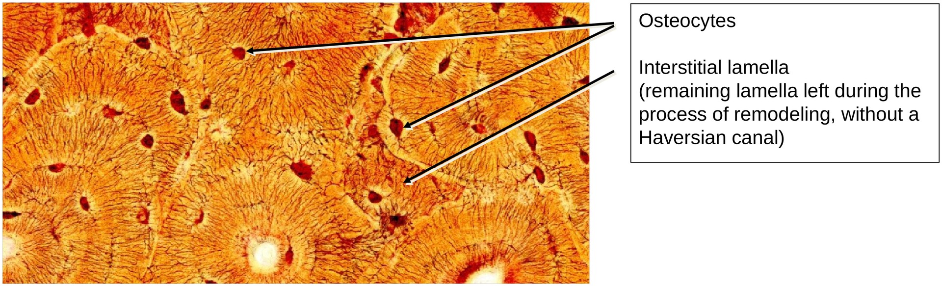
Between these osteons are irregularly shaped interstitial lamellae, often triangular or quadrangular in outline. These represent remnants of older osteons partially resorbed during bone remodeling.
Some osteons in this specimen contain large vascular spaces, where either osteoclastic resorption or new bone formation (osteogenesis) is taking place within the Haversian cavity.
Bone is a dynamic tissue, constantly undergoing remodeling, with concurrent resorption and deposition processes throughout life.
Volkmann’s canals are transverse or oblique channels that penetrate the circumferential and concentric lamellae, providing vascular and structural communication between adjacent osteons.
Osteocytes and Canaliculi (High Magnification):
The osteocytes are embedded between the concentric lamellae, lying within lacunae. Their cytoplasmic processes extend through minute bone canals (canaliculi), enabling diffusion-based exchange of nutrients and waste products with the blood vessels in the central Haversian canal.
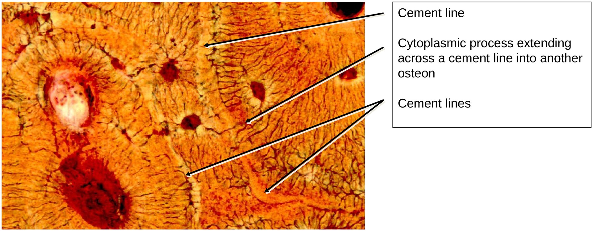
Between neighboring osteons are cement lines, marking the boundary between systems. These zones contain more ground substance and fewer collagen fibers.
Osteocytes within interstitial lamellae and circumferential lamellae are similarly nourished by diffusion through canaliculi, which connect with adjacent vascular channels. Exchange can also occur across cement lines, ensuring metabolic continuity between systems.
Tasks:
• Locate the inner and outer circumferential lamellae.
• Identify a large osteon and its Haversian canal.
• Observe the concentric lamellae surrounding the canal.
• Find examples of interstitial systems between osteons.
• Determine where the osteocytes are positioned relative to the lamellae.
• Trace several canaliculi in the peripheral area of an osteon, and identify any that cross the cement line into adjacent osteons or interstitial lamellae.
License
University of Basel
Downloads