DIGESTIVE ORGANS: ORAL CAVITY (ANATOMICAL MICROSCOPY)
18.13
Base of the tongue
Specimen:
SPECIMEN DETAILS:
Organ: Base of tongue with lingual tonsil
Origin: Human
Staining: Azan
METHOD AND SPECIMEN DESCRIPTION:
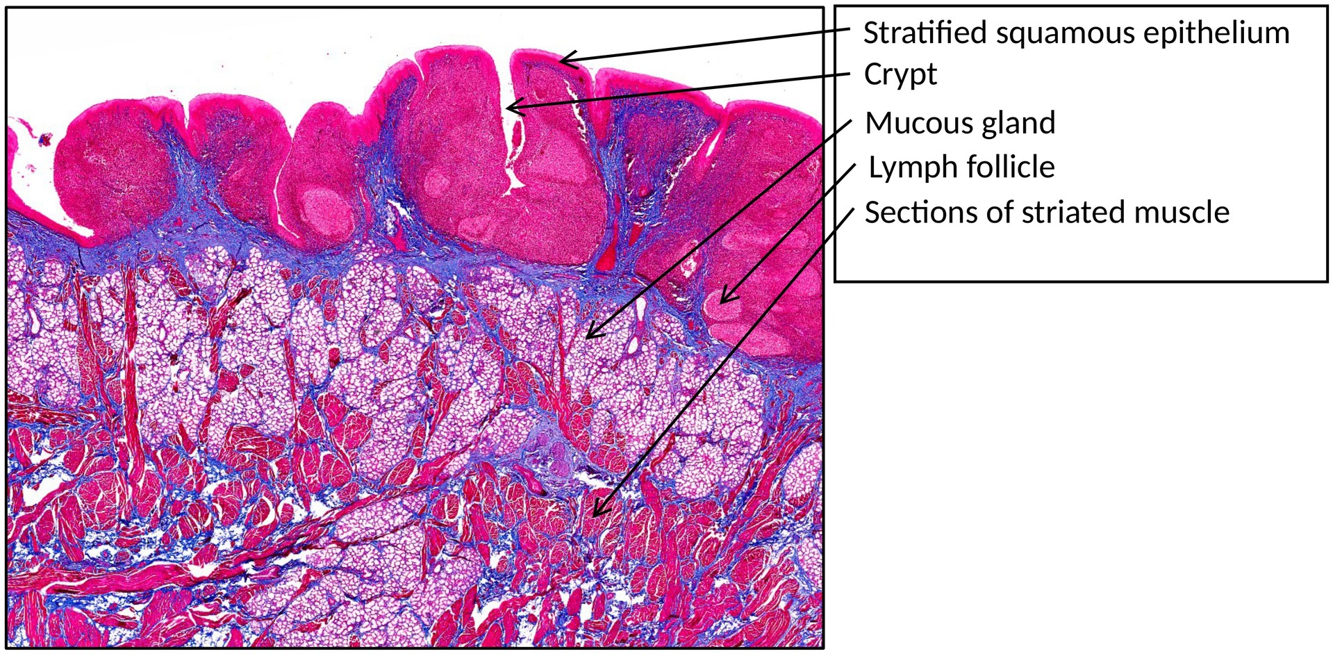
This is a normal sagittal section through the human base of the tongue, including the lingual tonsil. The Azan stain colors connective tissue blue, muscle fibers red, and cell nuclei dark purple, providing clear contrast between structural components.
OBJECTIVE OF THE EXAMINATION:
To understand the morphology and histological structure of the base of the tongue, with particular emphasis on the lingual tonsil and its lymphoid architecture.
SPECIAL FEATURES OF THE SPECIMEN:
General: The base of the tongue retains the typical muscular structure of the organ, characterized by interwoven bundles of striated muscle fibers arranged in longitudinal, transverse, and vertical planes.
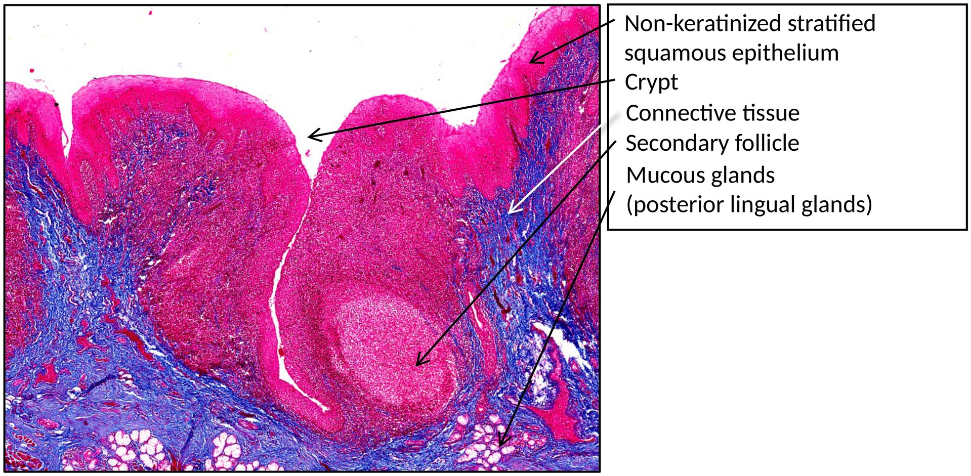
The surface epithelium is a non-keratinized stratified squamous epithelium, which transitions from the oral to the oropharyngeal region.
Posterior to the terminal sulcus, the lingual tonsil forms part of Waldeyer’s lymphatic ring, giving the posterior dorsal tongue a nodular (“hilly”) appearance.
Each lingual follicle consists of:
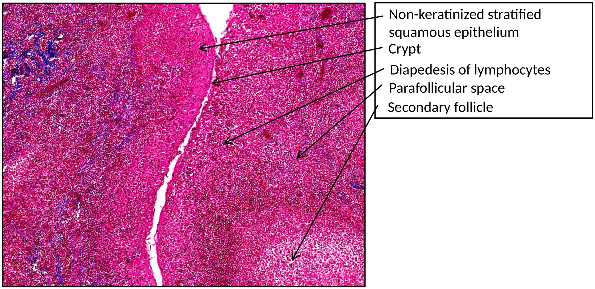
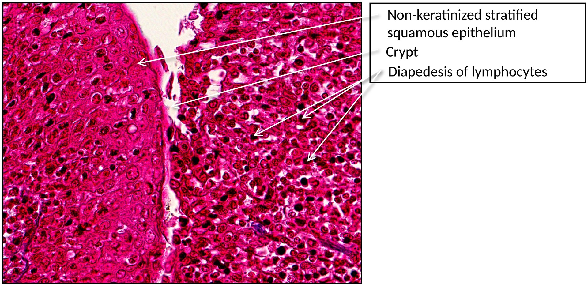
- One or several crypts (deep epithelial invaginations), lined by non-keratinized stratified squamous epithelium.
- Lymphoid tissue surrounding the crypts, containing primary and secondary lymphoid follicles.
- Parafollicular areas (T-cell zones) between follicles.
- Mucous glands located in the subepithelial connective tissue, whose ducts open into the crypt bases.
The epithelium often shows infiltration by lymphocytes migrating between epithelial cells (diapedesis), indicative of immune surveillance activity.
TASKS:
- Identify the non-keratinized stratified squamous epithelium of the lingual surface.
- Locate and describe the striated muscle bundles of the tongue.
- Examine the lingual tonsil, identifying:
- Epithelial crypts
- Secondary lymphoid follicles
- Parafollicular zones (T-cell areas)
- Observe sites of diapedesis, where lymphocytes migrate through the epithelial layers.
- Identify the mucous glands beneath the lymphoid tissue and their ducts opening into the crypts.
License
University of Basel