CARDIOVASCULAR ORGANS (ANATOMICAL MICROSCOPY)
14.5
Heart (cardiac conduction)
Specimen:
Specimen Details:
Organ: Heart
Origin: Sheep
Staining: Hematoxylin - Eosin (H&E)
Method and Specimen Description:
Standard histological section stained with the general Haematoxylin and Eosin (H&E) method.
Objective of the Examination:
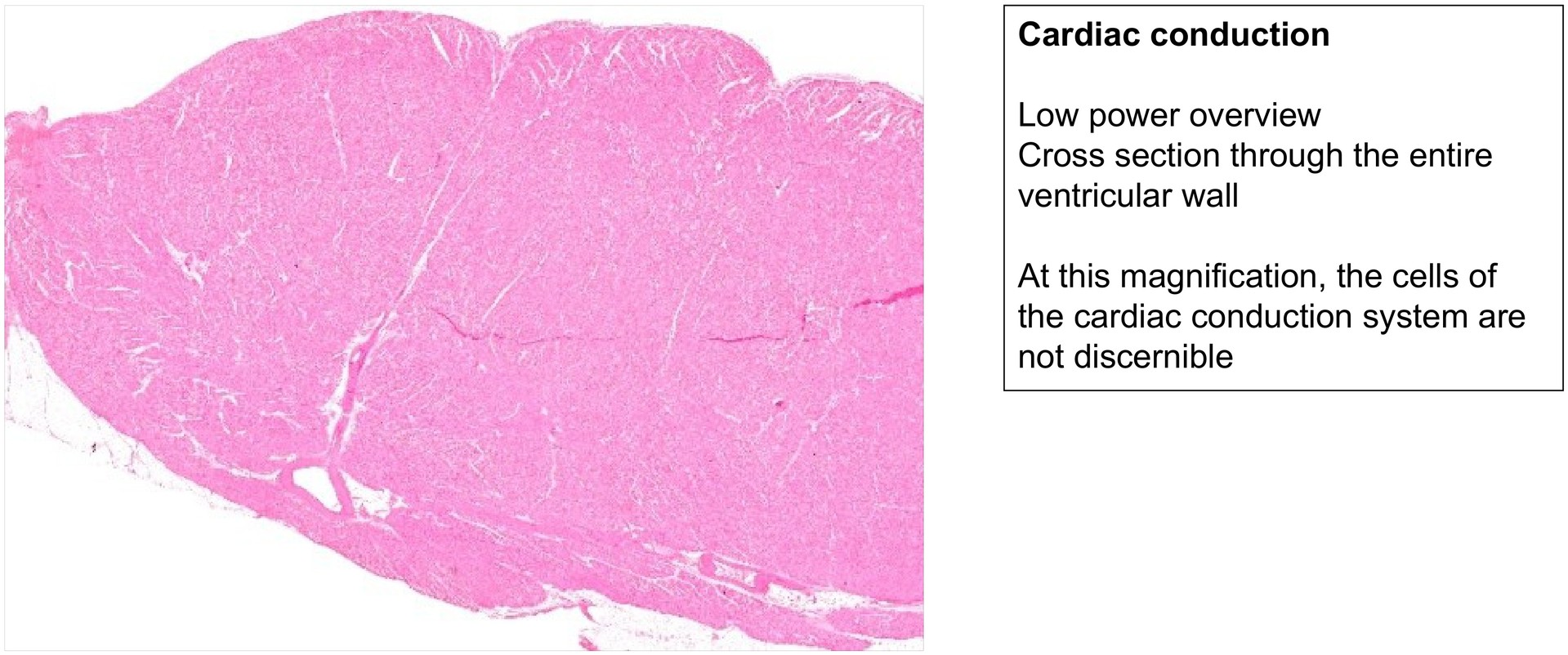
In addition to the working cardiac muscle, the heart contains a specialized type of cardiac muscle whose function is to generate and conduct electrical impulses. The differences in structure between the two cell types — those of the working myocardium and those of the conduction system — are pronounced. The aim of this preparation is to identify and understand the morphological characteristics of the conduction system cells.
Special Features of the Specimen:
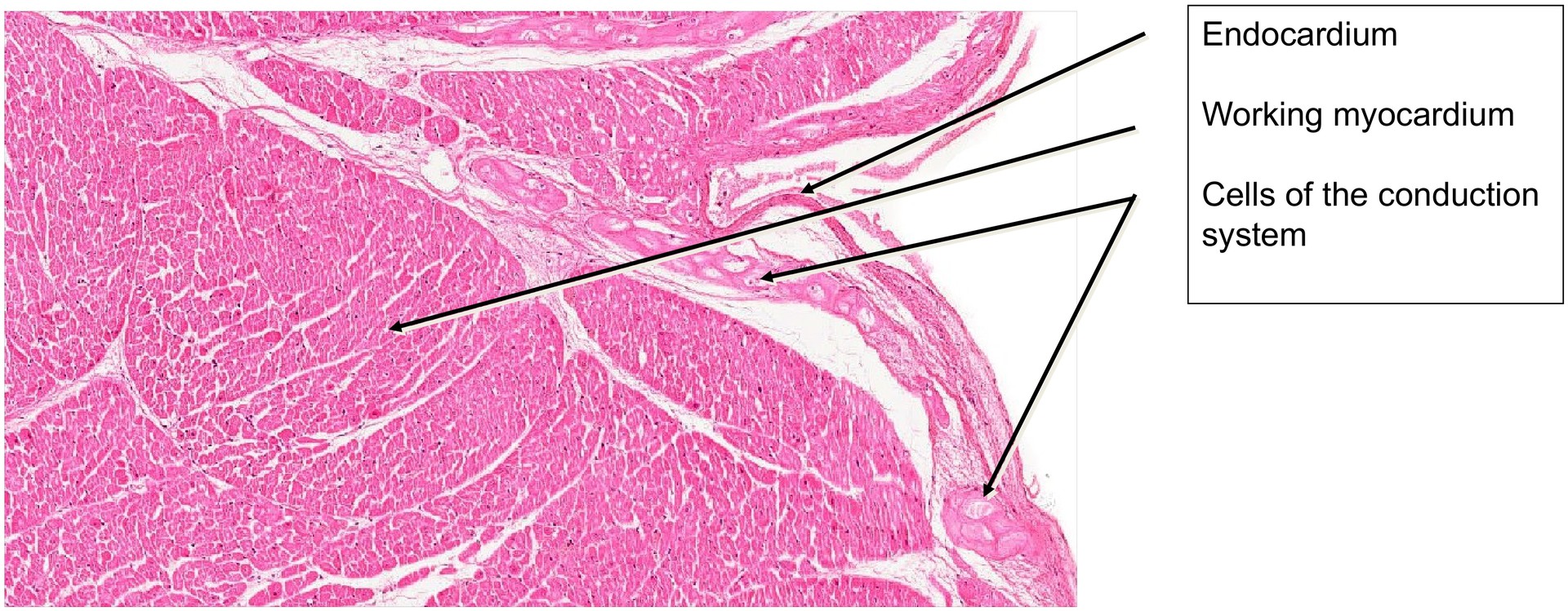
This specimen represents a longitudinal section through the interventricular septum. Immediately beneath the endocardium, the cells of the cardiac conduction system can be readily identified.
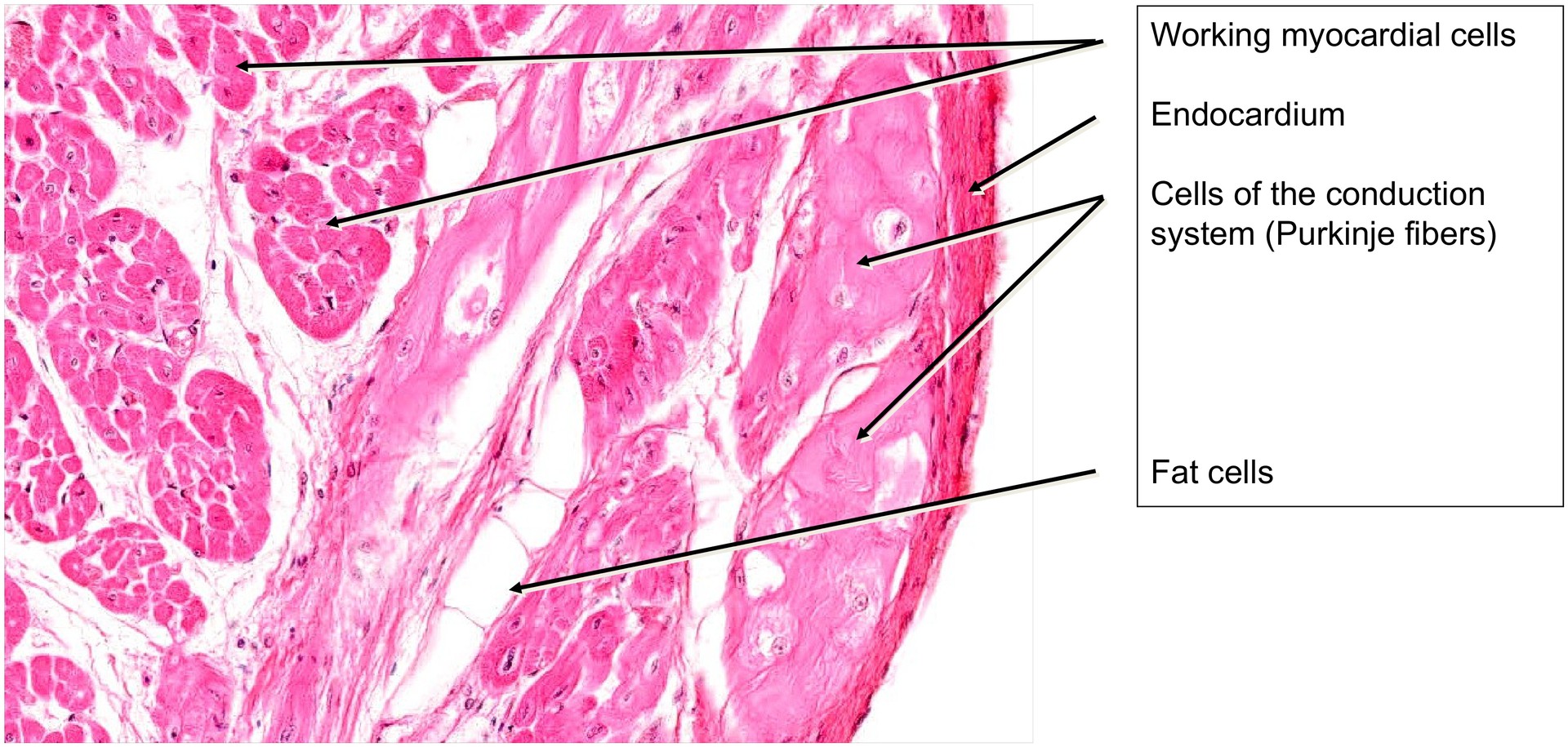
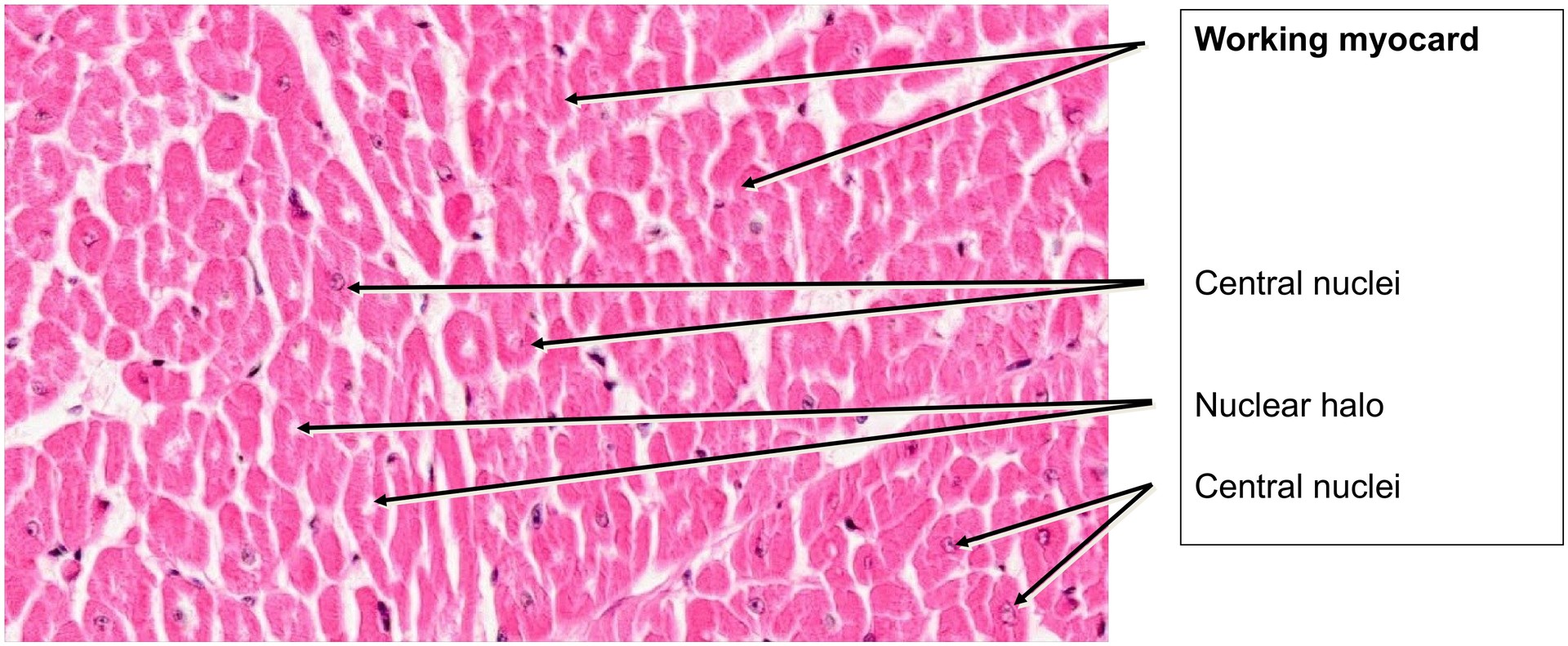
The cells of the conduction system are noticeably larger, contain fewer myofibrils, and are less intensely stained than those of the working myocardium. Their weaker staining is attributable to their high glycogen content, which is lost during tissue processing. The nuclei of these cells are large, and the cells are frequently binucleated.
The conduction system fibers seen here belong to the bundle of His, part of the cardiac conduction pathway (comprising the sinoatrial node, atrioventricular node, bundle of His, and Purkinje fibers).
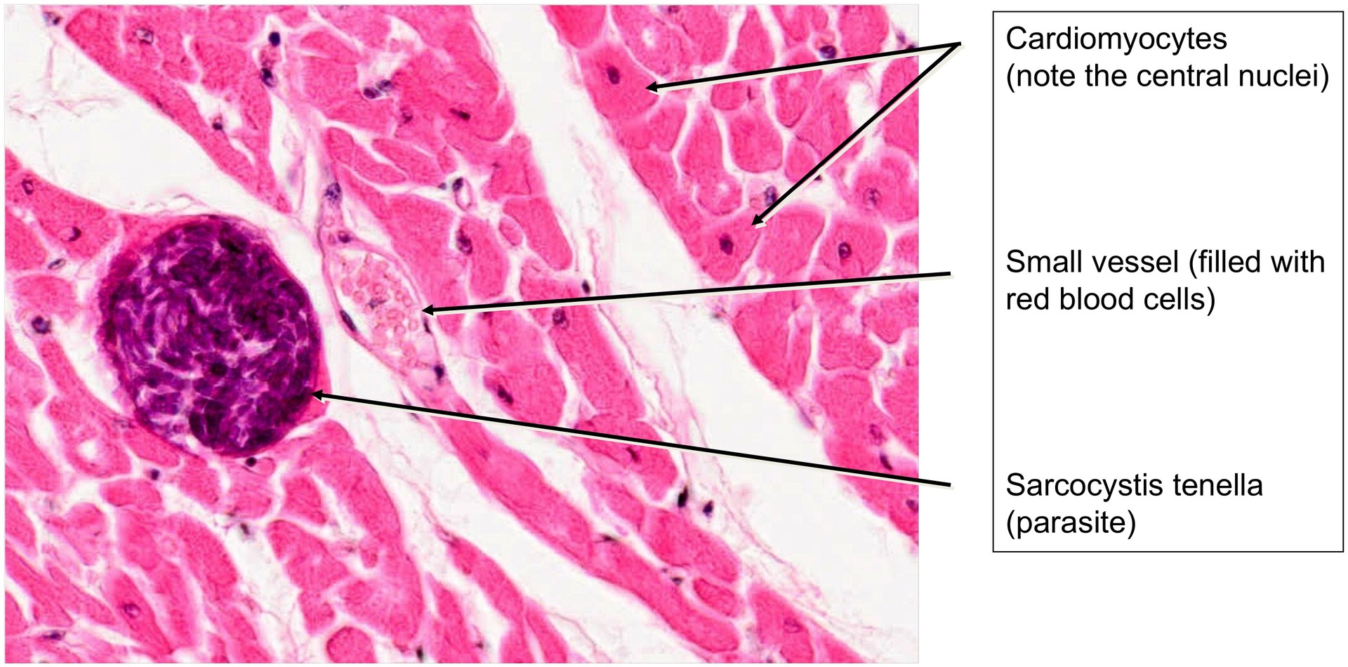
In this particular specimen, which originates from sheep, cysts of the parasite Sarcocystis ovicanis sive tenella can also be observed. This protozoan parasite, common in sheep and dogs, generally causes no harm in adult animals but may be fatal in lambs.
Tasks:
- Identify the majority of the tissue as working myocardium. Which histological criteria allow you to make this distinction?
- Examine the peripheral regions of the section for cells of the conduction system.
- Describe the structural differences between conduction system cells and working myocardial cells. Where are the conduction system cells typically located?
- Locate a cyst of Sarcocystis tenella and note its appearance as a foreign structure within the myocardium.
License
University of Basel
Downloads