MUSCULATURE (GENERAL HISTOLOGY)
6.5
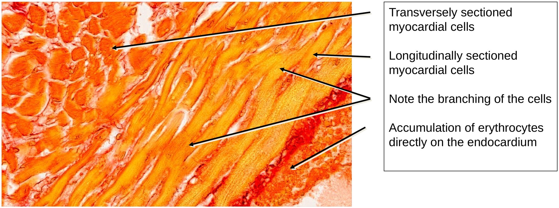
Cardiac muscle (trabeculae)
Specimen Details:
Specimen Details:
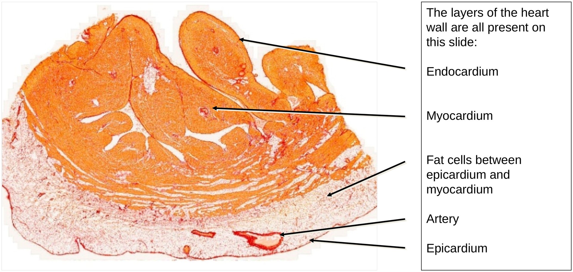
Organ: Heart wall
Origin: Human
Staining: Van Gieson
Method and Specimen Description:
This is a standard histological preparation stained with Van Gieson, which stains cardiac muscle fibers yellow and collagen fibers red, providing good contrast between muscle and connective tissue.
Objective of the Examination:
To study the structure of the heart wall and to identify the trabeculae carneae, which originate from the myocardium and project into the heart chamber lumen.
Special Features of the Specimen:
The different layers of the heart wall can be clearly distinguished: epicardium, myocardium, and endocardium. In some regions, univacuolar adipose tissue is present, which is a normal physiological feature.
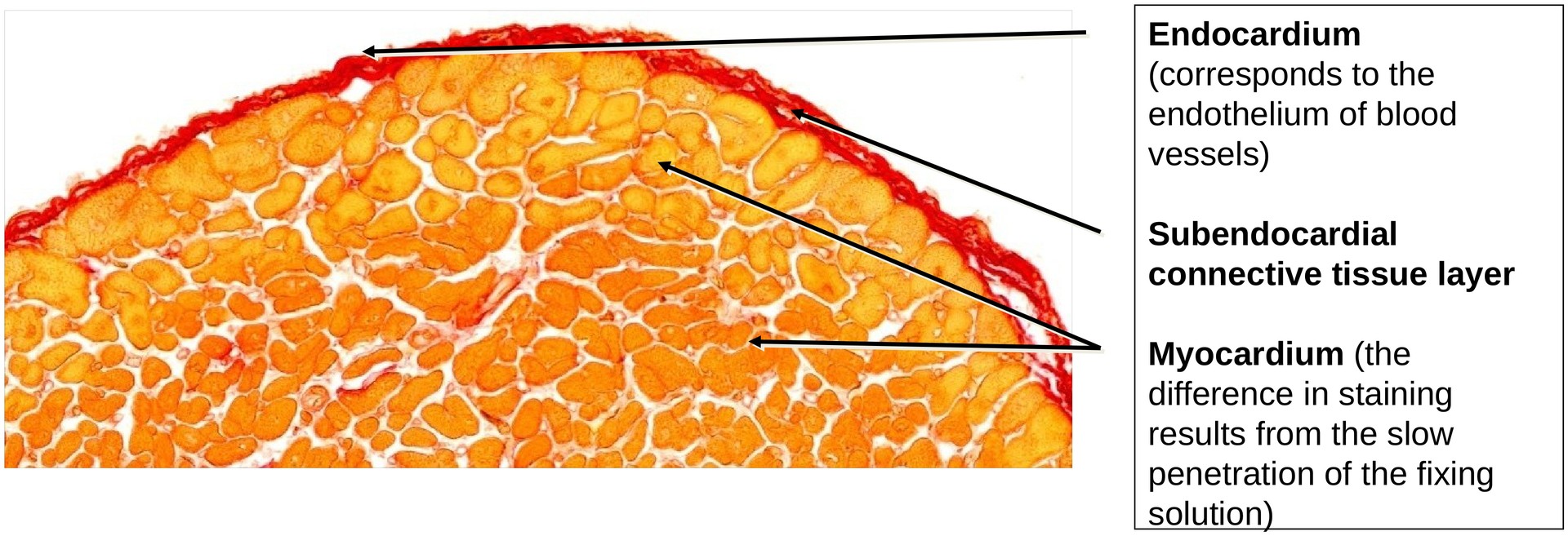
The endocardium consists of a single layer of endothelial cells resting on a variably developed layer of subendocardial connective tissue, which in turn lies directly upon the myocardium. At low magnification, the characteristic trabeculae carneae — muscular ridges that protrude into the ventricular lumen — can be readily identified.
Depending on the orientation of the myocardial fibers, the cardiac muscle cells appear either transversely or longitudinally sectioned.
-
In transverse sections, the centrally located nuclei and surrounding myofibril bundles are visible.
-
In longitudinal sections, the typical intercalated discs can be seen in some cells, particularly under higher magnification (×40).
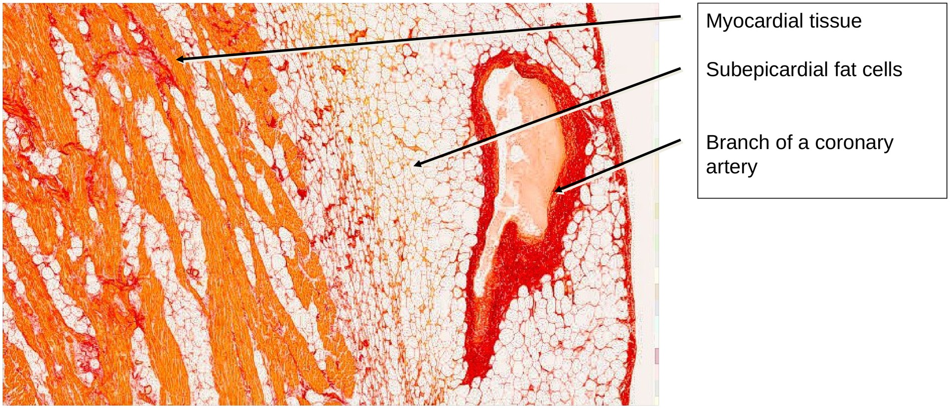
Externally, the myocardium is covered by the epicardium. In certain areas, a layer of adipose tissue is present between the epi- and myocardium. This is especially prominent near the coronary arteries and their branches, which are embedded within epicardial fat.
Tasks:
-
Identify the trabeculae carneae in the overview image.
-
Examine the individual layers of the heart wall:
-
Endocardium – What are the reddish-yellow rounded structures lying externally to it in some regions?
-
Myocardium – Identify and describe the typical characteristics of cardiac muscle cells and verify these in the specimen.
-
Epicardium – Describe the layer that lies between the epi- and myocardium.
-
-
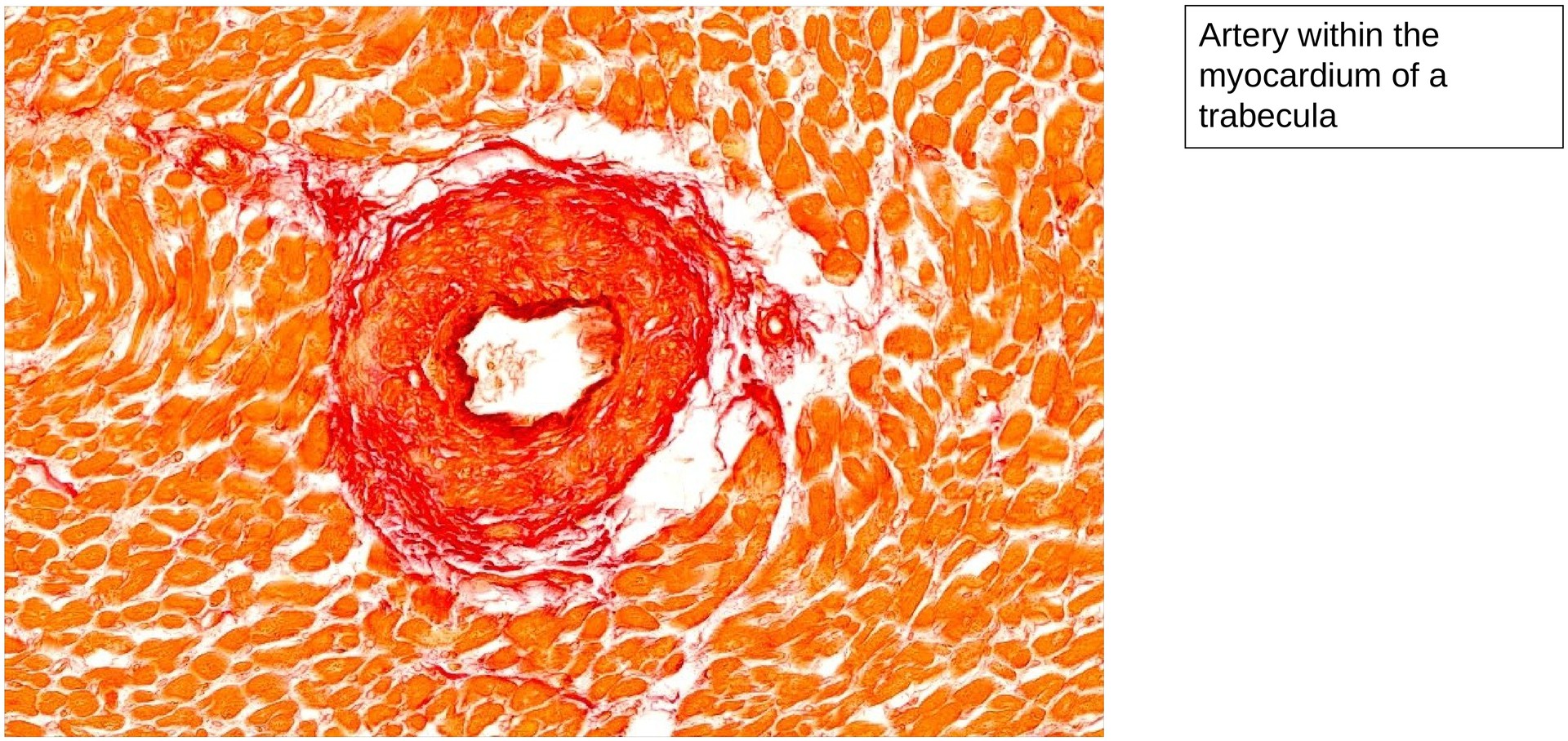
Observe the vascular supply of the myocardium, both via the larger coronary vessels and the capillary network.
License
University of Basel
Downloads