MUSCULATURE (GENERAL HISTOLOGY)
6.7
Skeletal muscle, transverse section (neck muscle)
Specimen Details:
Specimen Details:
Organ: Neck muscle
Origin: Human
Staining: Iron hematoxylin
Method and Specimen Description:
Iron hematoxylin stains primarily the cell nuclei and myofibrils. Differences in staining intensity are unavoidable during differentiation and are partly due to variations in enzyme content among the fibers, as well as fixation-dependent differences.
Objective of the Examination:
To recognize the organization of skeletal muscle through its connective tissue subdivisions, and to understand the cross-sectional appearance of skeletal muscle fibers.
Special Features of the Specimen:
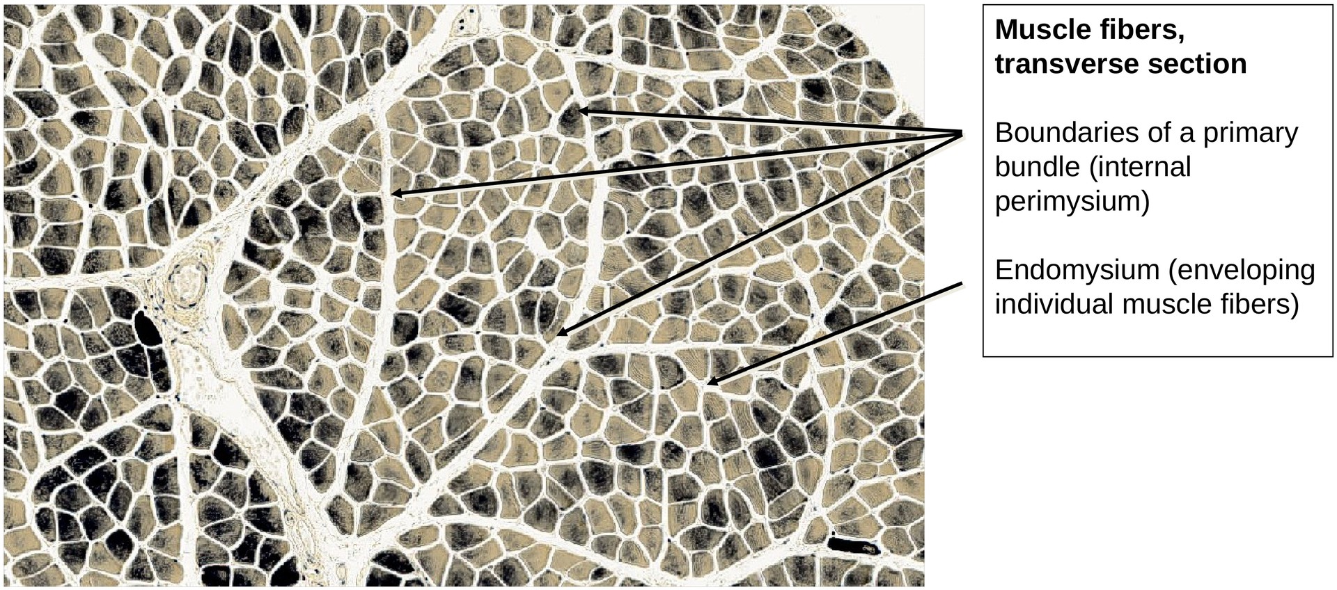
Structure of the Muscle
In cross-section, the muscle is subdivided by connective tissue strands into coarser and finer bundles of muscle fibers:
-
Perimysium – connective tissue surrounding groups of muscle fibers.
-
External perimysium (perimysium externum): surrounds secondary bundles.
-
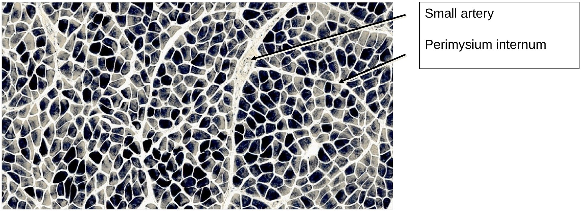
Internal perimysium (perimysium internum): surrounds primary bundles.
-
-
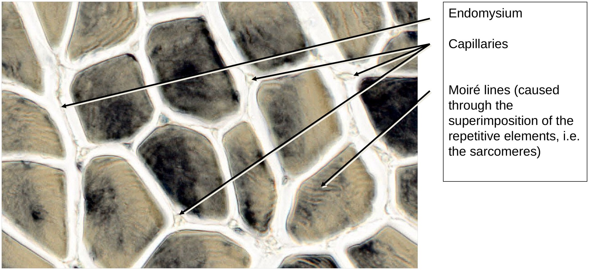
Endomysium – fine connective tissue enveloping individual muscle fibers.
-
Sarcolemma – composed of the plasmalemma, basal lamina, and a reticular connective tissue sheath.
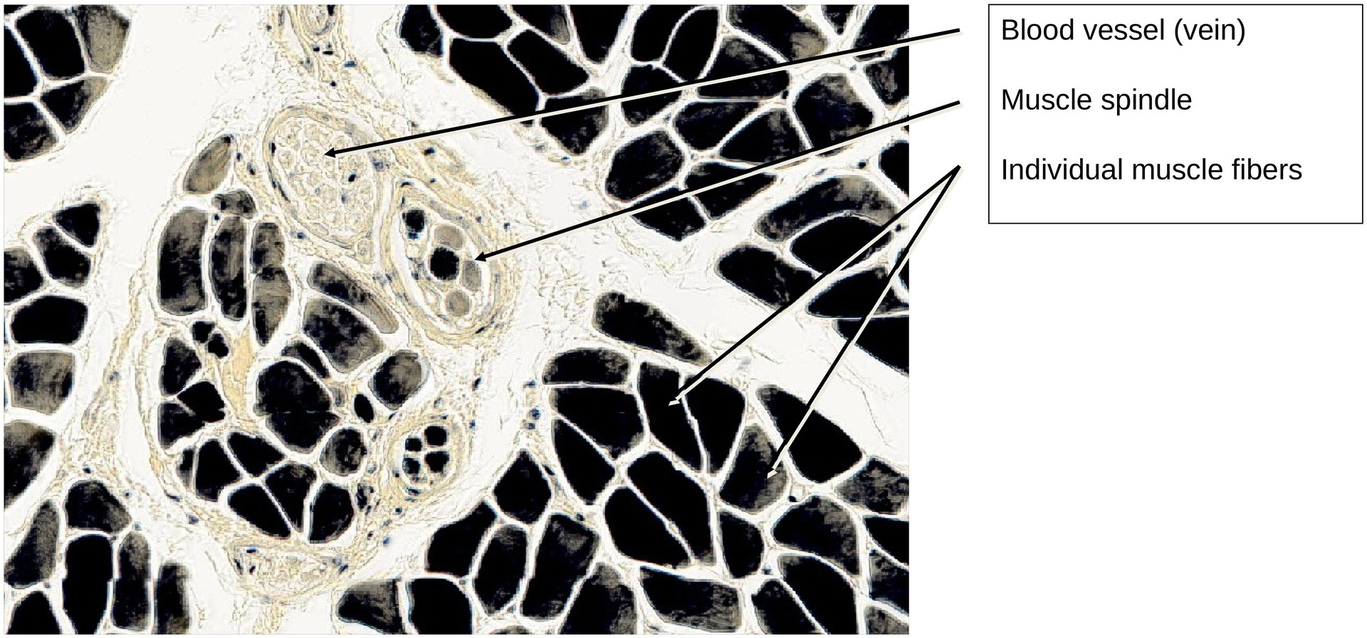
Within the perimysium, arteries and veins of various calibers can be observed, sectioned in different planes depending on their orientation.
Cross-section of Muscle Fibers
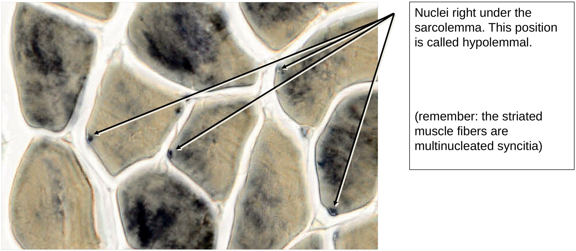
Muscle fibers appear as polygonal profiles in cross-section. Once a fiber containing a nucleus is located, the position of the nucleus can be determined relative to the sarcolemma (typically subsarcolemmal, or hypolemmal).
Due to slight tangential deviations in sectioning, intersecting striations may sometimes be seen under transmitted light, producing Moiré patterns. These optical effects result from interference between repetitive myofibrillar structures.
Tasks:
• Identify primary and secondary muscle bundles.
• Identify individual muscle fibres within the bundles.
• Observe the myofibrils, which give the sarcoplasm a fine granular appearance.
• Locate blood vessels (capillaries, arteries, and veins) within the perimysium.
• Note that the nuclei are positioned beneath the sarcolemma (hypolemmal).
License
University of Basel
Downloads