DIGESTIVE ORGANS: LIVER, GALLBLADDER, PANCREAS (ANATOMICAL MICROSCOPY)
20.2
Liver, Human
Specimen:
Specimen Details:
Organ: Liver
Origin: Human
Staining: Hematoxylin and Eosin (H&E)
Method and Specimen Description:
Normal histological section stained with H&E, serving as a general overview stain.
Objective of the Examination:
To study the lobular architecture of the human liver, with emphasis on: - the arrangement of hepatocyte cords, sinusoids, and central veins. - the portal triads (Glisson’s triads) and associated vessels. - and the vascular supply and drainage system.
Specific Features of the Specimen:
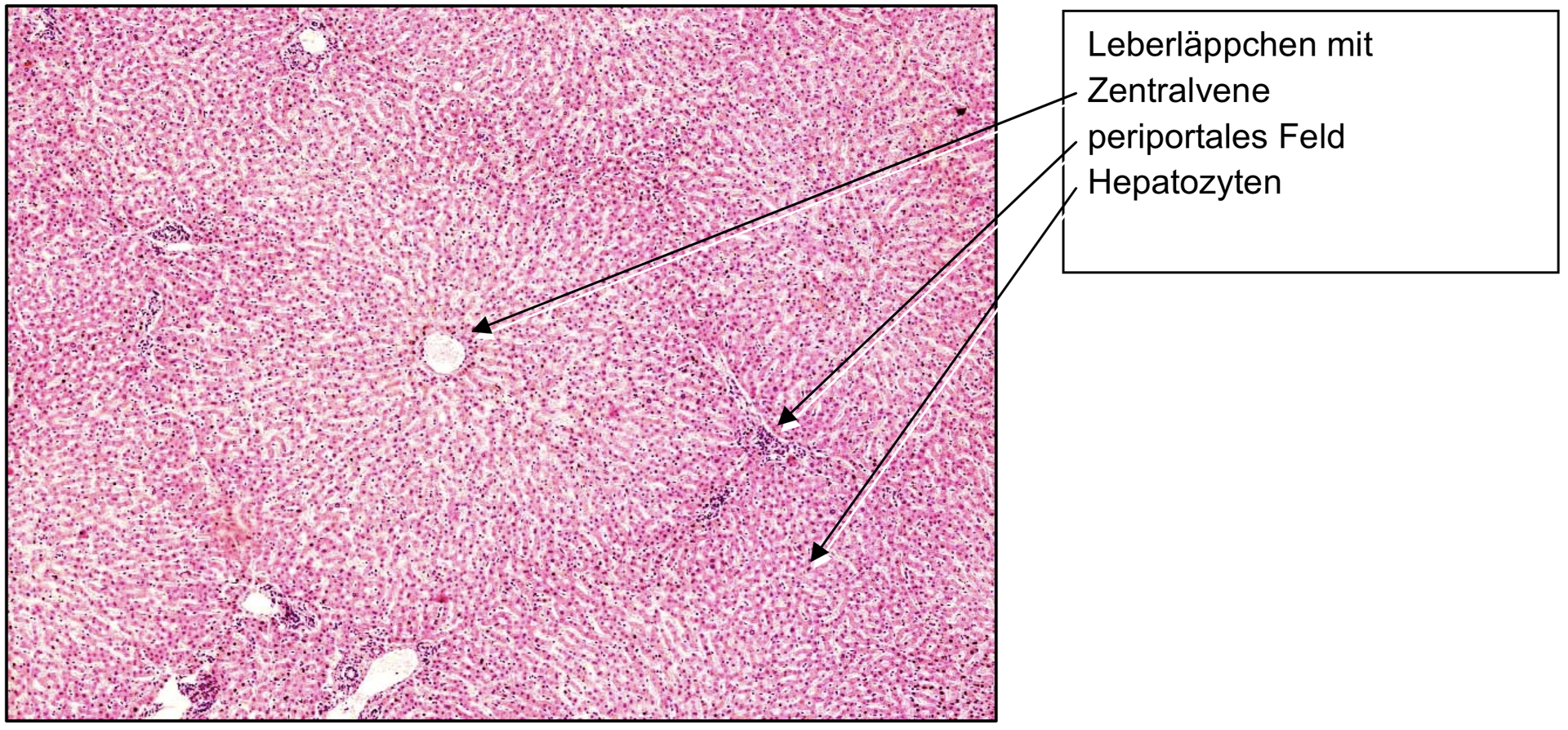
The lobular structure of the human liver is only schematically visible, as the interlobular connective tissue is poorly developed compared to other species (e.g. pig). Nonetheless, the lobular organization can be identified by the orientation of the hepatic cell plates and vascular structures.
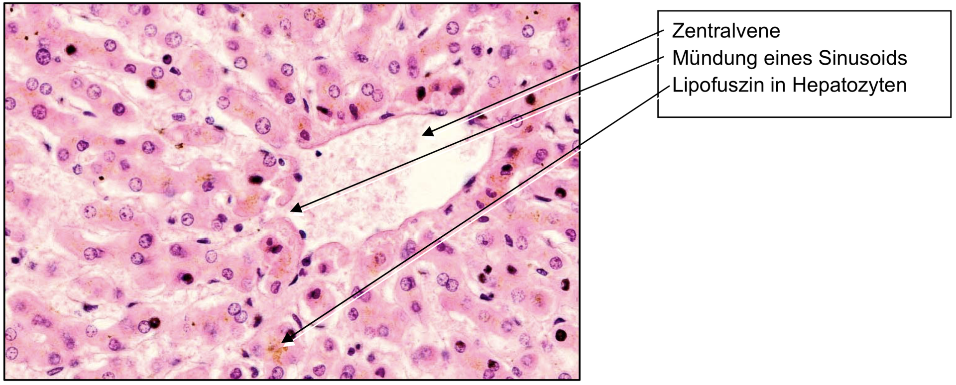
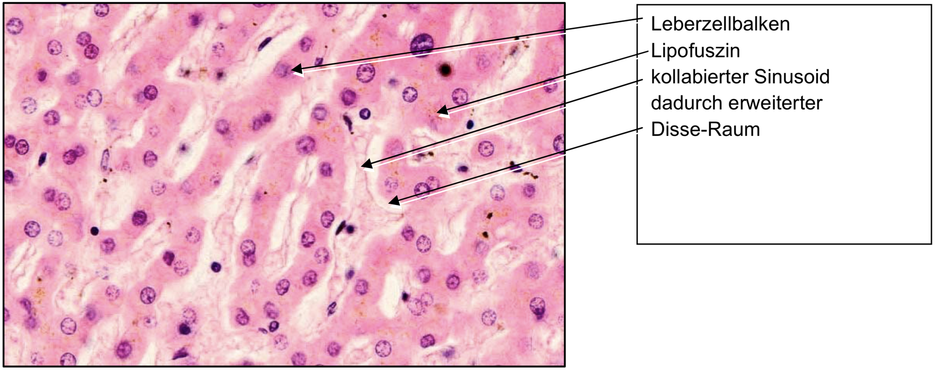
Each lobule is centered on a central vein, around which hepatocyte cords radiate outward. Between these cords run hepatic sinusoids, irregular capillary-like vessels that facilitate exchange between blood and hepatocytes.
The sinusoidal endothelium is fenestrated and separated from the hepatocytes by the space of Disse, which acts as an exchange interface between blood plasma and hepatocytes. The irregular, anastomosing course of the sinusoids prolongs blood transit time and enhances metabolic exchange.
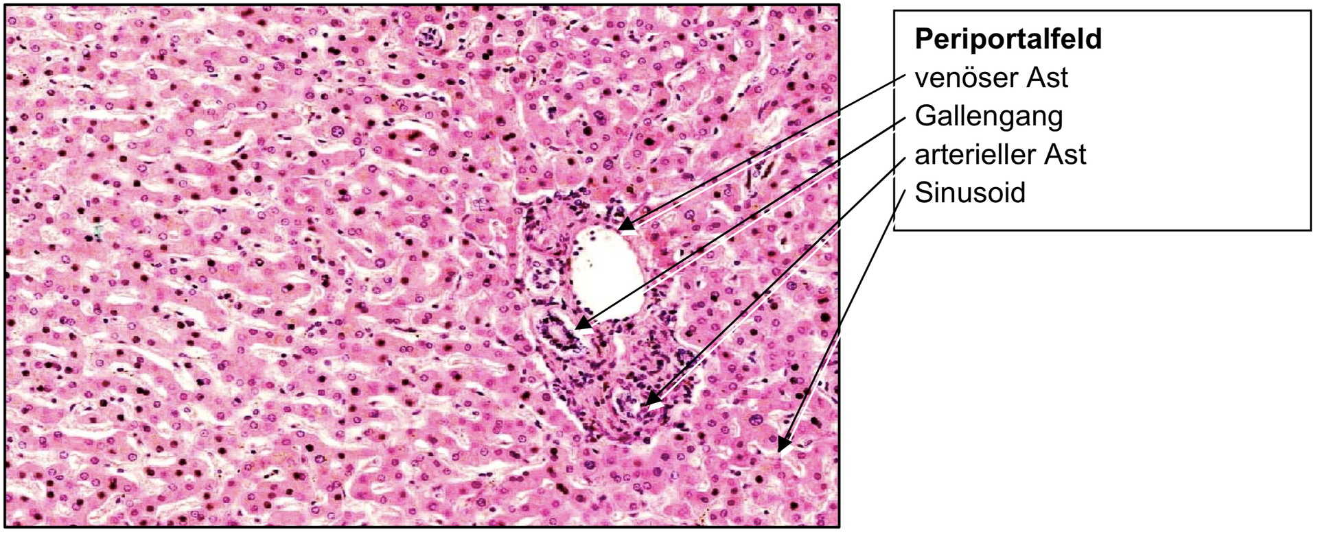
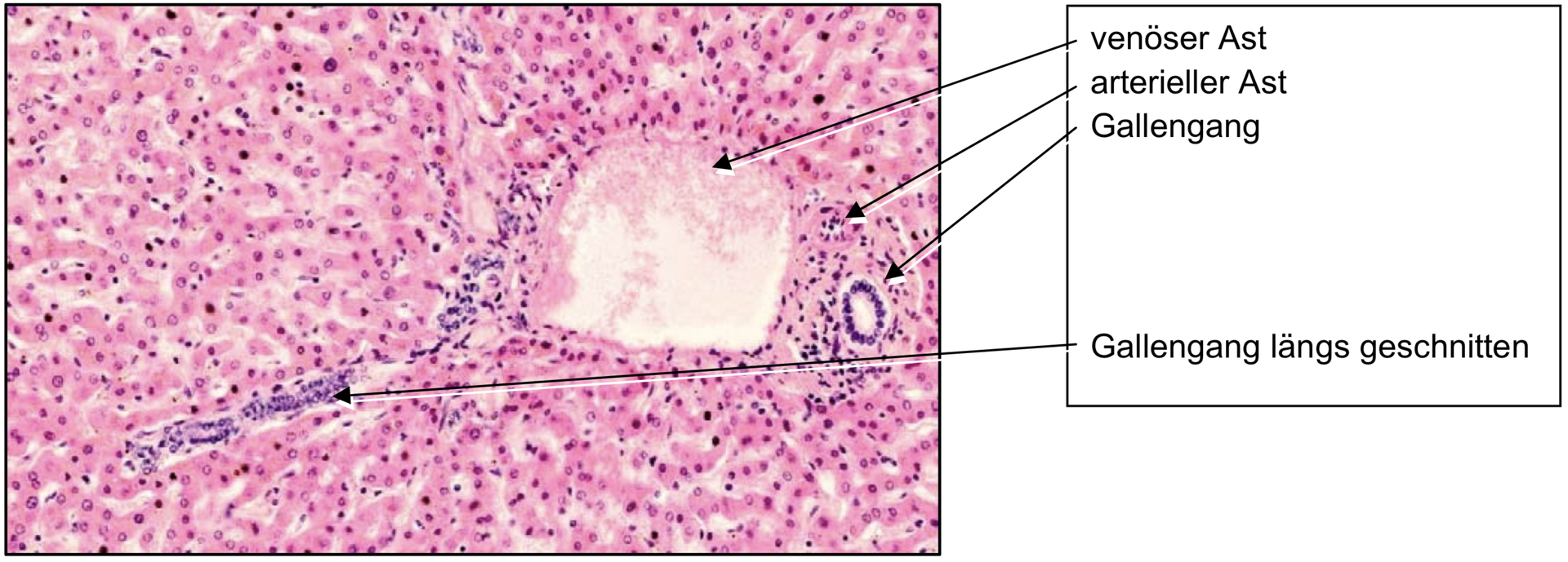
At the lobular periphery, periportal fields (portal tracts or Glisson’s triads) can be observed. Each portal field typically contains:
- a branch of the portal vein (thin wall, wide lumen),
- a branch of the hepatic artery (thicker wall, smaller lumen), and
- a bile duct (lined by cuboidal epithelium).
The portal vein delivers nutrient-rich blood from the gastrointestinal tract, while the hepatic artery supplies oxygenated blood. Both mix in the sinusoids, which drain into the central vein, and ultimately into the hepatic veins and inferior vena cava.
In the interlobular connective tissue, collecting veins can sometimes be found — these drain blood towards the hepatic veins.
Occasionally, hepatocytes contain a brownish pigment, identified as lipofuscin, a wear pigment indicating cellular ageing or metabolic activity.
Tasks:
- Locate the central veins and note their position at the center of each lobule.
- Trace the sinusoids, following their irregular, radial course from the periportal fields towards the central vein.
- Identify the space of Disse (visible as narrow gaps between sinusoidal endothelium and hepatocytes).
- Find the periportal fields (Glisson’s triads) and identify the three characteristic structures:
- Portal vein branch
- Hepatic artery branch
- Bile duct
- Differentiate between the arterial and venous vessels within the triad based on wall thickness and lumen size.
- Locate a collecting vein in the interlobular region.
- Observe any lipofuscin granules within hepatocytes.
License
University of Basel
Downloads