MALE REPRODUCTIVE ORGANS (ANATOMICAL MICROSCOPY)
11.3
Testes, adult 1
Preparation:
Preparation Details:
Organ: Testis
Origin: Human
Staining: Van Gieson
Method and Specimen Description:
normal histological section stained with Van Gieson, a connective tissue stain. This method colors collagen fibers red and muscle cells (e.g. vessel walls) and erythrocytes yellow, allowing clear distinction between the fibrous and muscular components of the testis.
Objective of the Examination:
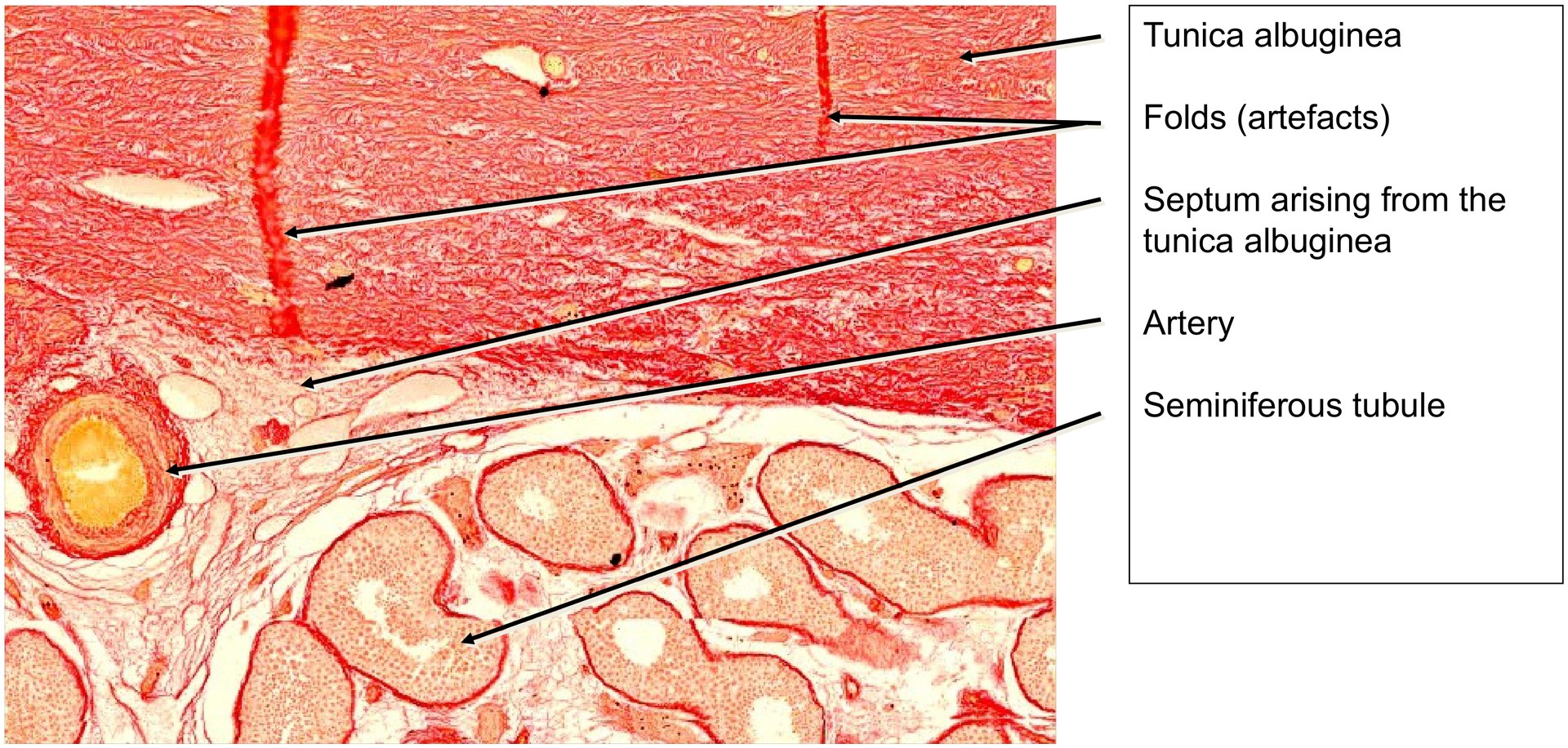
To study the adult human testis, including the tunica albuginea and the seminiferous tubules with their different stages of spermatogenesis.
Special Features of the Preparation:
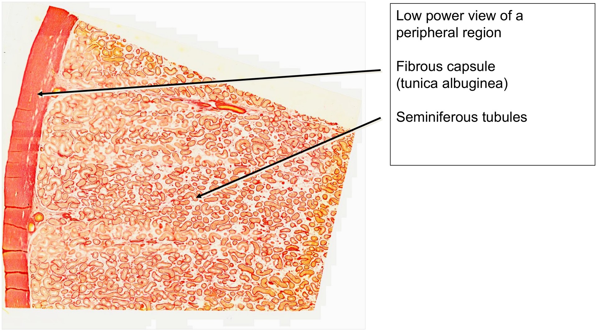
Even at low magnification, the dense fibrous capsule of the testis, the tunica albuginea, is readily visible. Together with the internal secretory pressure, it contributes to maintaining the relatively high intratesticular pressure necessary for the transport of sperm from the testis to the epididymis.
Due to differences in tissue consistency, the tunica albuginea may appear folded in the section — this is an artefact of preparation and does not occur in vivo. The seminiferous tubules are separated by incomplete septa, which extend inward from the tunica albuginea. Blood vessels are often seen between the capsule and the parenchyma, particularly at the points where septa penetrate deeper into the tissue.
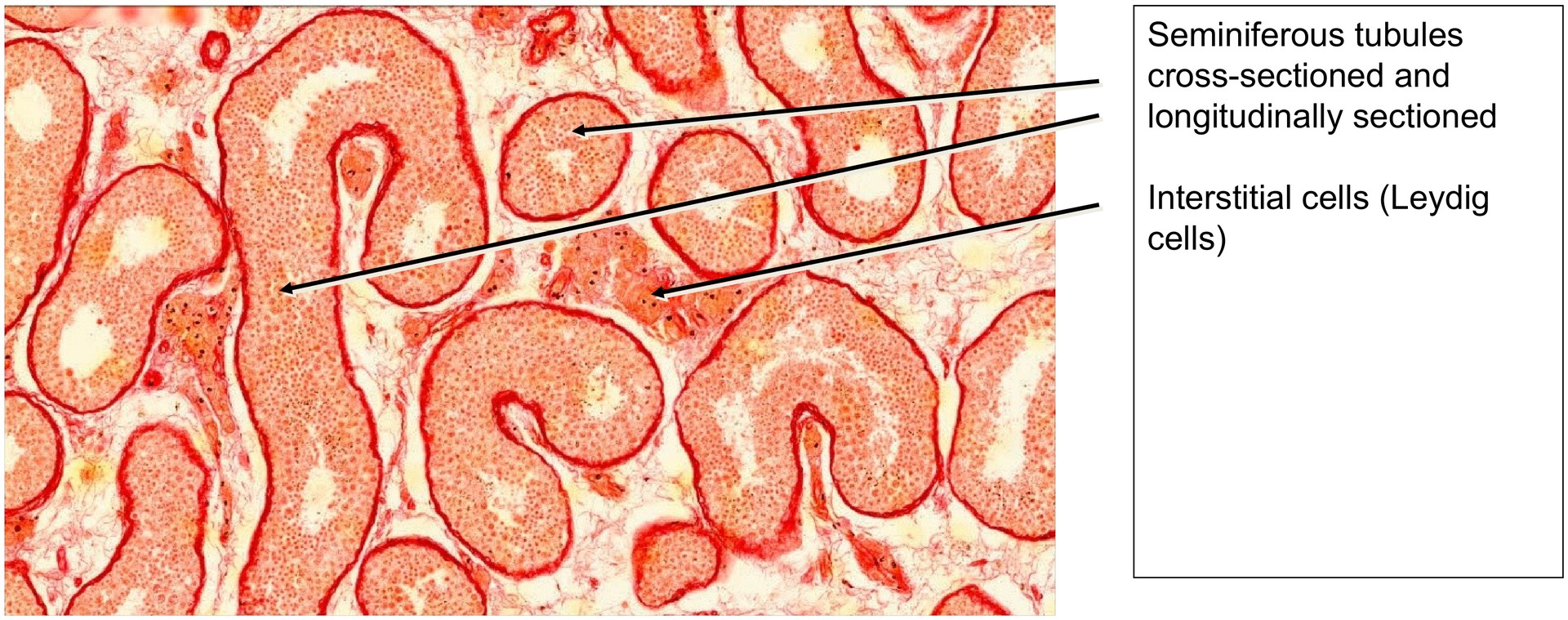
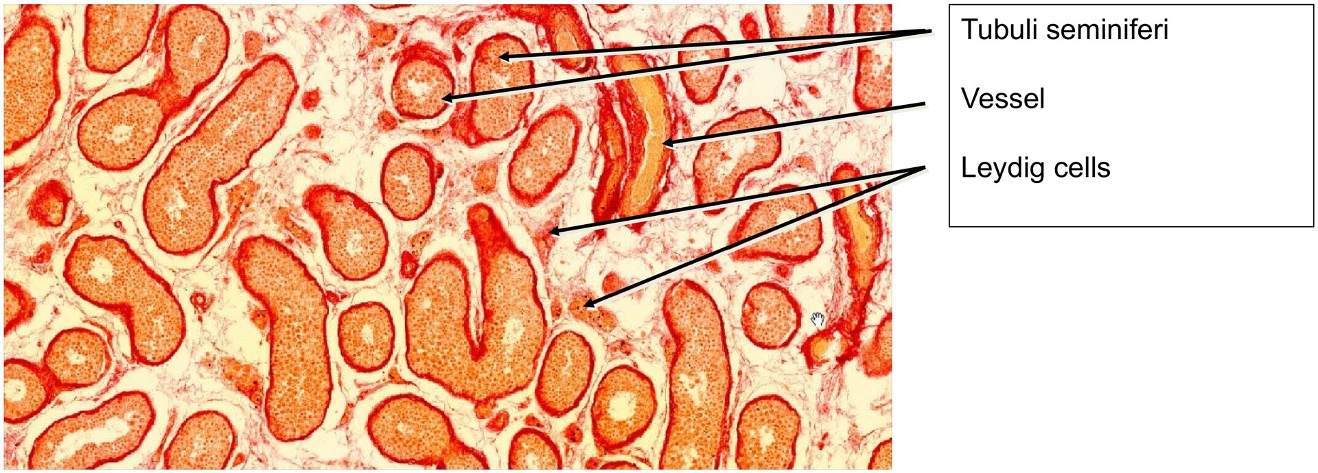
The seminiferous tubules are highly coiled, so their cross-sections appear in various orientations — transverse, tangential, or longitudinal. The Sertoli cells form the supporting epithelium of the seminiferous tubules, within which the germ cells at different stages of spermatogenesis are embedded.
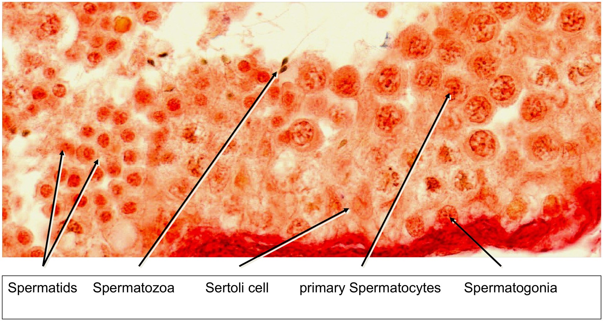
Sertoli cells are not always easy to distinguish in routine sections due to their indistinct cytoplasmic boundaries. Their nuclei are typically located in the basal third of the epithelium, and are oval to pear-shaped with a prominent nucleolus.
At the base of the epithelium, spermatogonia (types A and B) are found. Spermatogonia A divide mitotically to maintain the stem cell pool, while spermatogonia B enter meiosis to form primary spermatocytes (type I), which then undergo meiosis I to produce secondary spermatocytes (type II). The latter rapidly complete meiosis II to form spermatids, which then differentiate into spermatozoa (sperm) through spermiogenesis.
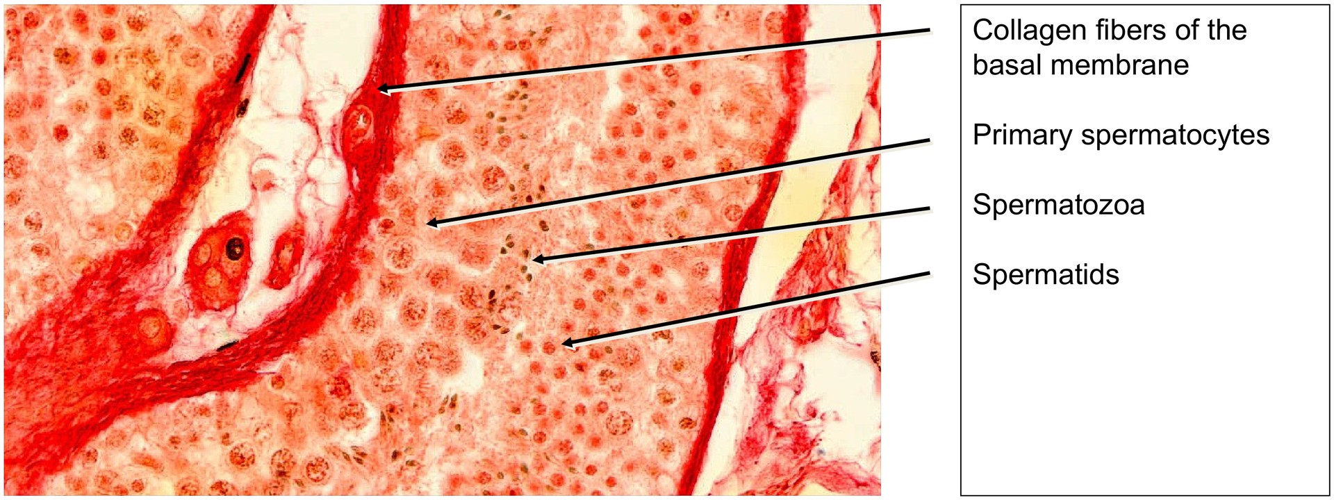
In this preparation, most spermatocytes present are primary spermatocytes, identifiable by their large, loosely structured nuclei. Secondary spermatocytes are rarely visible due to their brief lifespan. Spermatids are small, round cells close to the lumen, while spermatozoa are easily recognized by their slender, chromatin-dense heads projecting into the tubule lumen.
Each seminiferous tubule is bounded by a basement membrane reinforced with collagen fibers (stained bright red by Van Gieson). In the interstitial tissue between the tubules, clusters of interstitial (Leydig) cells are present. These cells are responsible for the production of testosterone, the principal androgen hormone.
Tasks:
• Identify the tunica albuginea and assess its thickness. What structural components can be recognized within it?
• Locate the septa arising from the tunica albuginea and trace one extending into the parenchyma.
• Examine a transverse section of a seminiferous tubule and identify the various cell types present.
• Locate spermatozoa within the lumen of the seminiferous tubules.
• Identify spermatids and distinguish them from other spermatogenic cells.
• Locate Leydig cells in the interstitial tissue. Why are they also referred to as interstitial cells? What is their function?
License
University of Basel
Downloads