DIGESTIVE ORGANS: ORAL CAVITY (ANATOMICAL MICROSCOPY)
18.11
Tongue Filiform papillae
Specimen
SPECIMEN DETAILS:
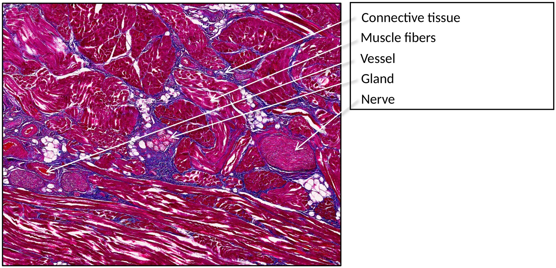
Organ: Tongue
Origin: Human
Staining: Azan
METHOD AND SPECIMEN DESCRIPTION:
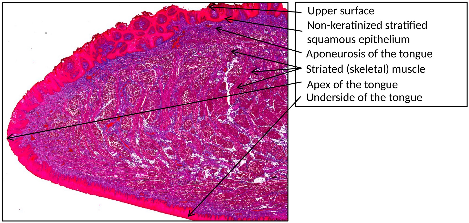
This is a normal sagittal section of a human tongue stained with Azan. In this staining, connective tissue appears blue, while muscle fibers appear red.
OBJECTIVE OF THE EXAMINATION:
To study the morphology and structure of the tongue, with a focus on the filiform papillae (papillae filiformes) and their relationship to the underlying tissues.
Special Features of the Specimen:
General: The tongue is a muscular organ covered by a mucous membrane. It is composed of:
- External muscles, which originate from bony structures and radiate into the tongue from various directions.
- Internal muscles, which are intrinsic to the tongue and arranged perpendicularly to each other, forming vertical, longitudinal, and transverse fiber bundles.
These muscle fibers interweave and attach to the lingual aponeurosis, a dense connective tissue layer that provides structural support. The mucous membrane on the dorsal surface of the tongue is firmly attached to the lingual aponeurosis.
On the dorsal and lateral surfaces of the tongue lie the lingual papillae, which serve distinct functions:
- Fungiform, foliate, and vallate papillae contain taste buds (gustatory papillae).
- Filiform papillae function primarily as mechanical receptors, aiding in the manipulation of food and providing a rough surface texture.
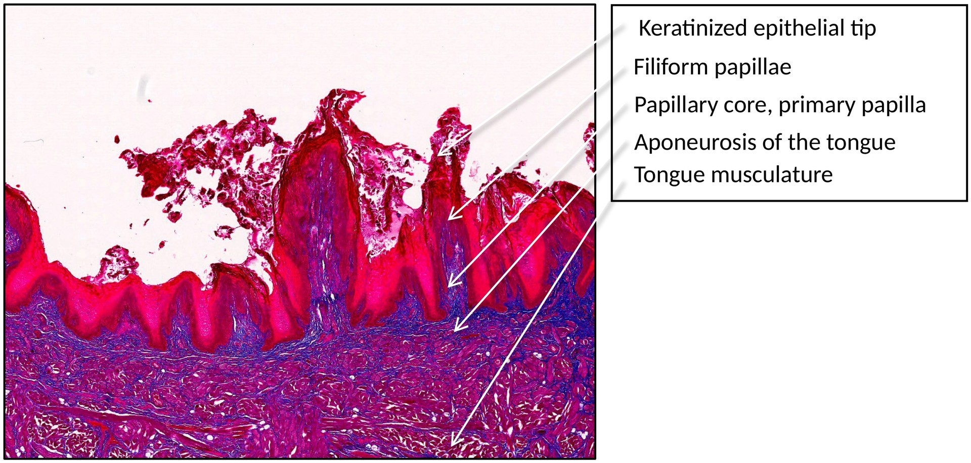
Filiform Papillae:
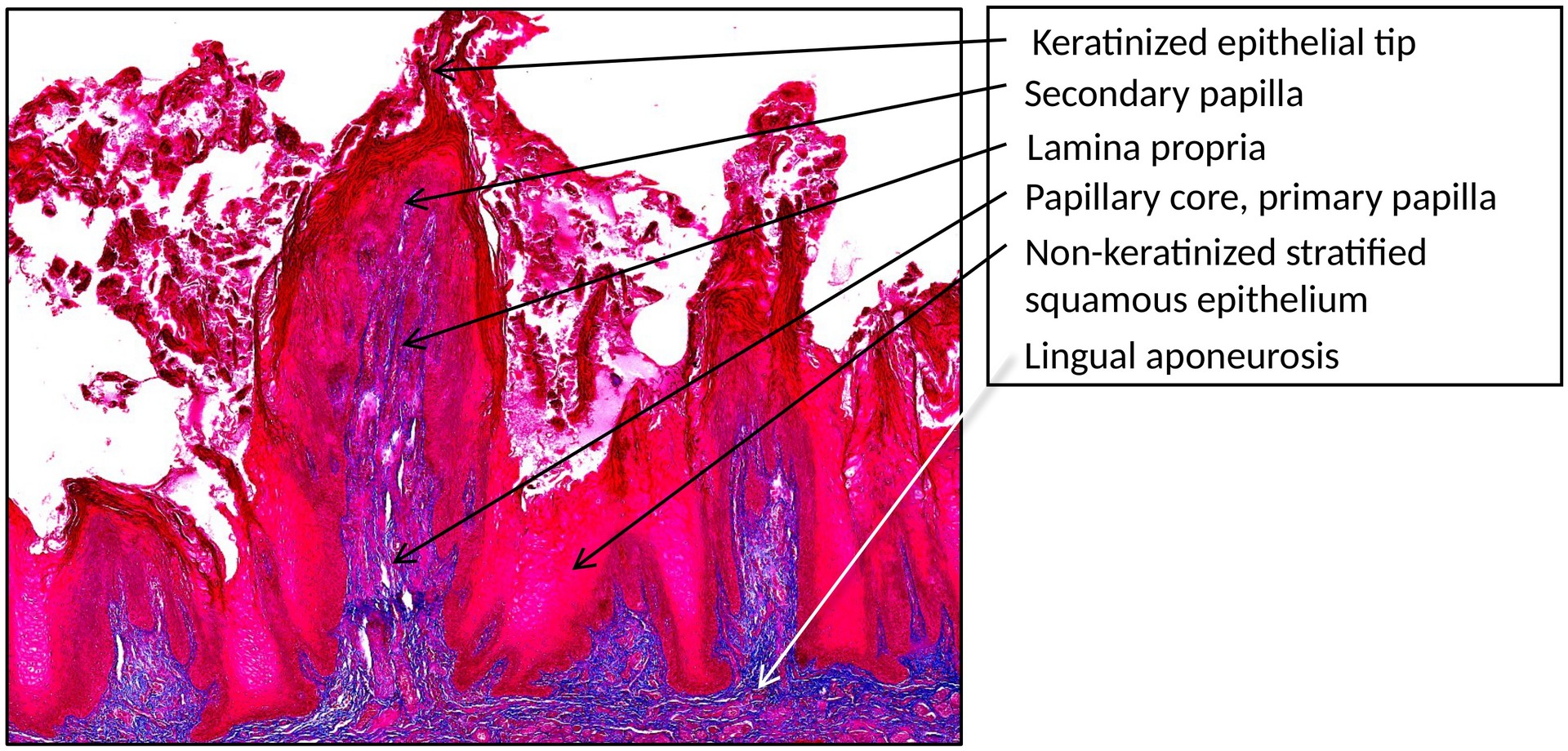
The filiform papillae are the most numerous type of lingual papillae. They are conical or thread-like projections of the mucosa that give the tongue its rough surface. Each papilla is covered by keratinized stratified squamous epithelium and supported by a connective tissue core (papilla core) derived from the lamina propria.
- The connective tissue projects upward into primary and secondary papillae, which interdigitate with the epithelium, strengthening the attachment between the mucosa and the underlying tissue.
- The keratinized tips of the filiform papillae may bend slightly posteriorly, contributing to the tactile sensation and aiding in food manipulation.
- Unlike the other papilla types, filiform papillae do not contain taste buds.
TASKS:
- Identify the dorsal and ventral surfaces of the tongue.
- Locate the filiform papillae on the dorsal surface.
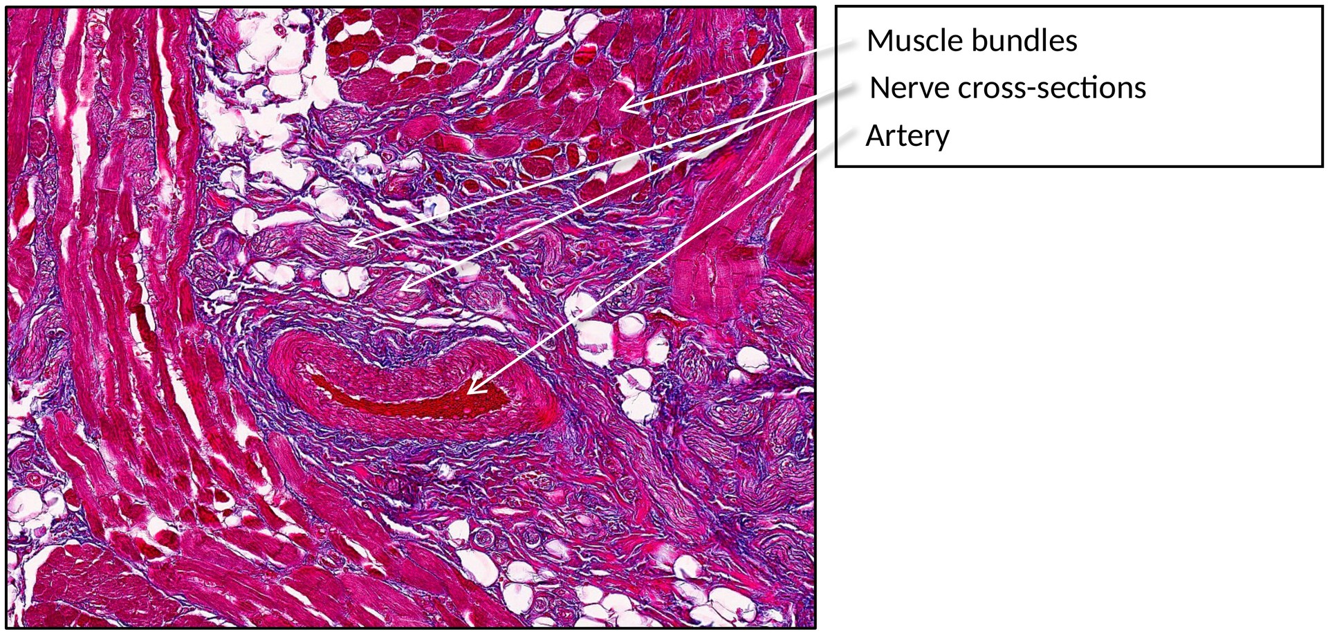
- Observe the lingual aponeurosis, striated muscle fibers, blood vessels, and nerves.
- Examine the filiform papillae in detail:
- Identify the epithelium (keratinized stratified squamous).
- Observe the primary and secondary connective tissue papillae.
- Identify the papilla core and lamina propria.
- Compare the keratinized surface of the filiform papillae with the non-keratinized mucosa in other tongue regions.
License
University of Basel