DIGESTIVE ORGANS: ORAL CAVITY (ANATOMICAL MICROSCOPY)
18.10
Taste bud (vallate papilla)
Specimen:
Specimen Details:
Organ: Tongue (area of the terminal sulcus)
Origin: Human
Staining: Van Gieson
Method and Specimen Description:
This is a normal histological section of the tongue stained with Van Gieson, in which:
- Muscle fibers appear yellow, and
- Connective tissue appears red.
Objective of the Examination:
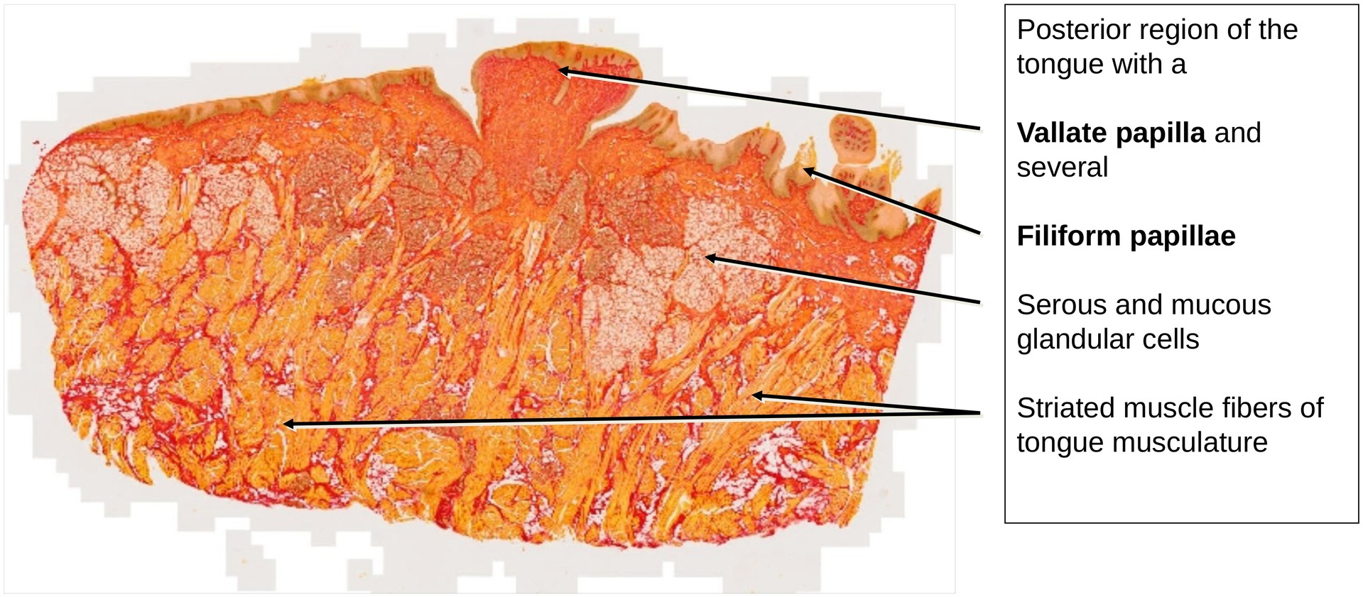
To study the vallate papillae, including their wall, groove, and excretory ducts opening into the papillary trench, as well as to observe the filiform papillae present in the same specimen.
Special Features of the Specimen:
At the posterior region of the tongue, near the terminal sulcus, the specialized epithelium forms the vallate papillae.\ A single vallate papilla is visible in this specimen.
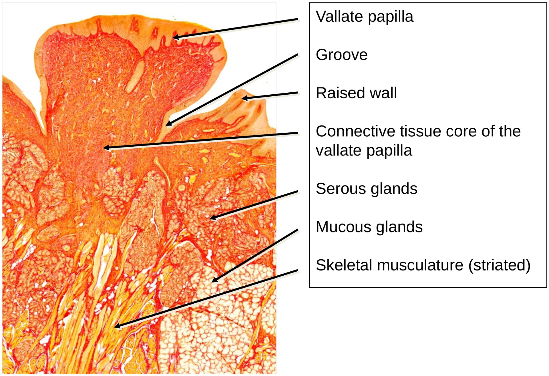
The vallate papilla is characterized by:
- A raised wall and a deep surrounding groove (trench),
- A broad connective tissue core, and
- The openings of excretory ducts from the serous glands (von Ebner’s glands) into the trench.
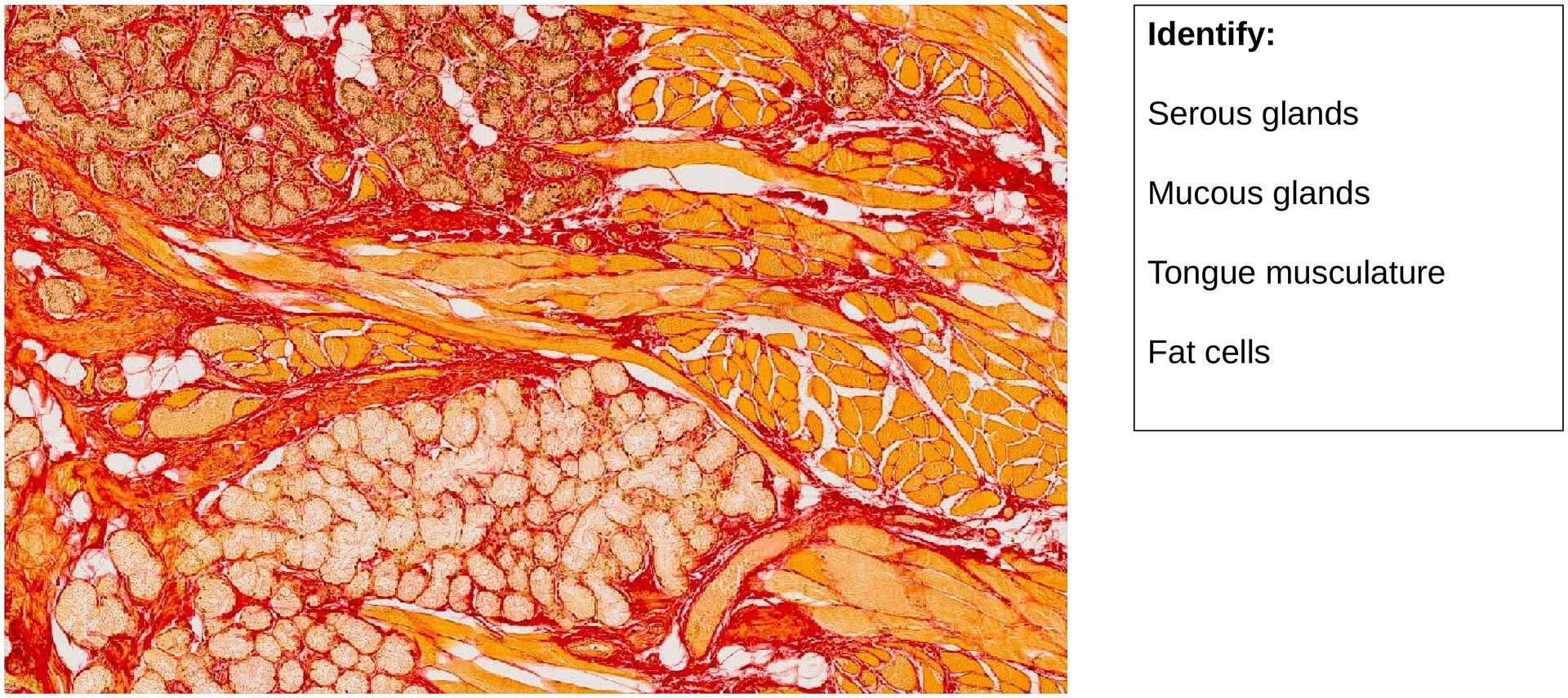
Although the duct openings are not sectioned directly in this specimen, several serous glandular cells can be observed nearby. Additionally, mucous glandular cells are also present in the deeper connective tissue.
On one side of the vallate papilla, a filiform papilla is visible. This papilla is smaller, conical, and keratinized, serving a mechanical rather than sensory function.
The muscle fibers of the tongue—particularly the vertical and longitudinal muscle bundles—are easily identifiable beneath the mucosa.
The epithelium of the tongue root is non-keratinized in most regions, except at the filiform papilla, where a yellow-stained stratum corneum (keratinized layer) can clearly be seen.
Tasks:
- Identify the vallate papilla, including its wall and surrounding groove.
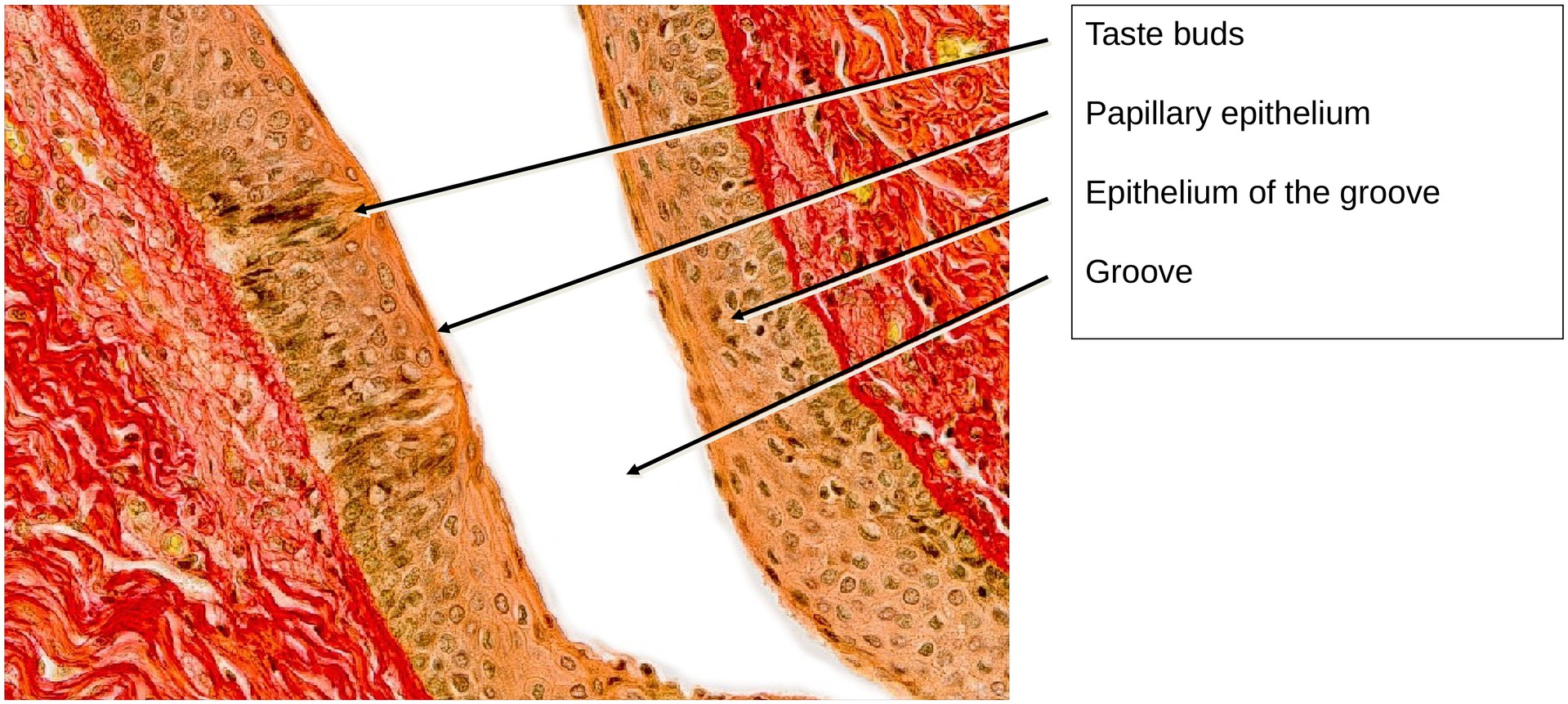
- Examine the lateral epithelium of the vallate papilla for taste buds.
- Note that taste buds are less distinct here than in the foliate papilla specimen.
- Identify the vertical and longitudinal muscle fiber bundles within the tongue tissue.
- Locate the filiform papilla present in this specimen.
- What are its distinctive histological features?
- Identify and distinguish between serous and mucous glandular cells.
License
University of Basel
Downloads