EPITHELIUM (GENERAL HISTOLOGY)
1.5
Transitional epithelium (urinary bladder)
Specimen:
Specimen Details:
Organ: Urinary Bladder
Origin: Cat
Staining: Hematoxylin - Eosin (H&E)
Method and Specimen Description:
This is a routine histological section of the urinary bladder stained with Hematoxylin and Eosin, providing an overview of cellular and tissue structures.
Objective of the Examination:
To examine the structure of the bladder wall, focusing on its multilayered transitional epithelium (urothelium), including the characteristic umbrella cells and the intracellular crusta.
Specific Features of the Specimen:
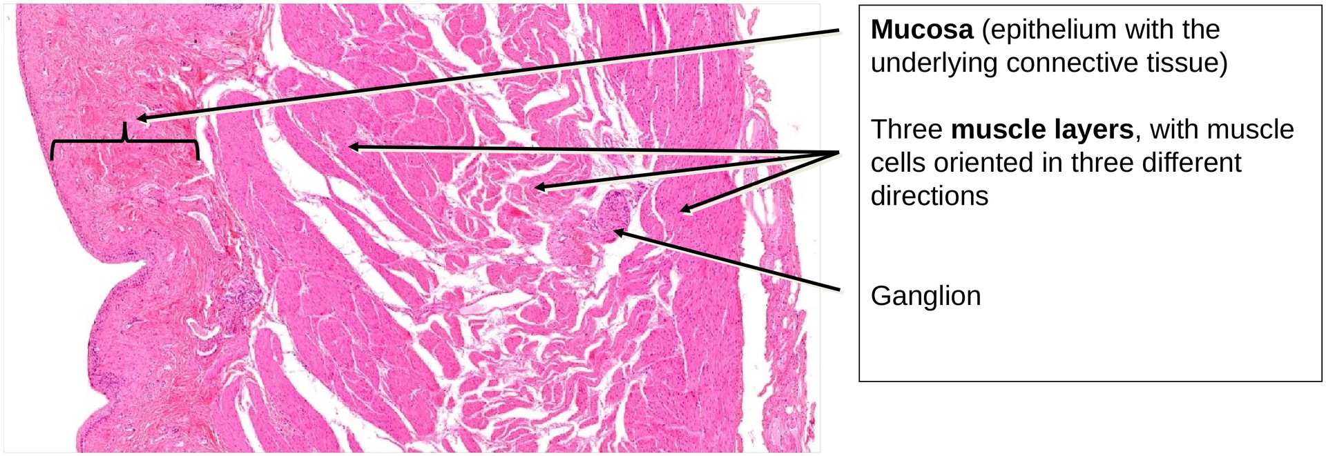
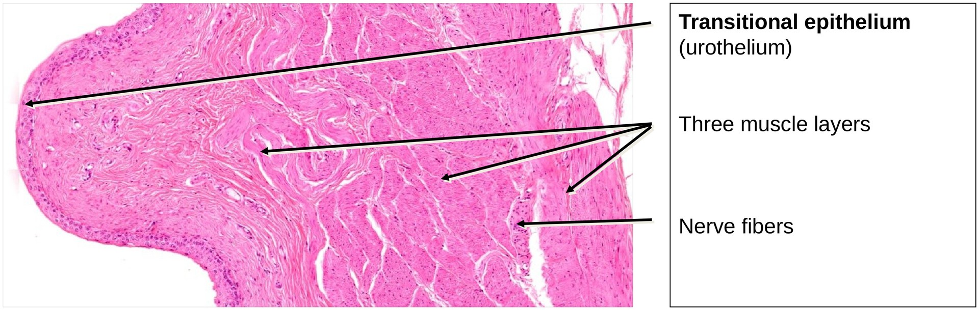
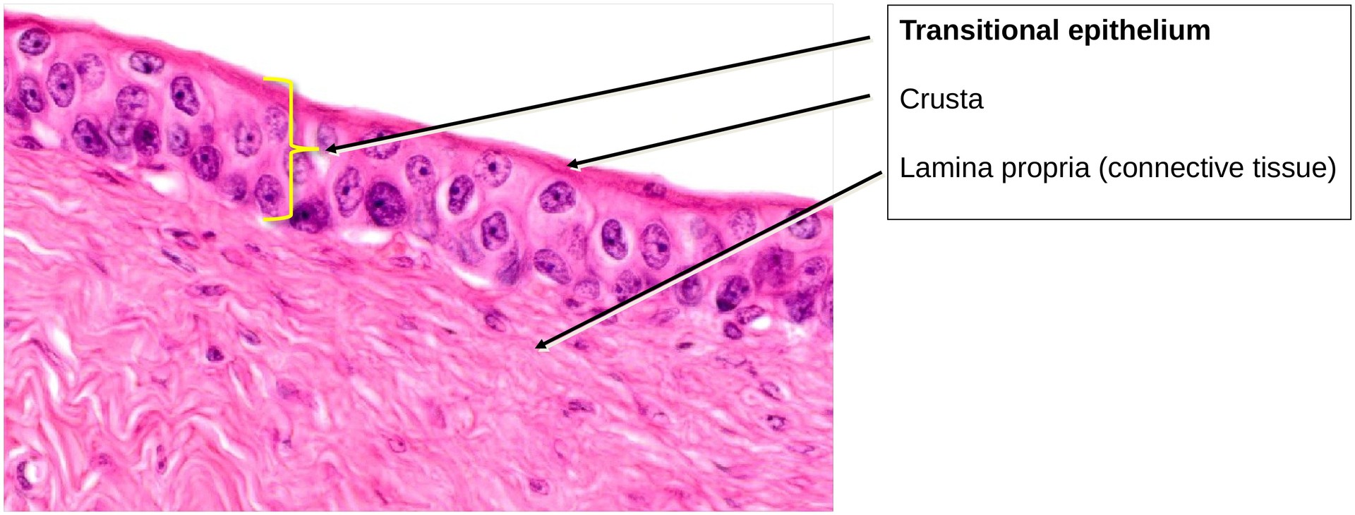
All portions of the urinary tract that conduct urine — the renal pelvis, ureters, urinary bladder, and urethra — are lined by transitional epithelium, also known as urothelium. This epithelium is stratified and uniquely adapted to withstand distension and exposure to urine.
A defining feature of the urothelium is the presence of large superficial umbrella cells. In humans (though not in this cat specimen), these cells are often binucleated and possess a distinct apical thickening, the crusta, which represents a cytoplasmic plaque rich in proteins (uroplakins) that form a protective barrier against the potentially harmful effects of urine. The crusta appears as a dark, dense rim along the luminal surface of the umbrella cell layer under the light microscope.
The mucosa of the bladder is highly folded, allowing for significant expansion during filling. These folds (rugae) flatten as the bladder distends.
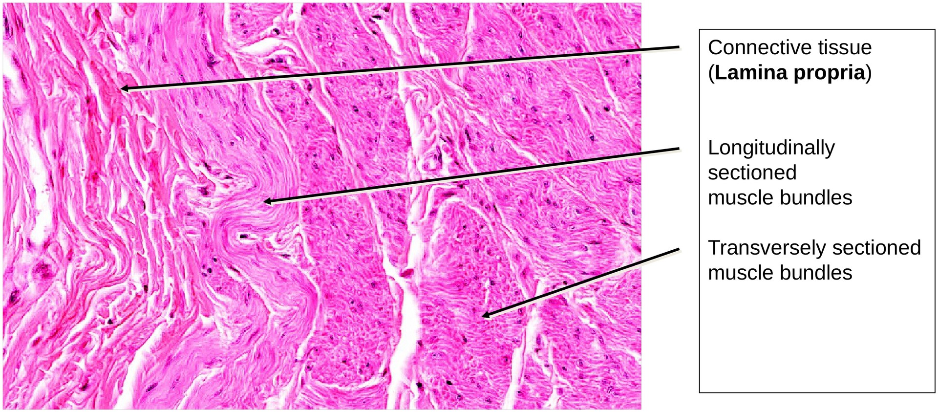
Beneath the epithelium lies the lamina propria, composed of dense connective tissue that may be considerably thick in some regions. It can be clearly distinguished from the underlying tunica muscularis, and contains a well-developed venous plexus, a common feature of the bladder’s vascular architecture.
The tunica muscularis, collectively known as the detrusor muscle, comprises three interwoven layers of smooth muscle:
-
An inner longitudinal layer,
-
A middle circular layer, and
-
An outer longitudinal layer.
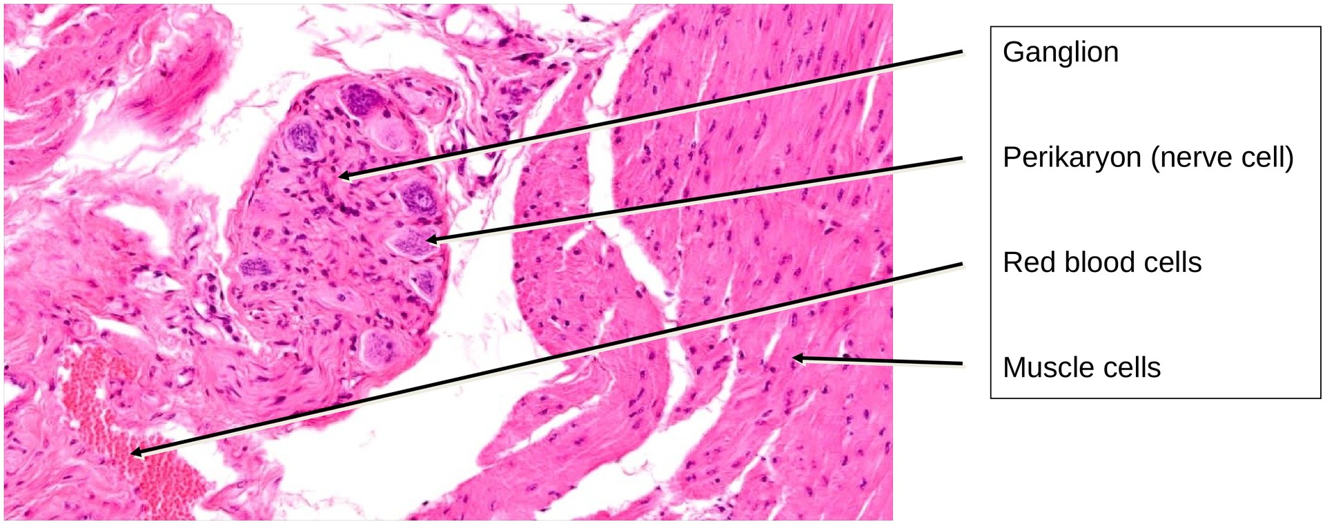
Between these muscle layers lies a prominent autonomic nerve plexus, containing ganglia and individual ganglion cells. The detrusor muscle is under parasympathetic control, which mediates bladder contraction during micturition.
Tasks:
• Obtain an overview of the section and identify the tunica mucosa, consisting of:
-
The epithelial lamina (urothelium), and
-
The lamina propria. Observe the relative thickness of the lamina propria.
• Identify the crusta within the umbrella cells of the transitional epithelium.
• Examine the tunica muscularis and attempt to recognize the three principal orientations of the smooth muscle bundles (note that not all are present in every region of the section).
• Locate ganglia, ganglion cells, and nerve fibers within the connective tissue between the muscle layers.
• Observe the pronounced folding of the mucosa, which diminishes as the bladder fills.
License
University of Basel
Downloads