DIGESTIVE ORGANS: GASTROINTESTINAL TRACT (ANATOMICAL MICROSCOPY)
19.6
Ileum, Human 2
Specimen:
Specimen Details:
Organ: Ileum
Origin: Human
Staining: Hematoxylin - Eosin (H&E)
Method and Specimen Description:
Normal histological section stained with an overview stain (H&E).
Objective of the Examination:
To study the microscopic structure of the human terminal ileum, especially the region containing Peyer’s patches, and to recognize its structural differences from other regions of the gastrointestinal tract (GIT).
Special Features of the Specimen:
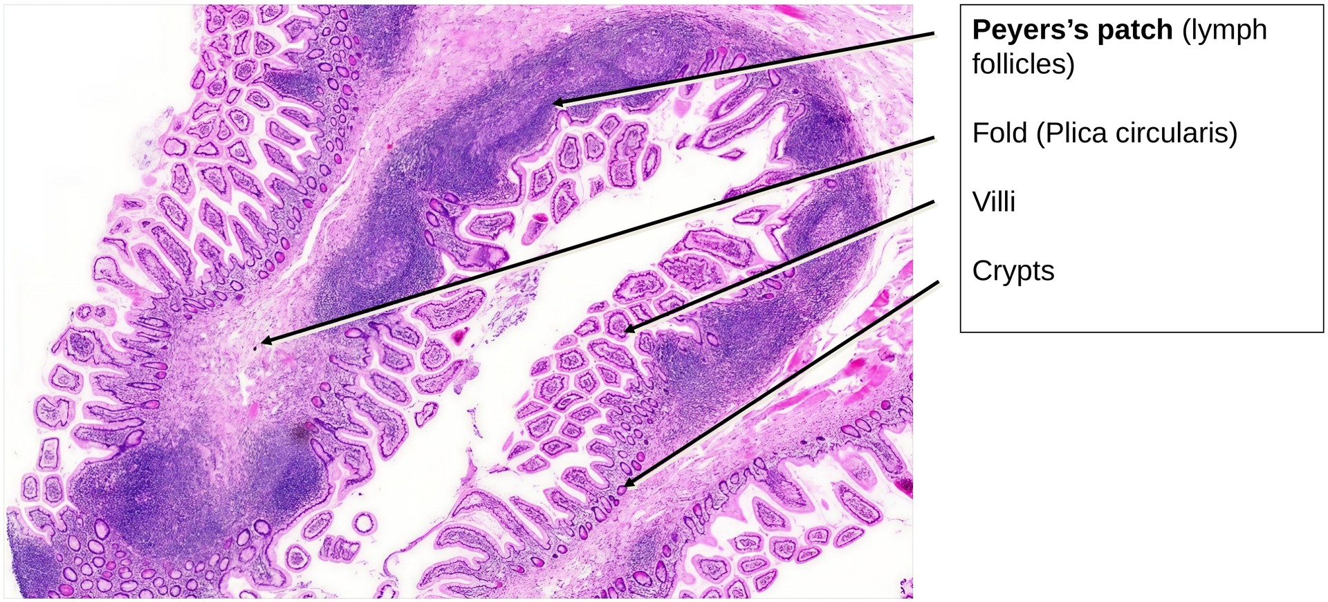
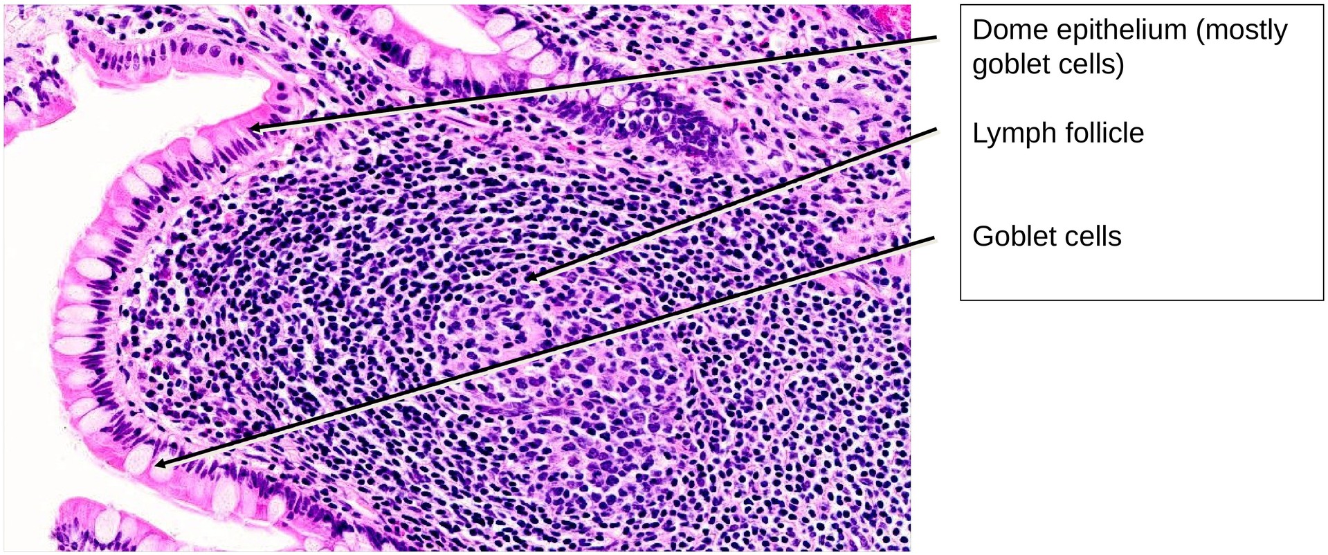
In contrast to the rabbit ileum, where the aggregated lymphoid follicles form a continuous layer (Peyer’s patches) on the antimesenteric side, in the human ileum these follicles are more irregularly distributed along the mucosal folds. Together, they form part of the gut-associated lymphatic tissue (GALT).
The epithelium covering the lymphoid follicles is termed the dome epithelium. Unlike the adjacent mucosa, this epithelium lacks villi and crypts. Within it, a few M cells (microfold cells) can be identified among the enterocytes; these specialized cells facilitate antigen transfer to the underlying immunocompetent lymphoid cells.
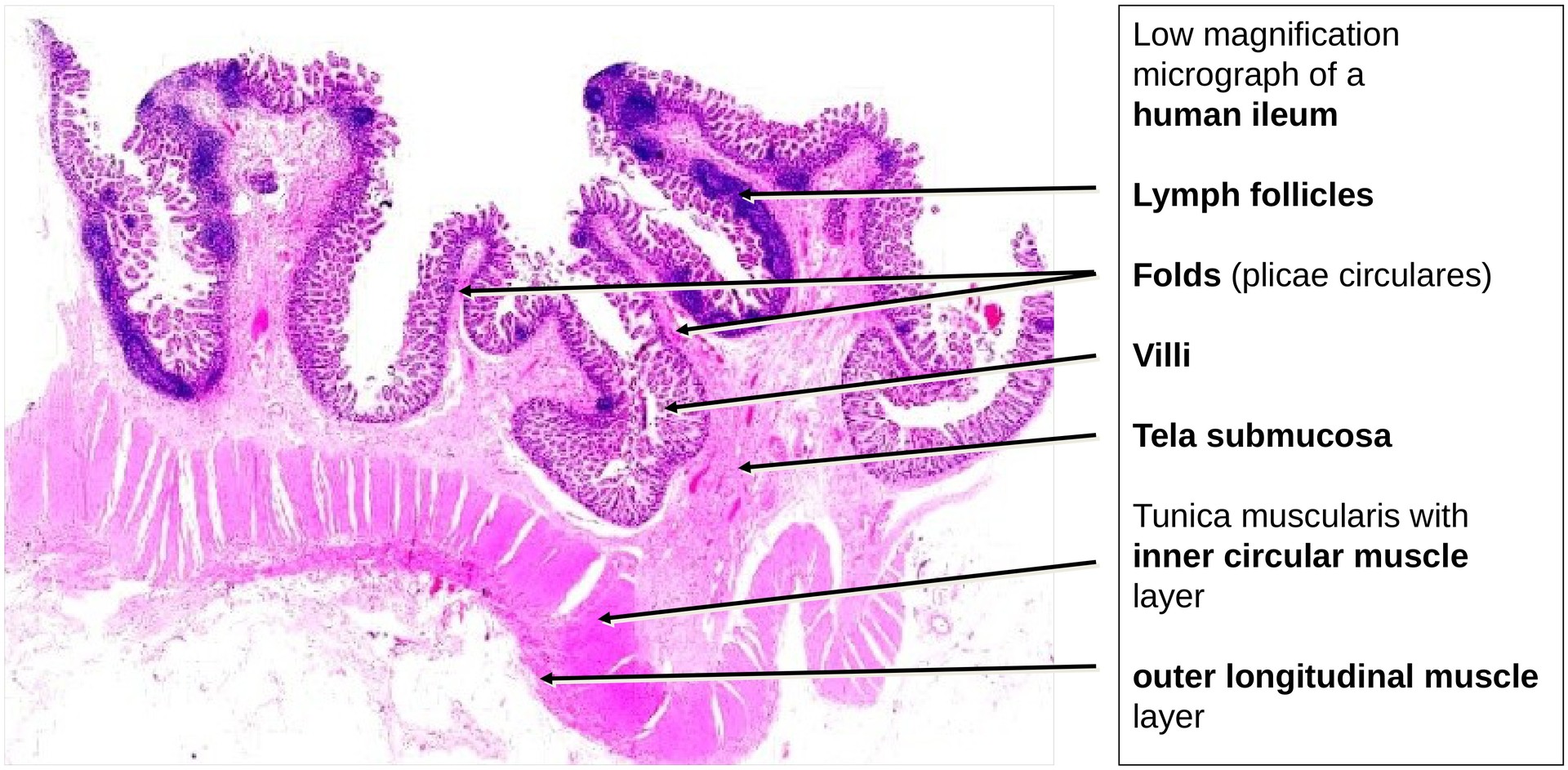
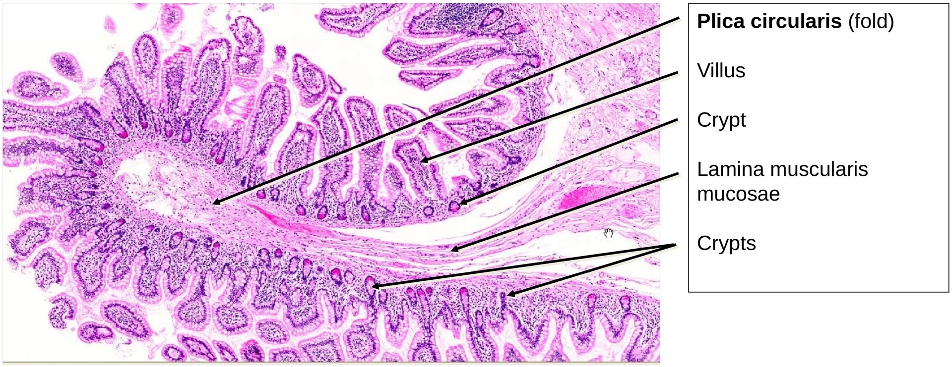
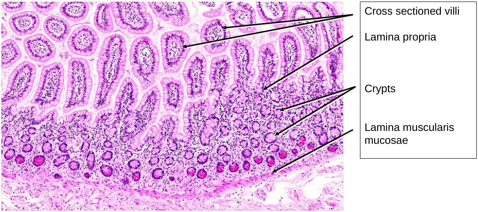
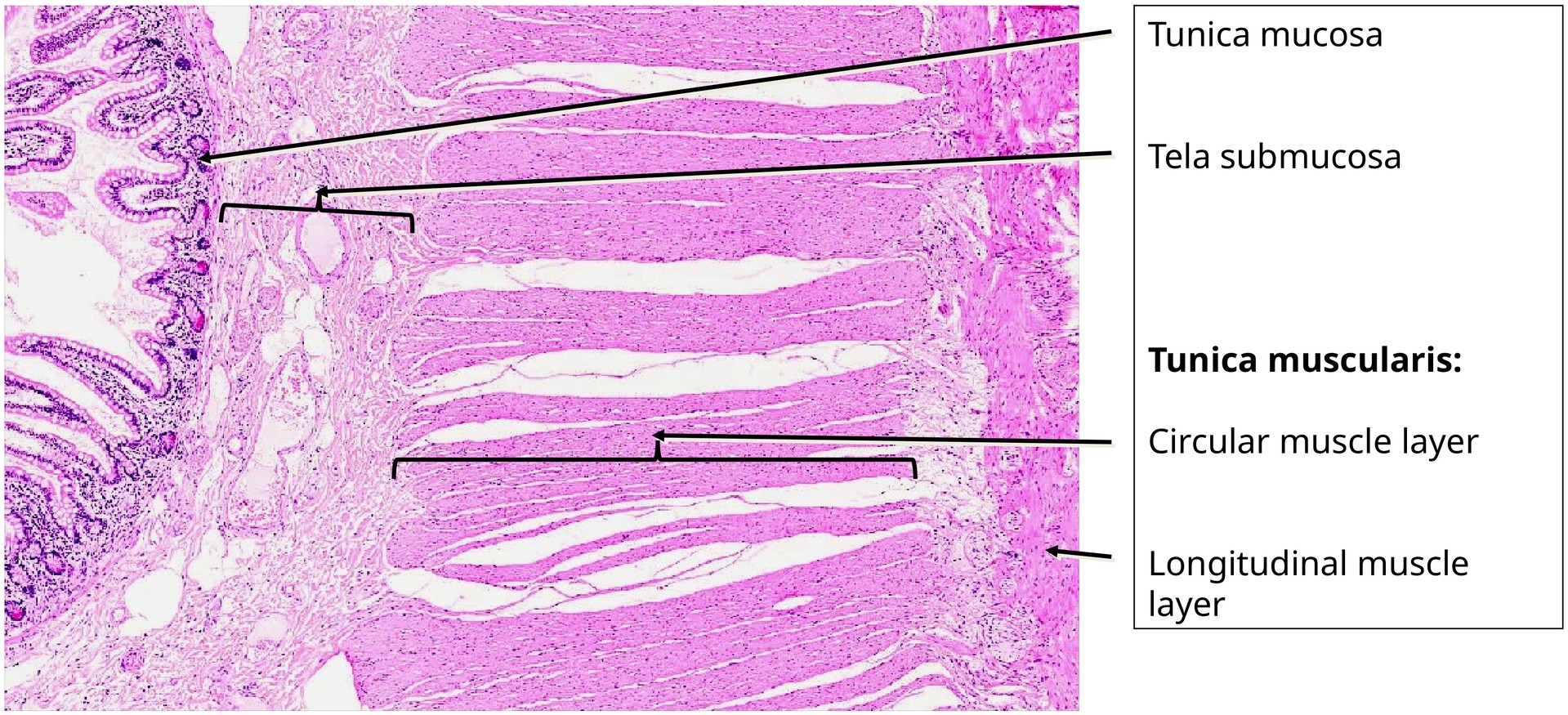
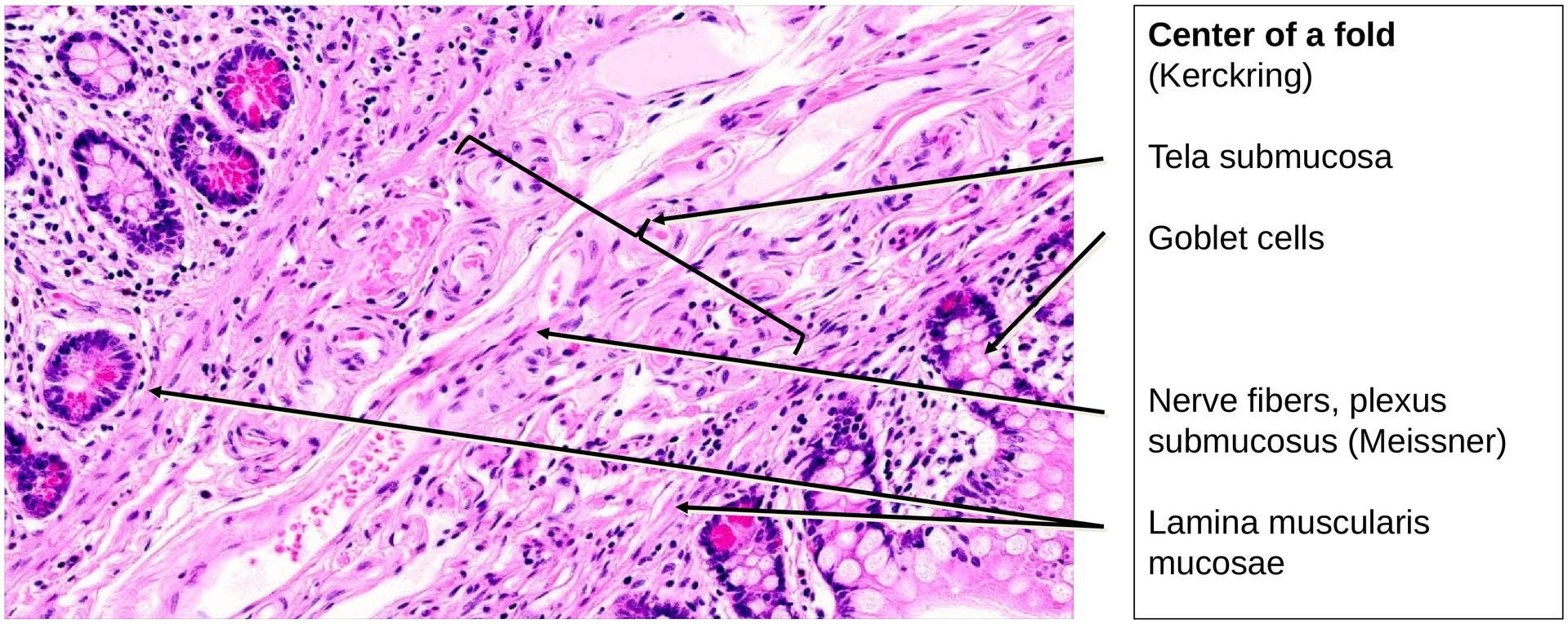
The general layered organization of the GIT wall is well preserved: - Tunica mucosa (with lamina epithelialis, lamina propria, and lamina muscularis mucosae). - The muscularis mucosae is relatively thin but extends into the circular folds and occasionally into the villi. - Tela submucosa, which appears robust but shows few identifiable elements of the submucosal plexus (Meissner’s plexus). - Tunica muscularis, composed of: - An inner circular layer (particularly prominent here). - An outer longitudinal layer, between which the myenteric plexus (Auerbach’s plexus) is clearly visible, containing ganglion cells and nerve fibers.
The circular folds (plicae circulares) are lower than those seen in the duodenum or jejunum. No glands are found in the submucosa (unlike Brunner’s glands in the duodenum).
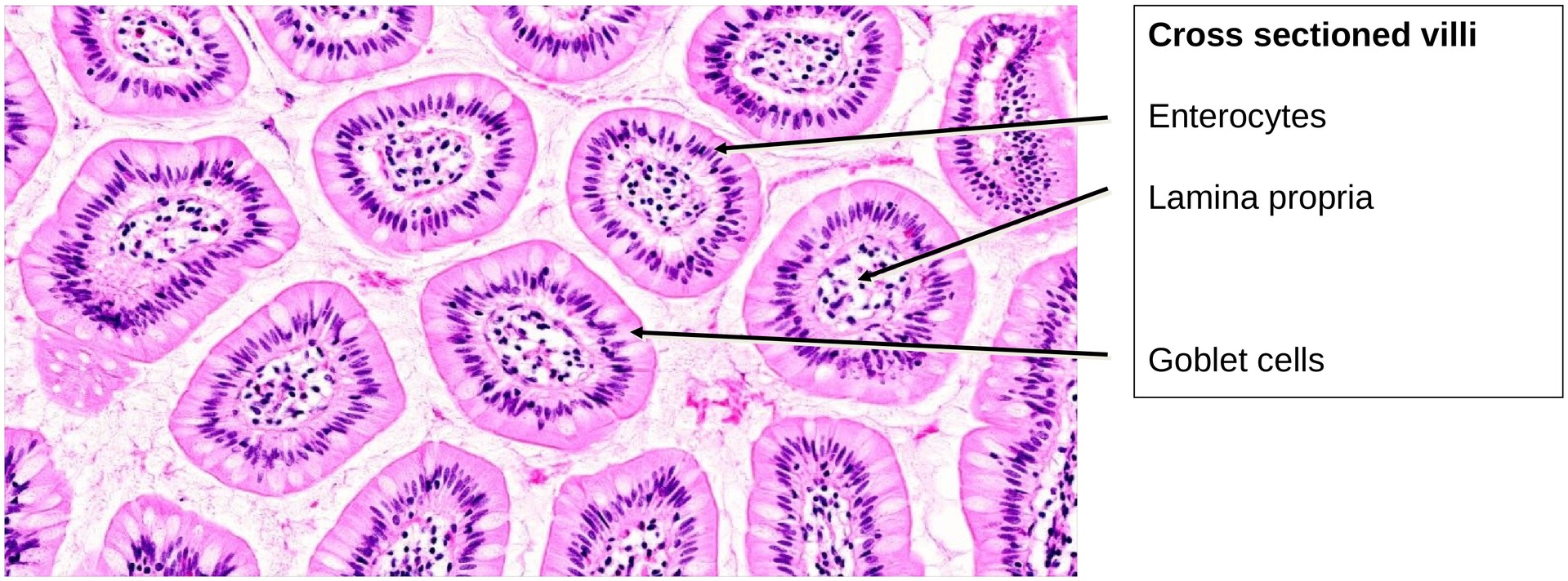
The epithelium of the villi shows numerous goblet cells, interspersed among the enterocytes bearing a distinct brush border. Between the villi, short intestinal crypts (crypts of Lieberkühn) extend into the lamina propria.
Many crypts contain Paneth cells, identifiable by their apical eosinophilic granules rich in lysozyme, which exert an antibacterial effect and help regulate the intestinal microbiota
Tasks:
- Identify the layers of the gastrointestinal wall in overview magnification:
- Tunica mucosa (lamina epithelialis, lamina propria, lamina muscularis mucosae)
- Tela submucosa
- Tunica muscularis
- Locate the circular folds and the villi projecting from them.
- Observe the brush border epithelium and note the abundance of goblet cells.
- Identify the brush border on the enterocytes.
- Assess the thickness of the muscularis mucosae and note its extensions into the folds and villi.
- Search for elements of the submucosal (Meissner’s) plexus within the submucosa.
- Identify the myenteric (Auerbach’s) plexus between the muscle layers.
- Locate the lymphoid follicles and observe their germinal centers (reaction centers).
- Search for M cells within the dome epithelium covering the lymphoid follicles.
- Note that in the area of the dome epithelium, villi and crypts are absent.
- Examine the crypts for Paneth cell granules and describe the function of Paneth cells (antibacterial activity via lysozyme secretion).
License
University of Basel
Downloads