LYMPHATIC ORGANS (ANATOMICAL MICROSCOPY)
15.7
Thymus, adult
Specimen Details:
Specimen Details:
Organ: Thymus
Origin: Human
Staining: Heamatoxylin - Eosin (H&E)
Method and Specimen Description:
Standard histological section stained with H&E for general structural overview.
Objective of the Examination:
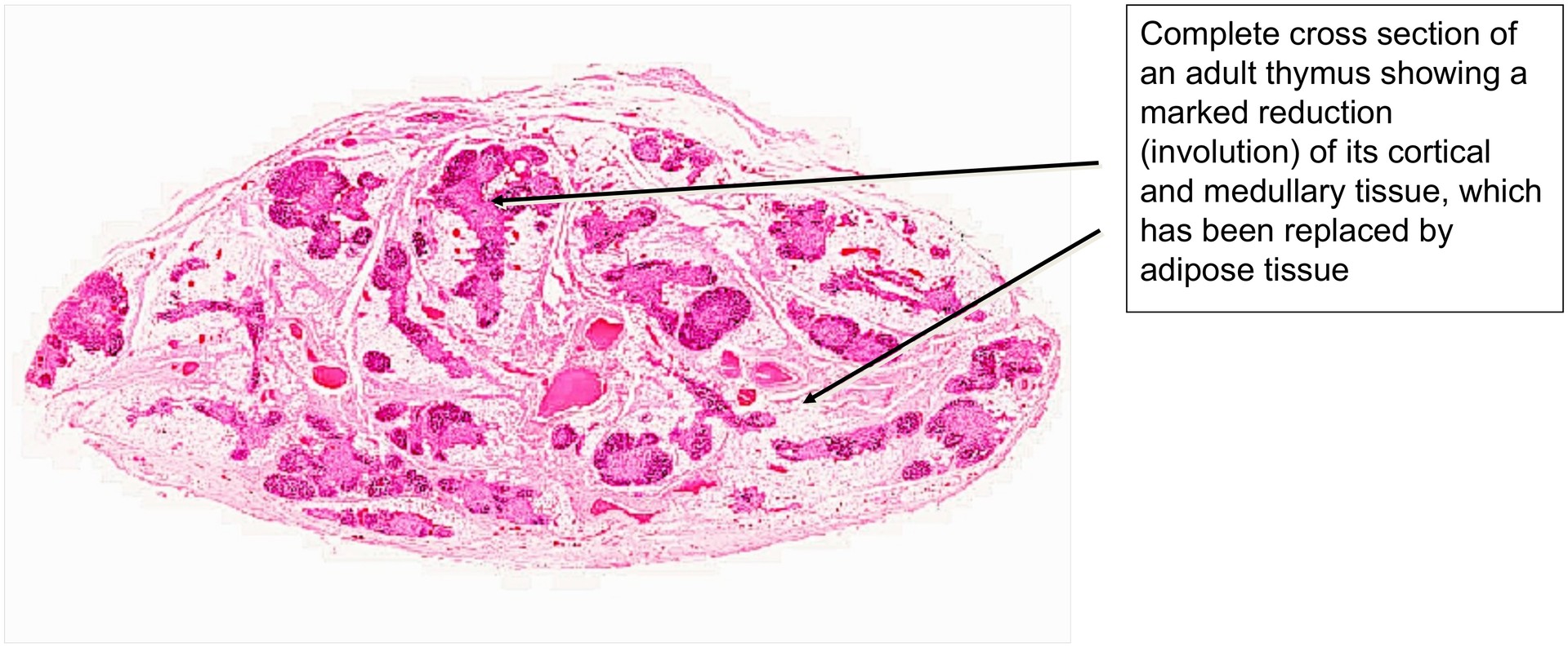
To study the adult thymus and the process of thymic involution, during which the functional parenchyma is progressively replaced by adipose tissue, ultimately forming a retrosternal fatty body.
Special Features of the Specimen:
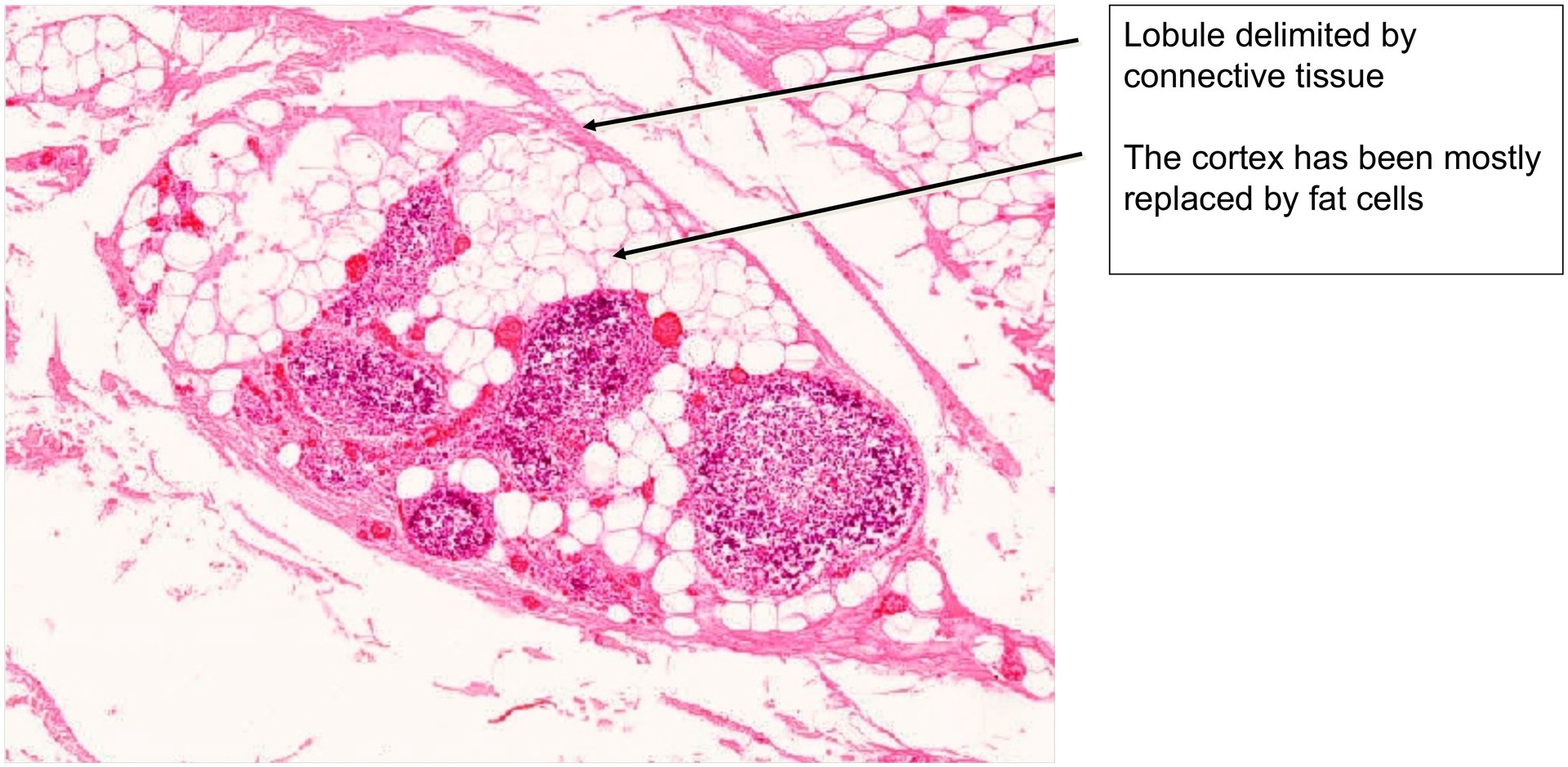
At low magnification, the adult thymus shows a marked reduction in its cortical substance, with a corresponding increase in univacuolar (white) adipose tissue. This replacement process transforms the thymus into a retrosternal fatty body.
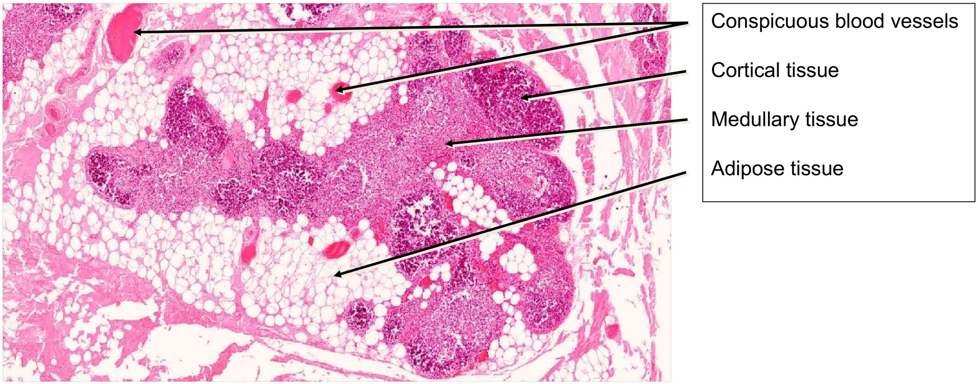
Both the cortex and medulla are reduced in size, although the medulla is relatively better preserved in adulthood. The original lobular architecture is still faintly recognizable but is heavily disrupted by infiltrating adipose tissue.
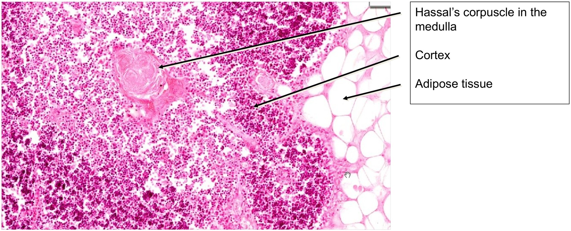
Within the remaining thymic parenchyma, Hassall’s corpuscles can still be identified in the medulla, although their number varies between individuals. In this specimen, only a few corpuscles are present. The physiological function of Hassall’s corpuscules still remains to be cleared.
Conversely, blood vessels are more conspicuous in the adult thymus than in the juvenile form, being visible both within lobules and in the interlobular adipose tissue.
Tasks:
- At low magnification, compare the proportions of cortex and medulla, and evaluate the extent of adipose tissue infiltration.
- Identify Hassall’s corpuscles within the medullary regions.
- Examine the vessels in both the medulla and the interlobular connective and adipose tissue, and assess their wall structure and organization.
License
University of Basel
Downloads