GLANDS (GENERAL HISTOLOGY)
3.2
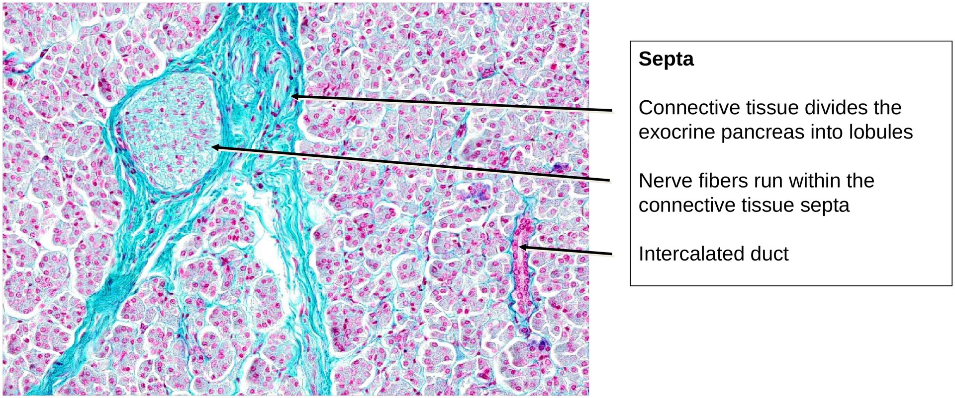
Pancreas, endocrine portion
Specimen Details:
Specimen Details:
Organ: Pancreas
Origin: Human
Staining: Fuchsin/Azocarmin/Fastgreen
Method and Specimen Description:
Routine histological section of the pancreas stained with a trichrome method (Fuchsin–Azocarmine–Fast Green), which distinctly differentiates the endocrine pancreatic islets (islets of Langerhans) from the surrounding exocrine tissue. The staining also permits partial distinction between two major endocrine cell types.
Objective of the Examination:
To study the structure and cellular composition of the endocrine portion of the pancreas and to distinguish it from the exocrine tissue.
Special Features of the Specimen:
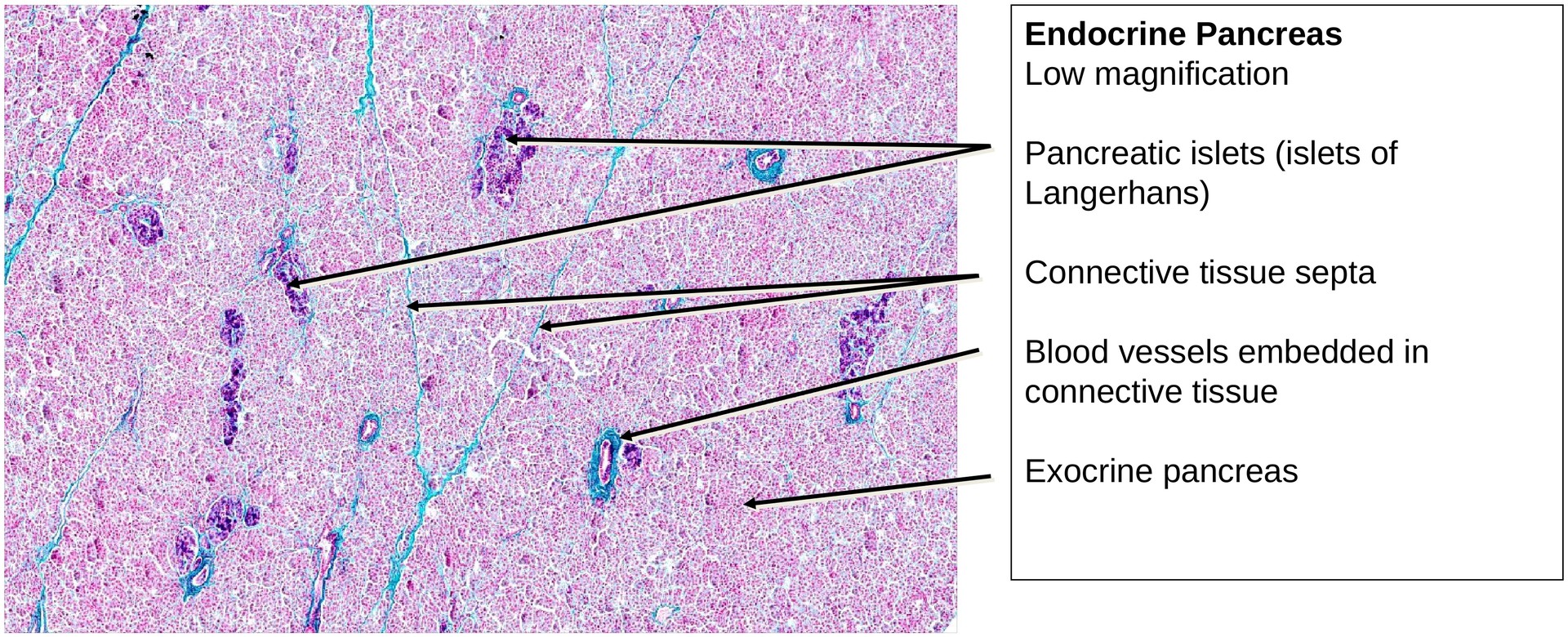
General (Low Magnification):
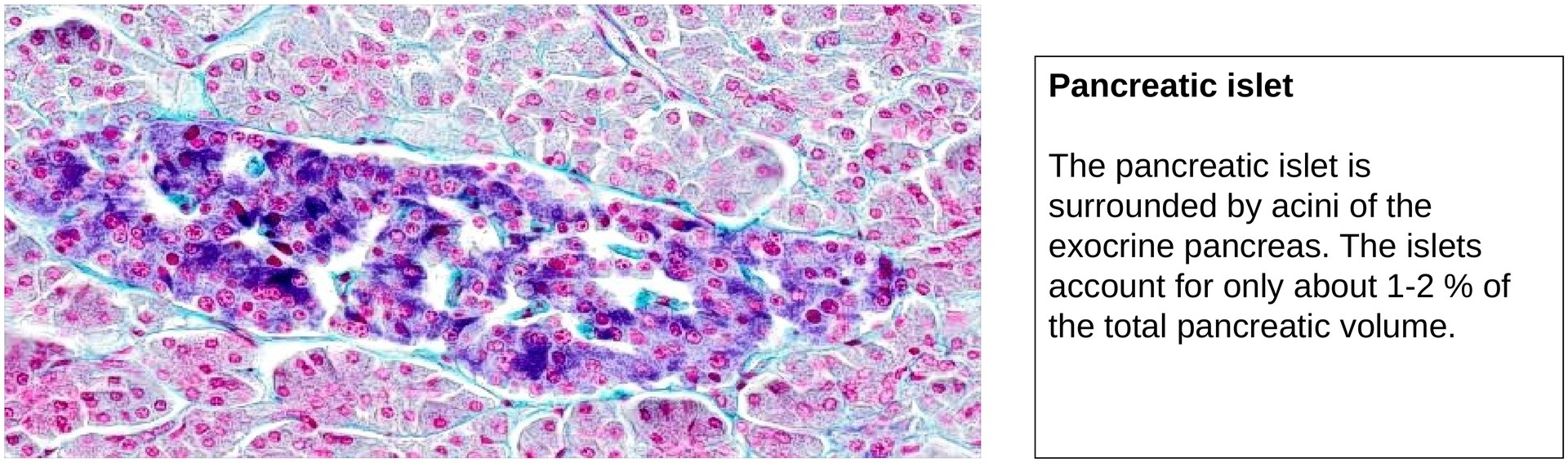
At low magnification, the islets of Langerhans appear as darker-staining, rounded or oval clusters of cells scattered throughout the lighter-staining exocrine parenchyma. Each islet is highly vascularized, with numerous thin-walled capillaries visible between the endocrine cells. Importantly, the islets lack excretory ducts, a key feature that differentiates them from the exocrine component.
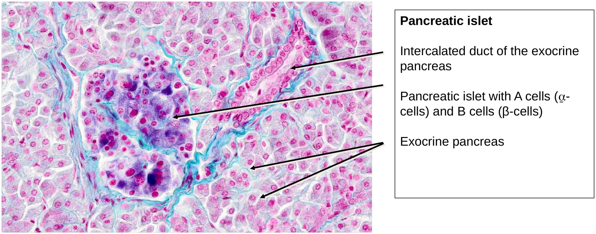
Medium to High Magnification:
With this staining method, at least two main cell types within the islets can be distinguished:
-
A cells (α-cells): Located predominantly at the periphery of the islets; they contain α-granules storing the hormone glucagon, which raises blood glucose levels.
-
B cells (β-cells): Found mainly in the central regions of the islets; they contain β-granules with insulin, which lowers blood glucose levels.
A third cell type, the D cells (δ-cells), occur sporadically. They secrete somatostatin, which inhibits both exocrine secretion and hormone release within the islet. However, this staining does not clearly differentiate the D cells from other types.
Further minor cell types (PP and ε cells) are not visible in this preparation.
The absence of excretory ducts is a defining morphological indicator of the endocrine function of the pancreas.
Tasks:
• At low magnification, locate and identify several pancreatic islets within the surrounding exocrine tissue.
• At higher magnification, examine individual islets and distinguish between A cells and B cells based on their staining characteristics and distribution.
• Evaluate the relative proportions of endocrine and exocrine tissue within the specimen.
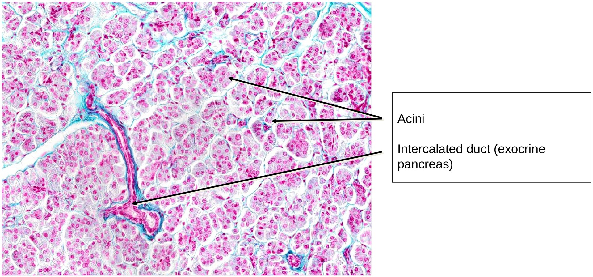
• Try to identify the pancreas based solely on the exocrine component (without relying on visible islets). Which histological features allow this identification?
License
University of Basel
Downloads