GLANDS (GENERAL HISTOLOGY)
3.5
Sublingual gland
Specimen Details:
Specimen Details:
Organ: Sublingual gland
Origin: Human
Staining: Hematoxylin - Eosin (H&E)
Method and Specimen Description:
Conventional histological section prepared using the general overview stain Hematoxylin and Eosin.
Objective of the Examination:
To study the microscopic structure of a predominantly mucous salivary gland and to recognize its characteristic secretory and ductal features.
Special Features of the Specimen:
General (Low Magnification):
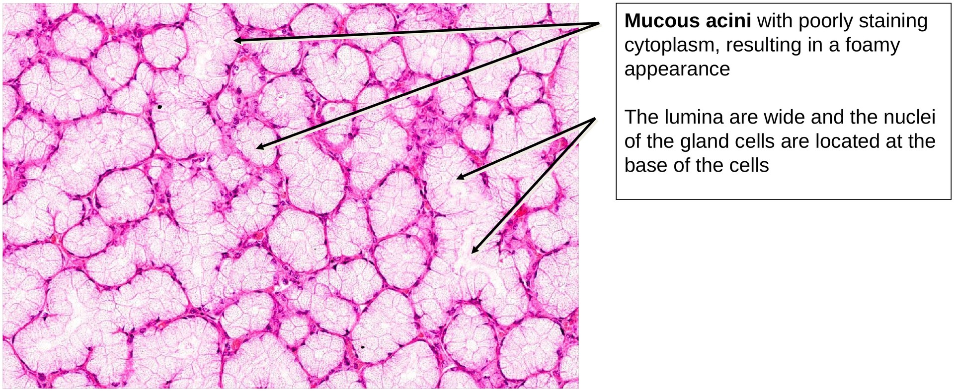
The sublingual gland appears uniformly structured but poorly stainable, reflecting its mucous nature. Most secretory units are mucous acini, which exhibit pale, foamy cytoplasm due to the presence of mucin.
Serous demilunes—crescent-shaped caps of serous cells on mucous acini described in textbooks—are only faintly visible in this specimen and are practically absent. Adipocytes (fat cells) are scattered throughout the glandular parenchyma, a common feature in major salivary glands.
Secretory Units:
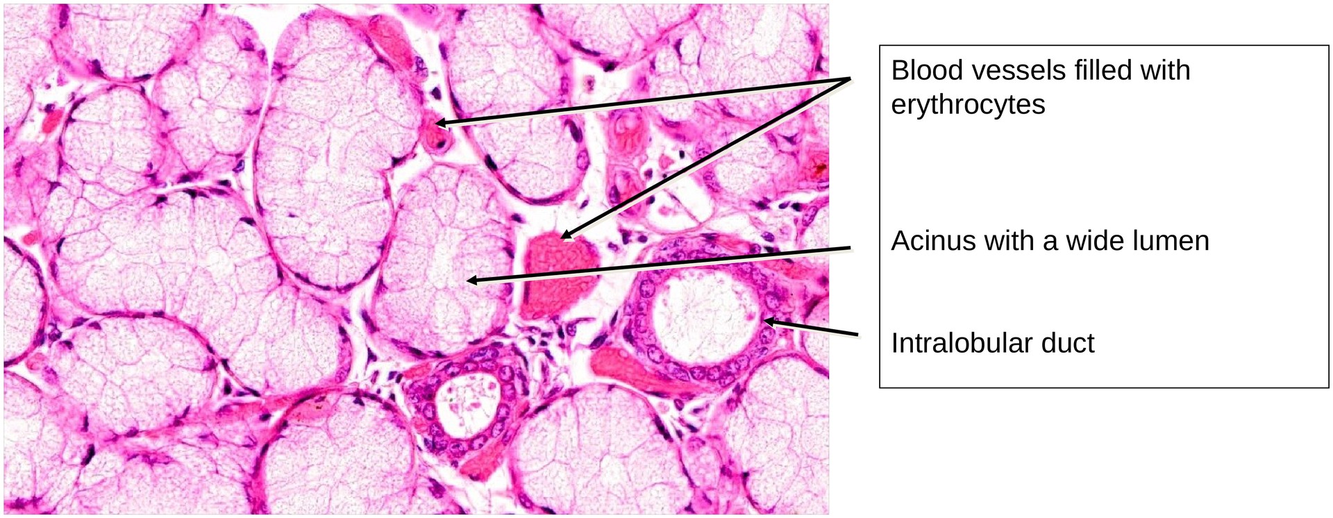
The mucous acini have large lumina, pale cytoplasm, and nuclei displaced towards the basal region of the cells. The nuclei appear flattened against the basement membrane. The mucous content, which does not stain well with H&E, gives the cytoplasm its foamy appearance.
Duct System:
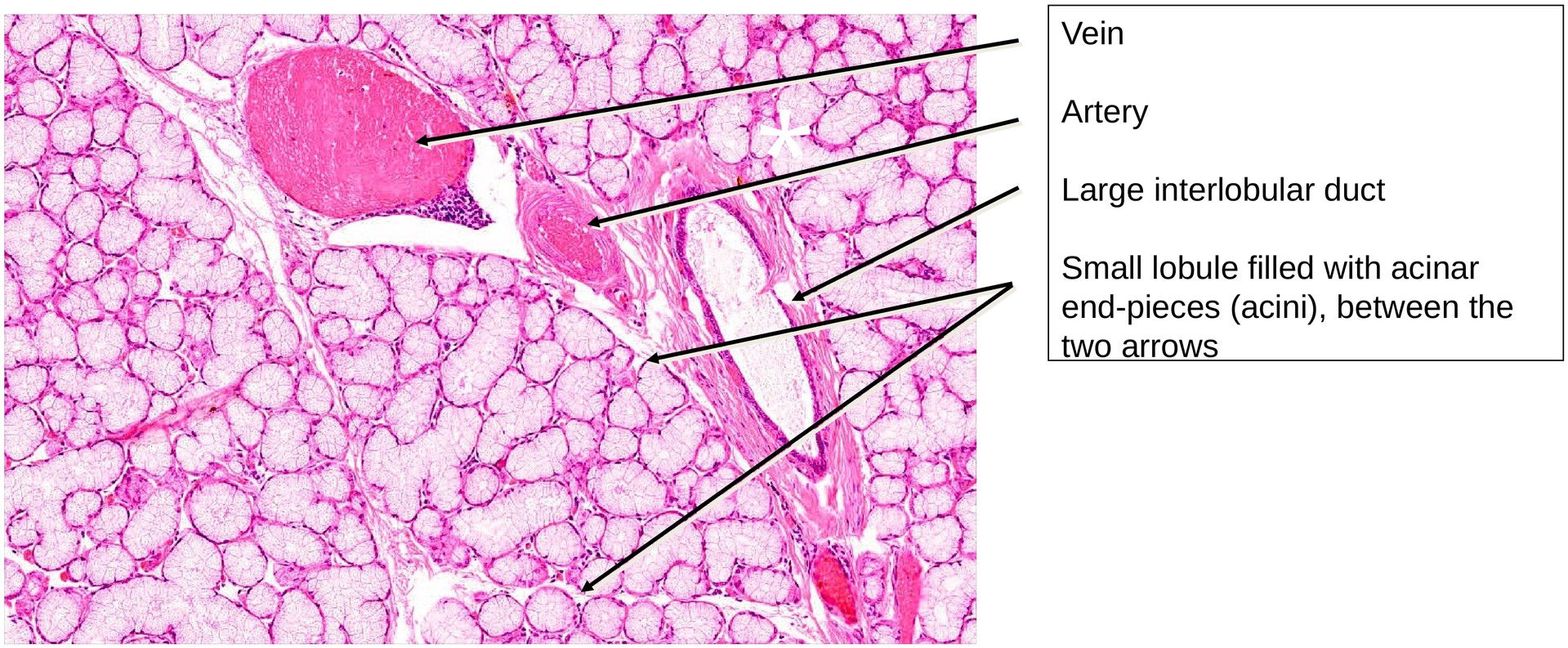
At medium magnification, it becomes clear that the ductal system is less developed than in purely serous glands such as the parotid.
-
Intercalated ducts are few and relatively short, as their narrow lumen is not well suited to the viscous mucous secretion.
-
Striated ducts are also short and infrequent, though occasionally present.
-
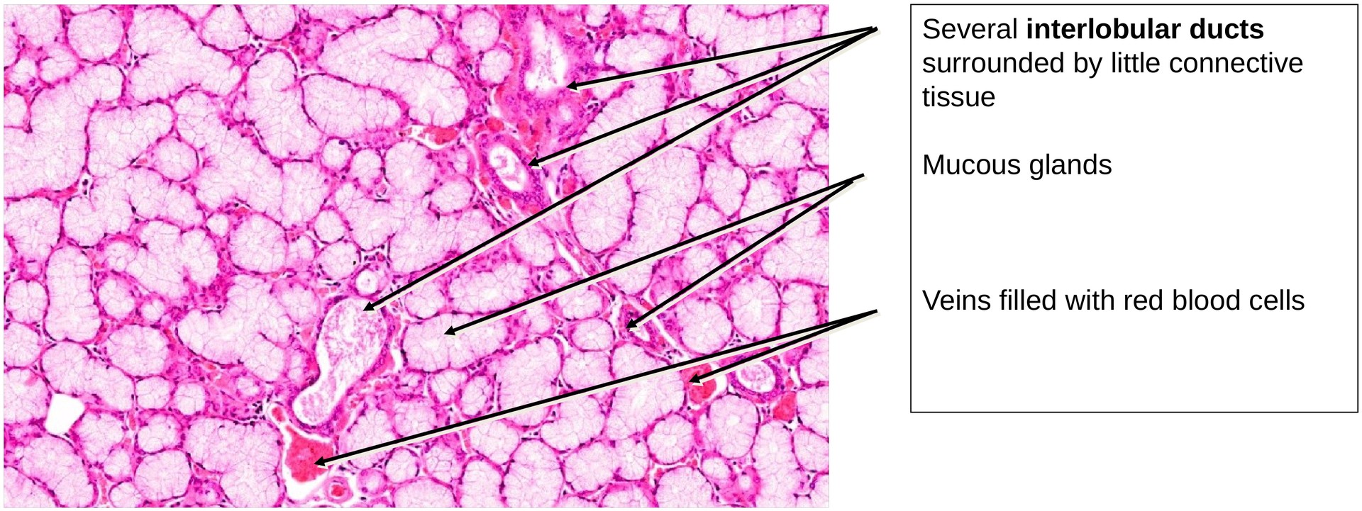
The larger interlobular excretory ducts, found in the connective tissue septa, are more numerous and can be easily identified by their cuboidal to columnar epithelium.
Within the lobules, smaller intralobular ducts connect to the mucous acini. The lumen of the glandular tubules is sometimes visible in cross-section.
Tasks:
• At low magnification, identify the excretory duct system and distinguish between intralobular and interlobular ducts.
• Determine which components of the duct system typically found in salivary glands are present or absent in the sublingual gland.
• Examine the structure of the acini and note the position and shape of the nuclei within mucous cells.
• Compare these features to those of a serous gland (e.g. the parotid gland) to understand functional and structural differences.
Summary Table – Key Features of the Sublingual Gland:
| Feature | Description |
|---|---|
| Type of secretion | Predominantly mucous (minor serous component) |
| Secretory unit | Mucous acini, occasionally with serous demilunes |
| Duct system | Few intercalated and striated ducts; well-developed interlobular ducts |
| Cytoplasmic appearance | Pale, foamy cytoplasm due to mucin |
| Nuclear position | Flattened, located basally against the basement membrane |
| Distinctive features | Poorly stainable with H&E, adipocytes scattered in parenchyma, minimal serous content |
License
University of Basel
Downloads