GLANDS (GENERAL HISTOLOGY)
3.7
Submandibular gland 1
Specimen Details:
Specimen Details:
Organ: Submandibular gland
Origin: Human
Staining: Haematoxylin - Eosin (H&E)
Method and Specimen Description:
Routine histological section stained with the general overview stain Hematoxylin and Eosin (H&E).
Objective of the Examination:
To study the microscopic structure of a predominantly serous gland of the sero-mucous type, including the organization of its ductal system.
Special Features of the Specimen:
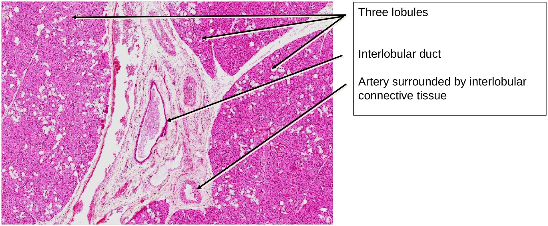
General (Low Magnification):
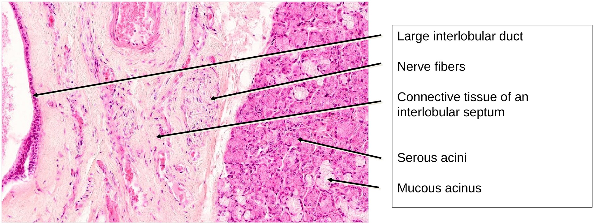
The submandibular gland is organized into distinct lobules, separated by interlobular connective tissue septa. Within these septa run the larger excretory ducts, as well as blood vessels and nerves supplying the gland.
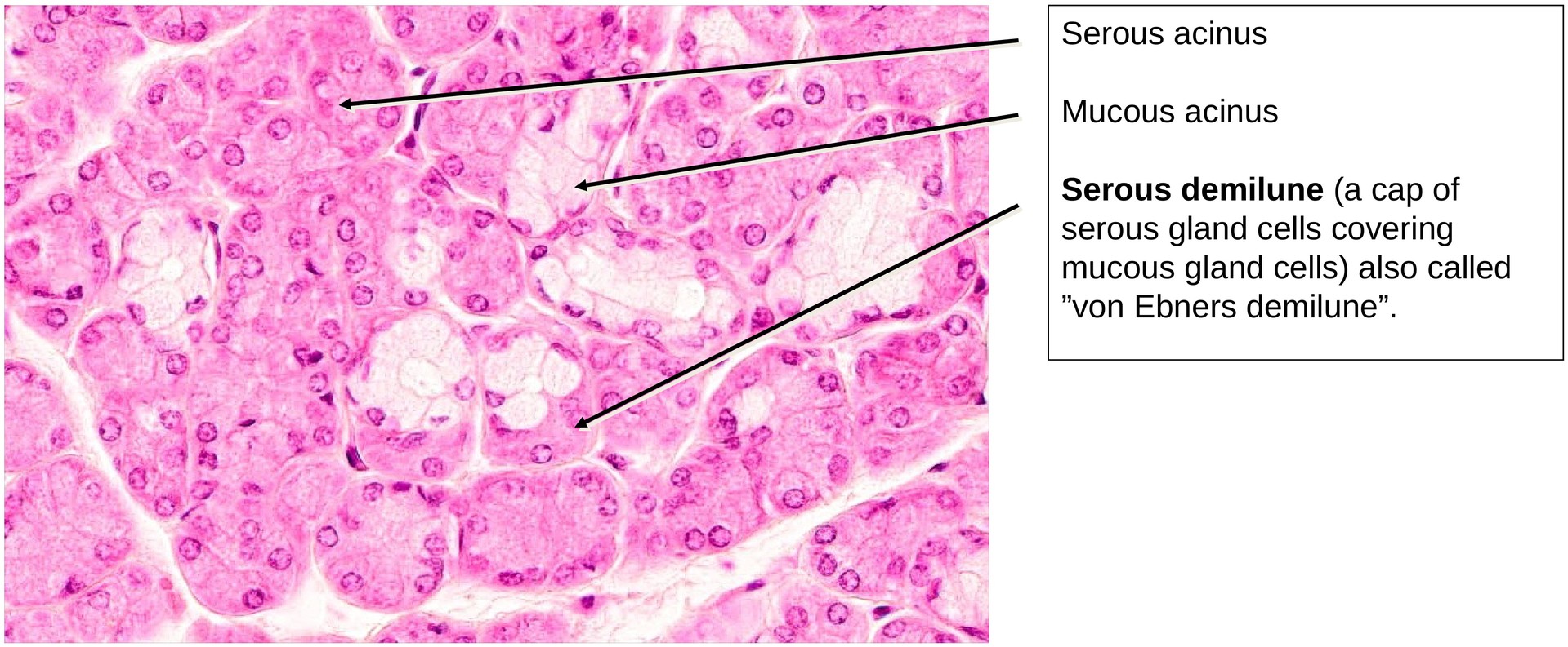
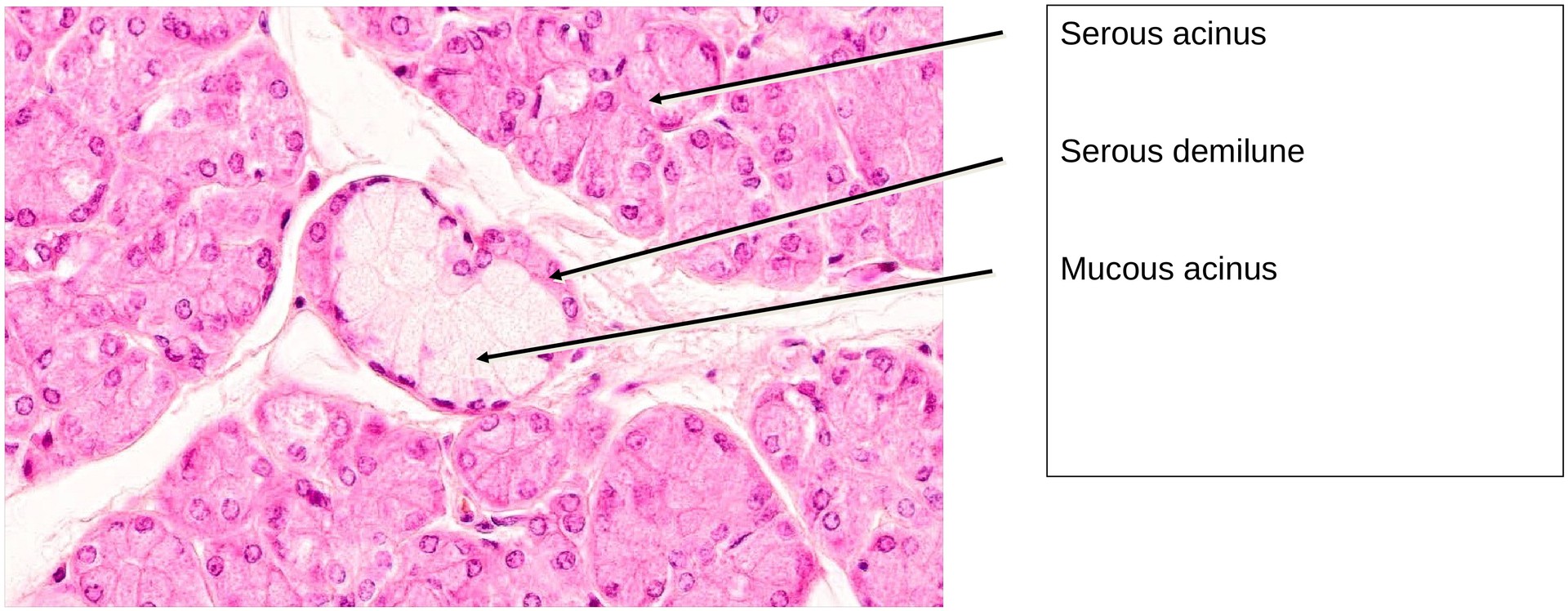
Even at low magnification, the mixed sero-mucous character of the gland is evident. The majority of secretory units are serous acini, but mucous tubules also occur. On the mucous tubules, serous demilunes—crescent-shaped caps of serous cells—can be identified. These are also known as von Ebner’s crescents, and they function to flush the viscous mucous secretion through the ducts.
Secretory Units and Duct System (Medium Magnification):
At higher magnification, the excretory duct system is clearly recognizable. As in the parotid gland, all components of a typical compound gland duct system are present:
-
Interlobular excretory ducts: Large ducts located within the connective tissue septa, lined by cuboidal to columnar epithelium.
-
Smaller interlobular ducts: Located between lobules, connecting with intralobular components.
-
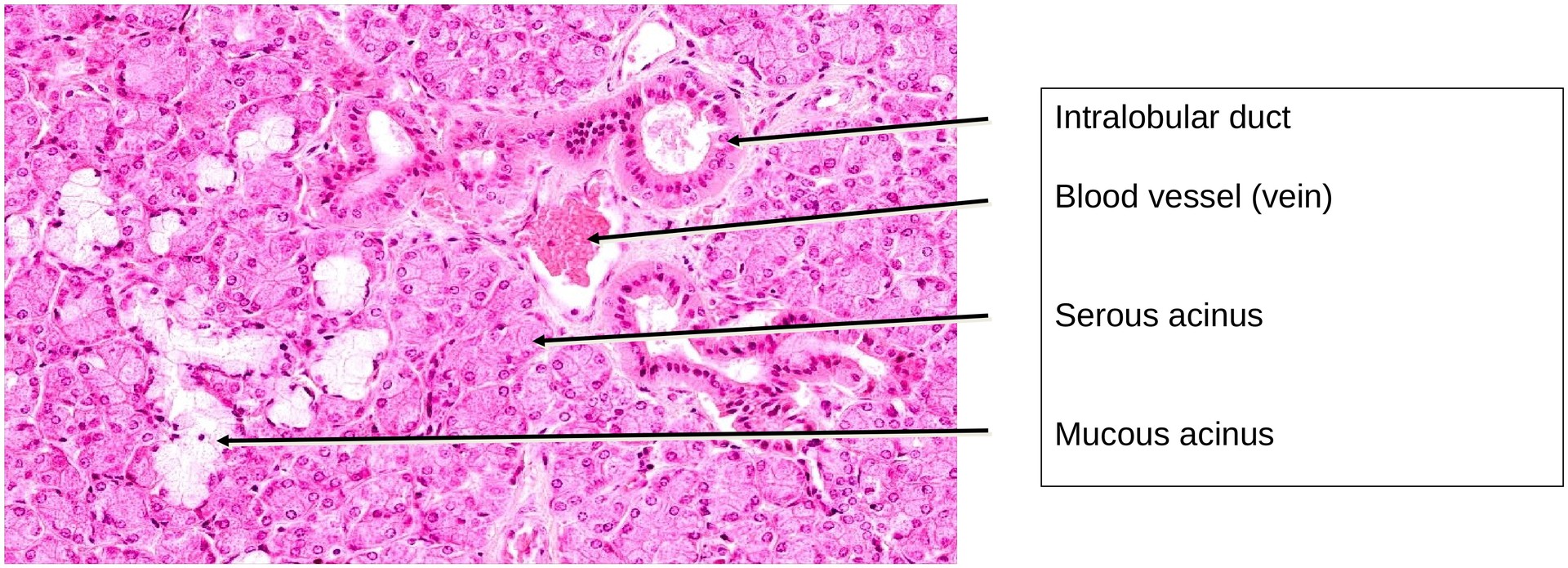
Intralobular ducts (striated ducts): Situated within lobules; lined by tall columnar epithelium showing basal striations formed by mitochondria in membrane infoldings.
-
Intercalated ducts: Narrow ducts connecting the acini to the striated ducts; they are short and infrequent in this gland, lined by low cuboidal cells.
-
Secretory end pieces: Mainly serous acini with round, basally located nuclei and well-staining granular cytoplasm. Mucous tubules appear paler and contain flattened basal nuclei.
Serous and Mucous Components:
The serous acini stain more intensely with H&E, reflecting their protein-rich secretion.\ The mucous tubules appear pale and foamy due to mucin, and their nuclei are compressed basally.\ The serous demilunes are often artefactual, resulting from shrinkage during fixation, but they still illustrate the close association between the two cell types in mixed glands.
Tasks:
• Obtain a general overview of the lobular structure of the submandibular gland.
• Compare serous acini and mucous tubules in terms of morphology and staining properties.
• Locate striated (secretory) ducts and note their position within lobules.
• Identify interlobular ducts and the blood vessels and nerves running alongside them in the connective tissue septa.
• Explain the presence and function of serous demilunes on mucous tubules.
• Determine which components of the excretory duct system are visible in this specimen.
Summary Table – Key Features of the Submandibular Gland:
| Feature | Description |
|---|---|
| Type of secretion | Mixed: predominantly serous, with mucous components |
| Secretory units | Serous acini; mucous tubules with serous demilunes |
| Duct system | All major duct types present (intercalated, striated, interlobular, excretory) |
| Cytoplasmic staining | Serous cells stain strongly (basophilic); mucous cells stain weakly (pale/foamy) |
| Nuclear position | Serous nuclei round and basal; mucous nuclei flattened and peripheral |
| Distinctive features | Mixed gland; von Ebner’s crescents; well-developed striated ducts; lobulated organisation |
License
University of Basel
Downloads