GLANDS (GENERAL HISTOLOGY)
3.3
Parotid gland
Specimen:
Specimen Details:
Organ: Parotid gland
Origin: Human
Staining: Hematoxylin - Eosin (H&E)
Method and Specimen Description:
Standard histological section of the parotid gland, stained with the general overview stain Hematoxylin and Eosin.
Objective of the Examination:
To study the microscopic structure of a serous, compound gland and to understand its duct system and lobular organization.
Specific Features of the Specimen:
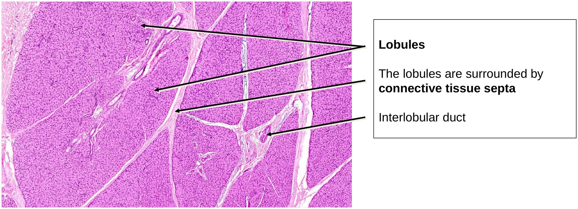
General (Low Magnification):
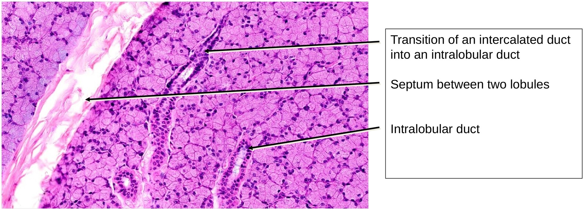
The parotid gland appears homogeneous and lobulated, divided by fibrous connective tissue septa that contain larger ducts, vessels, and nerves. The presence of ducts within these septa indicates the compound nature of the gland.
Glandular Structure:
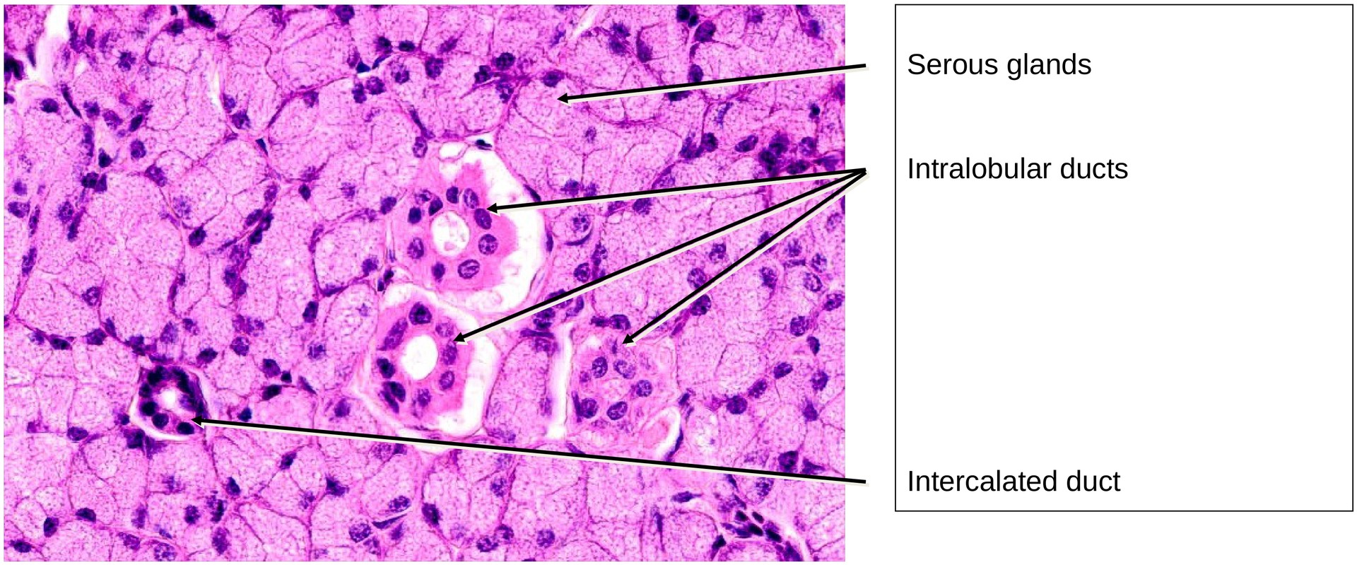
The secretory end-pieces are exclusively serous acini. Each acinus is roughly spherical, consisting of pyramidal cells with basophilic basal cytoplasm (due to abundant rough endoplasmic reticulum) and eosinophilic apical cytoplasm containing zymogen granules. The lumen is small and sometimes difficult to recognize.
Myoepithelial cells are present between the basal plasma membrane of the secretory cells and the basement membrane, assisting in the expulsion of secretion.
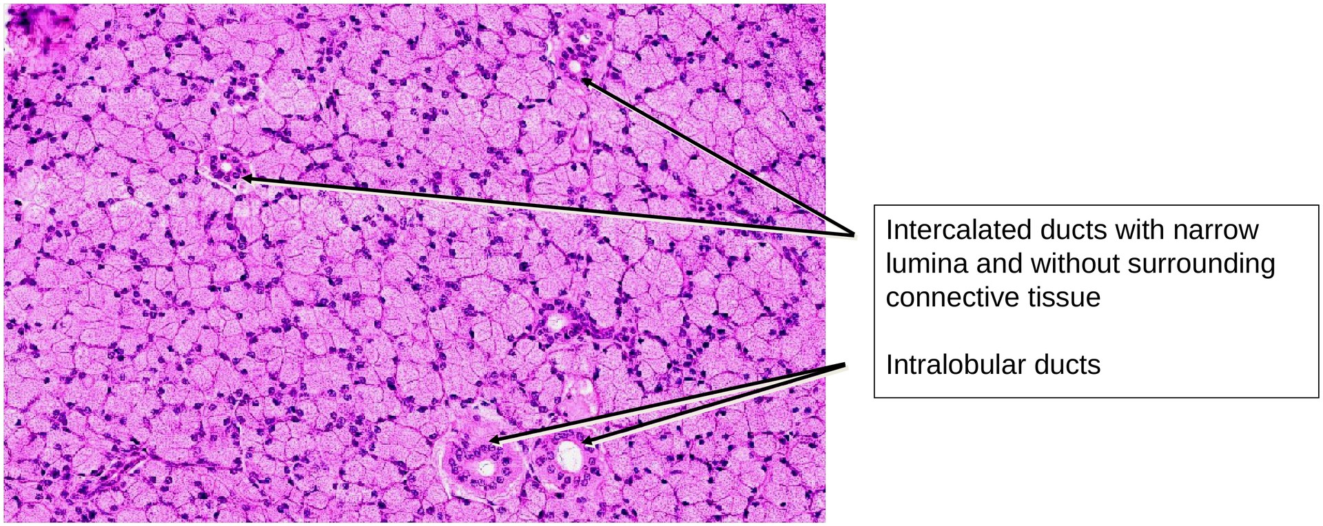
Duct System:
The parotid gland exhibits a well-developed duct system consisting of:
-
Intercalated ducts – Very narrow lumen, lined by low cuboidal epithelium. These ducts may contain myoepithelial cells and connect directly to the acini.
-
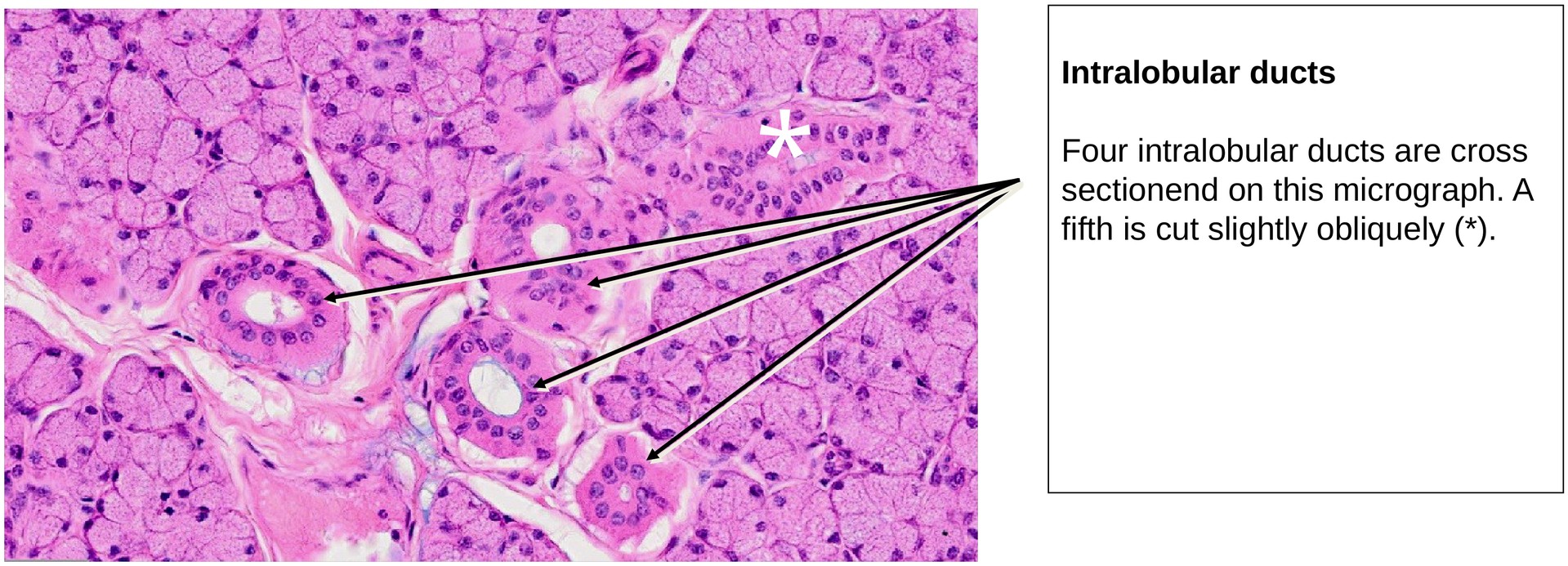
Striated ducts (intralobular ducts) – Lined by tall columnar epithelium. The basal striations are formed by infoldings of the basal membrane with aligned mitochondria, responsible for ion transport and modification of the saliva (secretion of potassium and bicarbonate, reabsorption of sodium).
-
Interlobular ducts – Located within the connective tissue septa between lobules; lined by columnar or stratified columnar epithelium and surrounded by connective tissue.
-
Main excretory duct (Stensen’s duct) – Lined by stratified squamous epithelium near its opening into the oral cavity.
Note: In the parotid gland, intercalated ducts are long, while striated ducts are relatively short compared to other salivary glands.
Tasks:
• At low magnification, obtain an overview of the lobular organization of the gland.
• Identify fibrous septa and locate larger ducts within them (interlobular ducts).
• Observe and describe the serous acini – their size, cell shape, and staining characteristics.
• Search for narrow intercalated ducts emerging from the acini and follow them into the striated ducts.
• Compare the epithelial types along the duct system and note their structural adaptations for secretion and modification
Summary Table – Key Features of the Parotid Gland:
| Feature | Description |
|---|---|
| Type of secretion | Purely serous |
| Secretory unit | Serous acini |
| Duct system | Intercalated → Striated → Interlobular → Main excretory duct |
| Myoepithelial cells | Present around acini and intercalated ducts |
| Connective tissue | Forms septa dividing the gland into lobules |
| Distinctive characteristics | Homogeneous structure, long intercalated ducts, short striated ducts, absence of mucous acini |
License
University of Basel
Downloads