NERVOUS SYSTEM (ANATOMICAL MICROSCOPY)
16.4
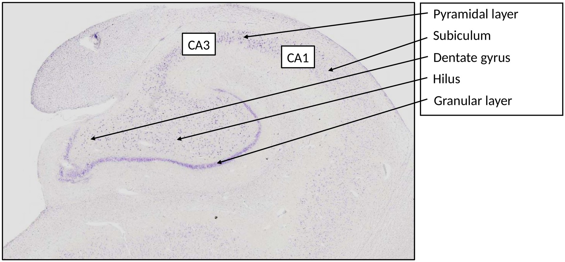
Hippocampus
Preparation:
PREPARATION DETAILS:
Organ: Hippocampus
Origin: Human
Staining: Cresyl Violet
METHOD AND SPECIMEN DESCRIPTION:
Cresyl violet is a basic dye that binds to acids, i.e. to RNA and DNA. It stains the chromatin of the cell nucleus, the nucleolus, and the rough endoplasmic reticulum (Nissl substance) blue–violet.
The hippocampus is an allocortical region of the forebrain and forms the largest part of the archicortex, representing a phylogenetically older type of cortex compared with the six-layered neocortex.
OBJECTIVE OF THE EXAMINATION:
To understand the morphology, subregions, and layer structure of the human hippocampus.
SPECIAL FEATURES OF THE PREPARATION:
The hippocampus forms a rolled, S-shaped gyrus that can be divided into two major subregions:
- Dentate gyrus
- Ammon’s horn (Cornu ammonis)
Both subregions possess a three-layered cortex enclosed by two relatively cell-poor molecular layers.
Dentate Gyrus:
- Layers:
- Molecular layer
- Granule cell layer
- Hilum (Hilus or polymorphic layer)
The hilus is filled with loosely arranged interneurons, which differ markedly from the pyramidal cells of CA3, though both form a continuous cell band. This region is sometimes referred to as the polymorphic layer.
Ammon’s Horn (Cornu Ammonis):
- Subdivided into CA3 and CA1 regions.
- CA1 continues into the subiculum, which in turn merges into the entorhinal cortex.
Layers of the Cornu Ammonis:
- Stratum oriens
- Stratum pyramidale
- Stratum radiatum
- Stratum lacunosum
- Stratum moleculare
- Stratum multiforme
The Stratum pyramidale contains densely packed pyramidal neurons, which form the principal output cells of the hippocampus.
The Stratum oriens lies beneath the pyramidal layer, and the alveus (containing efferent axons) forms the outermost layer adjacent to the white matter.
TASKS:
- Which cell type predominates in the dentate gyrus?
- From where do the moss fibers originate, and to where do they project?
- From where do the Schaffer collaterals arise, and where do they terminate?
- Try to determine the boundary between CA3 and CA1.
- Try to identify the transition between CA1 and the subiculum.
- Determine the position of the Stratum oriens and the alveus.
License
University of Basel