THE FOUNDATIONS OF COGNITION
3.4
Feature Creatures
Content: How do brains evolve to recognize specific perceptual features of stimuli? Research on cats has shown how they detect and respond to different frequencies and patterns. These findings provide key insights into how more advanced perceptual systems emerge.
Frog’s eyes filter what is meaningful even before the brain gets involved. How then does the brain represent and encode this visual information once it arrives from the eye?
To answer this, we turn to a foundational series of studies by David Hubel and Torsten Wiesel. Through single-cell recordings in the visual cortex of cats, they made key discoveries that fundamentally changed our understanding of the brain.
Implanting microelectrodes into the primary visual cortex (V1), they recorded the activity of single neurons while presenting different visual forms.
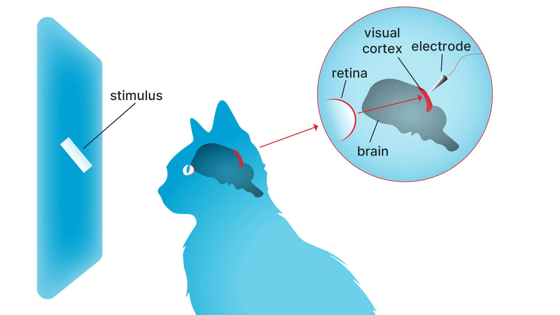
Experimental setup used by Hubel and Wiesel to study visual processing in the cat’s brain.
A visual stimulus, such as a moving light bar, is displayed on a screen while a microelectrode records electrical activity from individual neurons in the primary visual cortex.
At first, nothing seemed to work. Despite correct electrode placement, the neurons remained silent regardless of the stimuli shown. Eventually, after hours of trial and error, the researchers noticed a pattern. The cell only responded when the shadow of an edge on the glass slide moved across a specific area. But that wasn’t all: the shadow line also had to be at a very specific angle (a particular orientation). In every other case, the cell wouldn’t fire.
More accidentally than anything else, Hubel and Wiesel stumbled upon what they would later call a “complex cell” — one type within a hierarchy of visual neurons. A complex cell is a neuron that responds selectively to both orientation and motion. In their experiments, this cell fired only when a light bar was angled at 11 o’clock and moved up and to the right. Building on this finding, and across many experiments, Hubel and Wiesel mapped out a hierarchy in the primary visual cortex in great detail. They identified three main types of neurons:
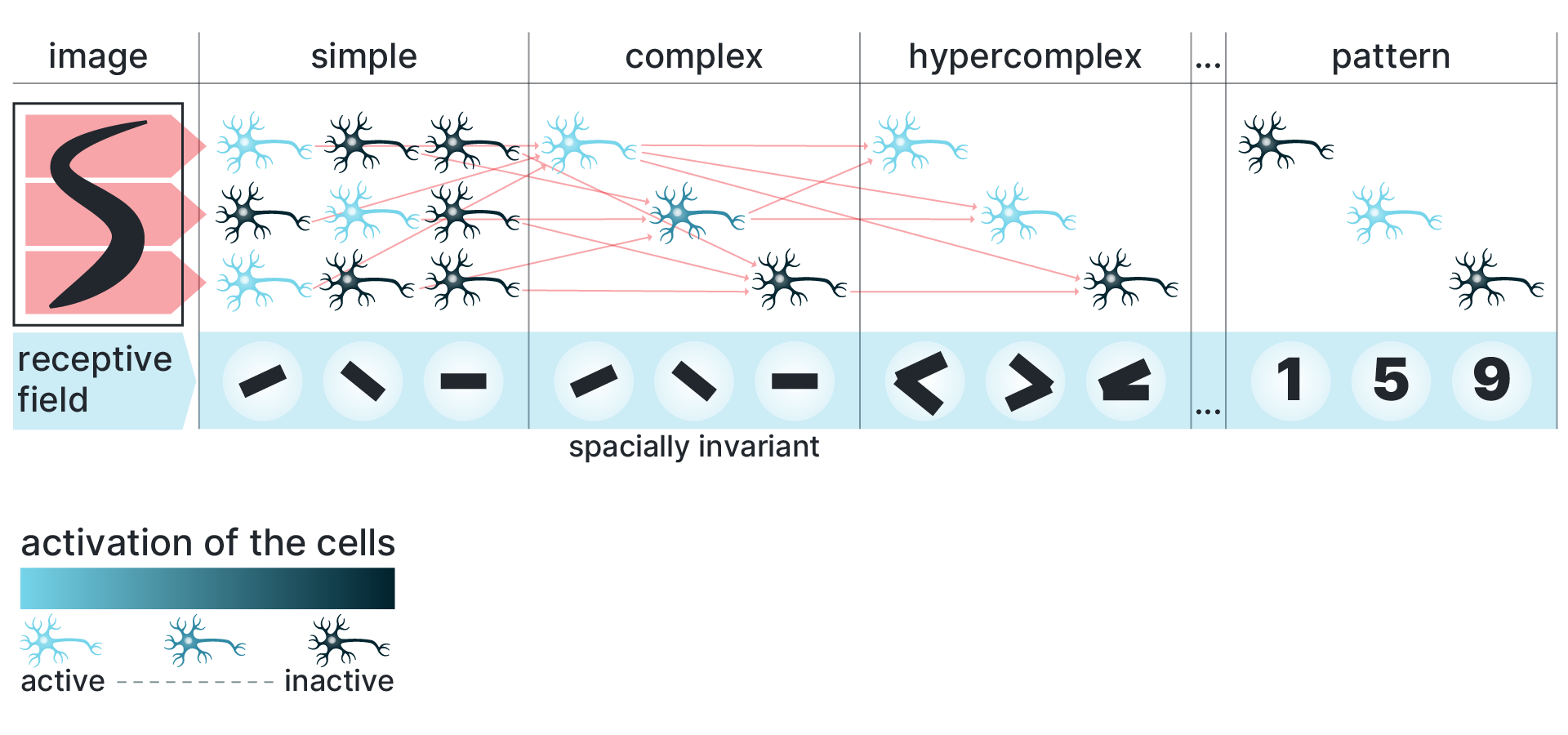
Hierarchical feature selectivity in the visual cortex.
Cell colors indicate activation strength for the stimulus shown below (red = strong response; darker tones = weaker or no response). The lines and shapes beneath each column depict the visual features that optimally activate each cell type, from simple oriented edges to more complex configurations. The final stage does not yet represent a single object identity. Instead, the activated features are compatible with multiple possible patterns (i.e. 1, 5 or 9)
In this diagram, each cell type is represented schematically to show what kind of visual stimulus it responds to. The lines and shapes beneath each cell indicate the stimulus features that elicit a response, while the cell’s position reflects how sensitive it is to stimulus location within the visual field. Importantly, as processing moves from simple to complex cells, responses become less dependent on exact position — a property known as spatial invariance.
· Simple cells: Fire in response to very specific shapes, such as lines or oriented edges, at precise locations within the visual field.
· Complex cells: Aggregate inputs from multiple simple cells. They respond to oriented stimuli across a broader area and are often sensitive to motion but less dependent on exact position.
· Hypercomplex (end-stopped) cells: Tuned for line endings, corners, or specific movement directions. Respond to more complex features, such as line endings, corners, or moving stimuli of specific lengths. These cells help detect more abstract or bounded visual features.
With these findings, Hubel and Wiesel showed that, in just a few steps, the visual system transforms the detection of individual photons into the perception of lines, edges, and motion. As a fundamental principle, from lower to higher processing stages, visual information flows in a feedforward manner through a hierarchy of increasingly complex feature detectors, each layer building upon the previous one. Simple cells detect edges and orientations; complex cells integrate them into motion and shape; and higher-order cells combine these elements into meaningful patterns.
Of course, we’re still far from recognizing the full richness of real-world visual objects, but it’s now easy to imagine how that happens: by sequentially integrating information along neural pathways to extract increasingly complex features. This is, in essence, the transformation from simple signals into structured, meaningful information that allows us to interpret and interact with the world.
Within the neuroscience community, those studying hierarchical feature analysis came to be playfully known as “feature creatures.” In formal recognition of their groundbreaking contributions, David Hubel and Torsten Wiesel were awarded the Nobel Prize in Physiology or Medicine in 1981.
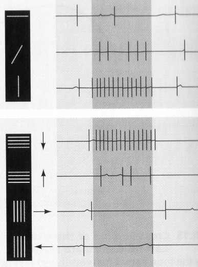
Hubel & Wiesel Recording of specialized neurons that only respond to specific orientation of a line.
By Jimmierock - Own work, CC BY-SA 3.0, https://commons.wikimedia.org/w/index.php?curid=18671256
Pushing the logic of hierarchical feature detection to its extreme leads to the idea of a neuron that would respond only to a single, highly specific stimulus. Jerry Lettvin coined the term “grandmother cells” to jokingly describe a hypothetical neuron that responds only to a highly specific and complex stimulus — such as the face of one’s grandmother.
In this logic, sensory processing proceeds through hierarchical stages, with neurons responding to progressively more complex features: from edges, to shapes, to objects, and eventually to unique individuals.
A “grandmother cell” would sit at the top of this hierarchy, firing only when your actual grandmother appears. But the term also served as a quiet warning: this line of thinking leads to a theoretical dead end, the idea that neural processing simply culminates in the identification of increasingly specific object features. While conceptually provocative, the idea is now largely viewed as an oversimplification. Object recognition is thought to rely on distributed representations, where patterns of activity across many neurons (not a single cell) encode the identity of complex stimuli. Still, the grandmother cell remains a useful metaphor for exploring the limits of neural specificity and the logic of hierarchical representation. Yet in the early 21st century, functional imaging technologies brought renewed attention to this debate. Researchers discovered neurons in the brain that responded selectively to specific individuals—such as Jennifer Aniston. This neuron, famously dubbed the “Jennifer Aniston neuron,” also responded to her Friends co-star, Lisa Kudrow. Similarly, a neuron tuned to images of Luke Skywalker also responded to Yoda. These findings revived an old question: do individual neurons represent specific objects, or do they participate in broader conceptual networks? The results led to the proposal that such neurons encode not isolated perceivable objects, but networks of semantically related concepts—so-called concept cells. These cells appear to bind perception, memory, and meaning, suggesting that we do not store every detail of experience, but instead structure memory through associative networks, where what we remember is shaped by what matters to us. Crucially, this highlights the interplay of memory and emotion. In this light, Lettvin’s notion of the “grandmother cell” appears more insightful than it may have seemed at the time. Perhaps that is why, even in science, the grandmother cell remains an idea we never quite forget.1
Author: Fabian Müller
Suggested Media
Watch the video under the following link if you want to see how Hubel and Wiesel’s experiment actually looked. Disclaimer for cat lovers: this historical footage involves invasive procedures that may be uncomfortable to watch, though they were standard practice in early neuroscience research. https://www.youtube.com/watch?v=IOHayh06LJ4
-
Cf. Quiroga, R. Q., Reddy, L., Kreiman, G., Koch, C. & Fried, I. Nature 435, 1102–1107 (2005). ↩